
Abstract
Nine wild American white ibis (Eudocimus albus) were found deceased or were moribund and subsequently euthanized within 24 hours after exposure to theatrical fog containing propylene glycol and triethylene glycol at a Halloween event at a zoological institution. Gross examinations revealed that all birds had congestion, edema, and hemorrhage throughout the lungs. Histologically, all birds had pathologic changes within the trachea and lungs indicative of acute respiratory insult. Microscopic changes in the trachea included segmental to diffuse epithelial attenuation with loss of cilia, alternating with regions of goblet cells filled with abundant mucus. In the lungs, all birds had perivascular edema and degenerative changes to the epithelium lining primary and secondary bronchi including hypereosinophilia and apical cytoplasmic blebbing of bronchial epithelial cells. In addition, in the lungs of 4 birds with longer intervals between exposure and death, there was granulomatous pneumonia, heterophilic perivascular cuffing, and multifocal bronchial epithelial necrosis. Propylene glycol and triethylene glycol were detected in lung and kidney tissues by gas chromatography-tandem mass spectrometry (GC-MS/MS). Traces of oxalic acid were found, along with presence of glycolic acid. While exposure to aerosolized glycols has been shown to cause irritation and minor degenerative changes to the respiratory epithelium in laboratory animals and humans, this study represents a unique investigation into the first reported incidence of acute inhalation toxicity and death following exposure to aerosolized glycol-containing theatrical fog in birds.
Keywords
Glycols like triethylene glycol (TEG) and propylene glycol (PG) are organic compounds belonging to the alcohol family, characterized by 2 hydroxyl (OH) groups attached to adjacent carbon atoms. 24 PG (1,2-propanediol) is known to be one of the least toxic of the glycols. 24 It is used as an automotive antifreeze, an organic solvent, a vehicle for drug delivery, and as an additive in cosmetics and processed food.4,15,24 Metabolism commences within 2–4 hours post-exposure with the majority of PG and metabolites excreted within 24–48 hours.4,15 In mammals, following ingestion, PG is rapidly absorbed from the gastrointestinal tract and a portion is metabolized by the liver to lactic acid by alcohol dehydrogenase and further metabolized to pyruvic acid, while part is eliminated unchanged in the urine. 24 In all acute animal studies, large doses are required to demonstrate any effect, and lethality was demonstrated only at very high oral doses with a median lethal dose (LD50 [lethal concentration killing half of animals]) of 20–30 g/kg; however, when ingested in large quantities, PG can cause central nervous system depression and lactic acidosis. 15 With high level chronic inhalation exposure, PG has been observed to act as a dehydrating agent on peripheral tissues leading to nasal hemorrhage. 24 In laboratory rodent studies, prolonged exposure to aerosolized PG has resulted in goblet cell hyperplasia and degenerative changes to the upper respiratory tract.14,15,25
TEG is a colorless, odorless, viscous liquid, used as a plasticizer in air sanitizer products, and as a desiccant for natural gas and air conditioning systems.2,14 TEG is often used in conjunction with PG as a base for smoke or fog machine fluid in the entertainment industry.2,14 As a polymer of ethylene oxide, initiated by water or ethylene glycol condensation, TEG may be considered one of the simplest polyethylene glycols. 7 Similar to PG, a portion of polyethylene glycols are metabolized by alcohol dehydrogenase in the liver; however, the majority is excreted unchanged in the urine.2,3 Toxicity decreases substantially with increasing molecular weight, and TEG is generally thought of as safe with very low toxicity, based on laboratory animal studies (LC50 of TEG aerosol is thought to be above 4400 mg/m3).1,2,7,14,23 Though uncommon, toxicity associated with polyethylene glycols includes central nervous system depression, serum hyperosmolality, metabolic acidosis, and renal failure.2,22,29 With chronic high aerosol exposure to TEG, laboratory rodents exhibit signs of respiratory disease, including coughing, wheezing, and shortness of breath.2,3,14
Though PG and TEG toxicoses have not been described in avian species, much less by an aerosol route, it is well known that avian species are uniquely sensitive to airborne irritants and toxins due to specific anatomic and physiologic features of their specialized respiratory tract.16,22 In contrast to mammals, birds maintain continuous circulation of oxygenated air through their respiratory tract and have an exceptionally thin blood-gas barrier, causing increased sensitivity to inspired toxicants and making the avian respiratory surface more liable to damage from airborne toxicants.12,22 Birds’ sensitivity to toxicants is further increased by their small size, rapid metabolic rate, and low body fat content.12,16
In this study, a unique gas chromatography-tandem mass spectrometry (GC-MS/MS) methodology was developed to detect glycol compounds in lung and kidney tissue. In addition to toxicologic analysis, this study provides a comprehensive description and interpretation of the associated gross and histopathologic findings in nine ibis following exposure to aerosolized PG and TEG.
Materials and Methods
Ibis were numbered from 1 to 9 based upon the time of the exposure event to when they were found dead/euthanized with case 1 dying soonest after the exposure event, and case 9 being the last case to die in the series. Over the course of 24 hours following exposure to the fogging agent, five ibis were found dead (cases 1, 5, 6, 8, and 9) and four were euthanized by administration of intravenous Euthasol solution (cases 2, 3, 4, and 7), which contains 18% PG. Necropsies performed on each of the nine deceased ibis at the submitting zoological institution on October 22, 2021. For each bird, samples from major organs were fixed in 10% neutral-buffered formalin, routinely processed for microscopic examination, stained with hematoxylin and eosin, and reviewed by several veterinary pathologists. In addition, the lungs of 10 ibis that died of non-respiratory-related disease were examined grossly and histologically. Pooled tracheal tissue from all birds stored at −80° was tested by polymerase chain reaction (PCR) for highly pathogenic avian influenza.
Fresh-frozen kidney and lung samples from the ibis that died following exposure to glycol-containing fog were submitted to the Michigan State University Veterinary Diagnostic Laboratory for GC/MS-MS toxicologic analysis for glycol compounds and metabolites. Toxicologic analysis was performed on the lung and kidney from each bird. In the initial preparatory step, a 3 g of sample of lung or kidney was combined with 2 ml of Milli-Q (Millipore, Billerica, MA) water and homogenized in a Precellys Evolution homogenizer (Bertin Technologies, Redondo Beach, CA). The resulting homogenate was split between separate glass tubes each with an additional 4 ml of Milli-Q water for acid (pH = 3.0 glycine buffer) and basic (pH = 10.0 glycine buffer) extractions. Twelve milliliters of acetonitrile (Honeywell, Burdick and Jackson, Muskegon, MI) and 5 g of sodium chloride (VWR, Radnor, PA) were mixed in the tubes and centrifuged in a Beckman GS-6 centrifuge (Beckman Coulter, Brea, CA) at 4000 rpm for 15 minutes, after which the acetonitrile top layer was transferred to a QuEChERS cleanup tube (2.0 mg magnesium sulfate, JT Baker, Phillipsburg, NJ; 300 mg octadecyl, Sigma Aldrich, St. Louis, MO; 300 mg Bondesil, Agilent, Santa Clara, CA). Each extract tube was then washed with an additional 12 ml of acetonitrile and combined with the initial extract. Twenty microliters of dimethylformamide (Sigma Aldrich, St. Louis, MO) was added to acidic and basic preparations. Solvent volumes were reduced under a stream of nitrogen gas to approximately 10 ml, placed in a −80°C freezer for 1 hour, and centrifuged at 4000 rpm for 15 minutes. Next, the organic solvent layers were filtered through Acrodisc filters (Pall Corporation, Port Washington, NY) into new tubes. Solvents were evaporated to dryness, and the extracts were reconstituted in 150 μl of acetonitrile. Twenty-five microliters of kidney or lung extracts were combined with 10 μl anhydrous pyridine (Sigma Aldrich) and 25 μl N-methyl-N-tert-butyldimethylsilyltrifluoroacetamide plus 1% tert-butyldimethylchlorosilane (MTBSTFA + 1% tBDMCS: Sigma Aldrich) for t-butyldimethylsilyl (tBuDMS) derivatization in separate autosampler vials equipped with 200 μl glass inserts at 70°C–75°C for 45 minutes.
A method specific for glycol-related compounds was developed for GC-MS/MS. Analysis was restricted to tBuDMS derivatives of the base extract of each sample type. The Agilent 7890/7000 GC-MS/MS (Agilent, Santa Clara, CA) was equipped with a 15 m × 250 μm × 0.25 μm Phenomenex (Torrance, CA) ZB-CL Pesticides-1 capillary column connected via a T connector to a 30-m column of the same type. The column flow rates were 1.2 and 1.4 ml/min He, respectively. Following splitless 1 μl injection, the oven temperature was programmed as follows: 70°C held for 2 minutes, then 5°C/minutes to 180°C for 0 minutes, then 120°C/minutes to 300°C for 5 minutes, for a total 30-minute runtime. The inlet was at 210°C initially, held for 20 minutes, then programmed at 200°C/minutes to 280°C for 12 minutes. Column 1 flow was reversed at the end of the analysis for backflush. A multiple reaction monitoring method was designed, and the MS acquisition parameters are listed in Supplemental Table S1. Standards were configured as 1, 5, 10, 25, and 50 ppm in acetonitrile and derivatized for tBuDMS derivatives in the same manner. Quantitation was achieved by interpolation on a standard curve with the aid of Agilent Mass Hunter software. Compound identity was assured by comparison of sample retention times to those of standard compounds, as well as by verification that qualifier multiple reaction monitorings occurred at the same retention time and in the same specific ratios to the quantifier ions (Supplemental Figure S1).
Twelve random samples submitted to our lab for routine GC-MS analysis and not expected to contain any of the target compounds were also tBuDMS-derivatized in the same manner and analyzed to assure the ability of the GC-MS/MS method to distinguish negatives. These were dog and cat liver samples. These samples were negative for ethylene glycol, PG, diethylene glycol, and TEG. Two of 12 showed traces of plant product (oxalic acid) as well as the ethylene glycol metabolite glycolic acid.
All birds in this study were wild and died naturally or were found obtunded and euthanized due to poor prognosis.
Results
Theatrical fog containing TEG and PG was used at a Halloween event that occurred at a zoological institution the evening of October 21, 2021. Following suspected natural exposure to the fog, three mature wild American white ibis (Eudocimus albus) were found recumbent on the ground that evening and were hospitalized. One died overnight, and the and the two others were unresponsive and euthanized the next day (October 22, 2021). Throughout the day on October 22, 2021, four additional ibis were found deceased, and two more were found unresponsive and were euthanized. A total of one male and eight female ibis were necropsied October 22, 2021 between 11:45
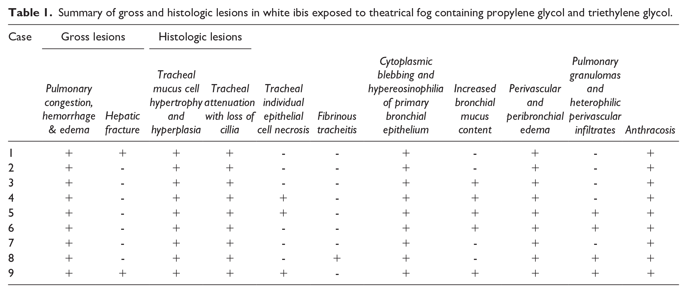
A summary of necropsy and histologic findings is available in Table 1. All ibis carcasses were in good nutritional condition and had pulmonary congestion, hemorrhage, and edema (Fig. 1). Two individuals (cases 1 and 9) also had evidence of acute hepatic fracture (presumably from falling from the roosting site).
Summary of gross and histologic lesions in white ibis exposed to theatrical fog containing propylene glycol and triethylene glycol.
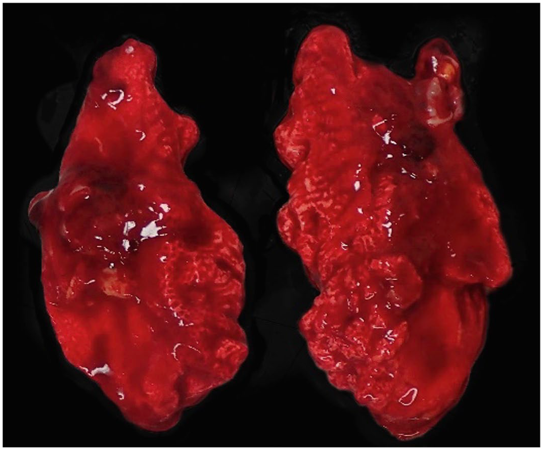
Postmortem lung lesions from an American white ibis, representative of the congestion, hemorrhage, and edema observed in the lungs of all nine birds that died or were found moribund and euthanized within 24 hours following exposure to glycol-containing theatrical fog. Case 6.
Histologically, all birds exhibited pathologic changes within the trachea and lungs. In the trachea of all ibis, there were multifocal regions of hypertrophy and hyperplasia of tracheal alveolar mucus glands characterized by increased numbers of goblet cells and increased mucus content both within cells and along the apical surface of mucosa. These regions alternated with regions of coalescing (cases 1–4) to extensive (cases 5–9) attenuation of the tracheal epithelium with loss of cilia (Fig. 2a). In three birds (cases 4, 5, and 9), there was rare individual cell necrosis of the tracheal epithelium. In case 8, the apical surface of the tracheal epithelium was lined by a thick band of eosinophilic fibrillar material (fibrin) with occasional interspersed syncytial cells and moderate numbers of heterophils and fewer lymphocytes and plasma cells expanding the lamina propria (Fig. 2b).
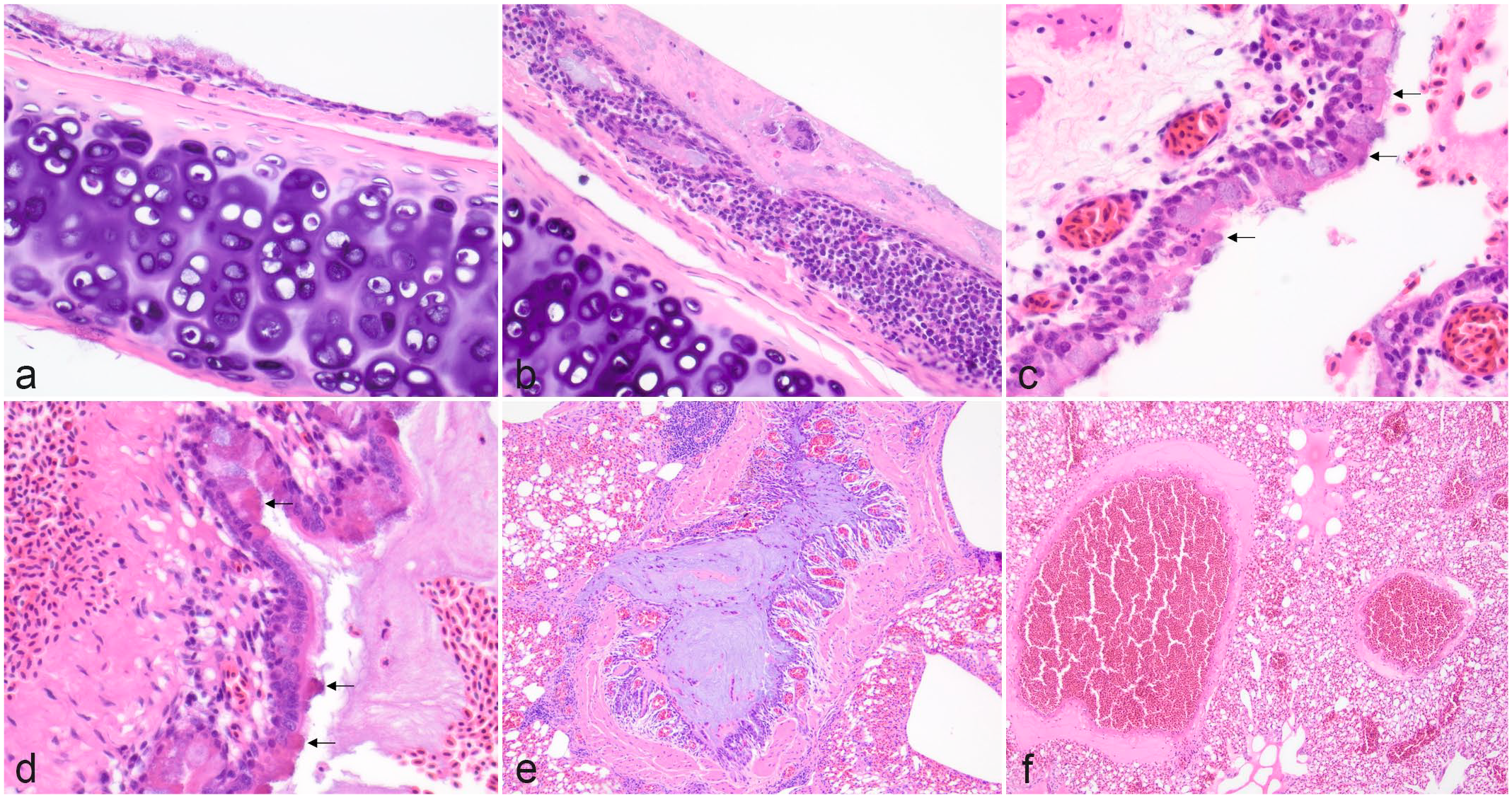
Histopathologic changes to the respiratory epithelium in the trachea and lungs of American white ibis exposed to glycol-containing theatrical fog. Hematoxylin and eosin. (a) Trachea with alternating regions of epithelial attenuation, loss of cilia, and mucous cells filled with mucus. Case 9. (b) Tracheal epithelium lined by a thick band of eosinophilic fibrillar material (fibrin) with syncytial giant cells. The lamina propria is expanded by moderate numbers of heterophils and fewer lymphocytes and plasma cells. Case 8. (c) Bronchus with peri-bronchial edema and hypereosinophilia and blebbing of the apical cytoplasm of bronchial epithelial cells (black arrows) with occasional nuclear fragmentation and goblet cells filled with mucus. Case 3. (d) Bronchus with peri-bronchial edema, blebbing and hyperesoinophilia of the apical cytoplasm of the bronchial epithelium (black arrows), and goblet cells filled with abundant mucus and intraluminal mucus. Case 9. (e) Secondary bronchus plugged by mucus with embedded fibrin, karyorrhectic nuclear debris, and heterophils. Case 6. (f) Edema surrounding medium caliber pulmonary vessels and filling a tertiary bronchus. Case 2.
In primary bronchi in all cases there was multifocal to coalescing, hyalinosis (hypereosinophilia), and blebbing of the apical cytoplasm of bronchial epithelial cells (Fig. 2c, d). In all individuals, there was increased mucus within the lumen of bronchi, varying from strands of wispy basophilic material lining the apical epithelial surface of primary bronchi (cases 3–6, and 9) (Fig. 2d) to mucus that completely filled primary or secondary bronchi (cases 6 and 9) (Fig. 2e). In all birds, there was perivascular and parabronchial edema (Fig. 2f).
In cases 5, 6, 8, and 9, there were occasional pulmonary granulomas composed of macrophages and fewer heterophils surrounding central foci of hypereosinophilic amorphous material and occasional fragments of green refractile material (Fig. 3a, b). These granulomas were adjacent to parabronchi and small vessels and were occasionally associated with necrosis of the bronchial epithelium (Fig. 3c). In cases 5, 6, 8 and 9, heterophils and fewer lymphocytes and plasma cells surrounded medium caliber blood vessels adjacent to regions of granulomatous inflammation (Fig. 3d). Grocott’s methenamine silver and Zeihl Neelsen acid-fast stains of sections of lung did not reveal any fungal or mycobacterial organisms within granulomas. Mild anthracosis was noted surrounding bronchi in all birds. For all birds, there were no additional gross or histopathological findings of significance in tissues examined including the heart, lungs, brain, liver, spleen, kidney, adrenal glands, thyroid glands, and gastrointestinal tract. In the control ibis, the lungs were light pink grossly, and histologically did now show evidence of pulmonary edema, perivascular infiltrates, granulomatous inflammation, or epithelial degeneration. All ibis tested negative for highly pathogenic avian influenza by PCR on pooled tracheal tissue.

Histopathologic lesions in the lungs of American white ibis exposed to glycol-containing theatrical fog. Hematoxylin and eosin. (a) Multifocal granulomas with dense perivascular aggregates of heterophils and fewer mononuclear inflammatory cells in the lungs. Case 9. (b) Pulmonary granulomas comprised of epithelioid macrophages often surrounding fragmented refractile material and perivascular inflammation. Case 9. (c) Bronchus with necrosis of the bronchial epithelium and syncytia formation containing fibrin and cellular debris. Case 6. (d) Medium caliber pulmonary vessels are surrounded by large numbers of heterophils and fewer mononuclear inflammatory cells. Case 9.
Quantitative results of glycol analysis by GC-MS/MS for kidney and lung tissue are shown in Table 2. Concentrations of PG were above the limit of quantitation of 1 ppm (μg/g) in the lungs of three birds (cases 3,4, and 7). Concentrations of TEG were above the limit of quantification of 1 ppm (μg/g) in the lungs of eight birds (cases 2–9) and kidneys of eight birds (cases 1, 2, and 4–9). Concentrations of glycolic acid were above the limit of quantification of 1 ppm (μg/g) in the lungs of six birds (cases 4–9) and kidney of seven birds (cases 1 and 4–9). Concentrations of oxalic acid were above the limit of quantification of 1 ppm (μg/g) in the lung of case 2 and kidney of case 1. Neither ethylene glycol nor diethylene glycol were detected in samples of lung or kidney.
Quantitative results for propylene glycol, triethylene glycol, ethylene glycol, diethylene glycol, glycolic acid, and oxalic acid acquired by GC-MS/MS in lung and kidney tissue from nine American white ibis (Eudocimus albus). Results were derived with a limit of quantification of greater than or equal to 1 μg/ml.
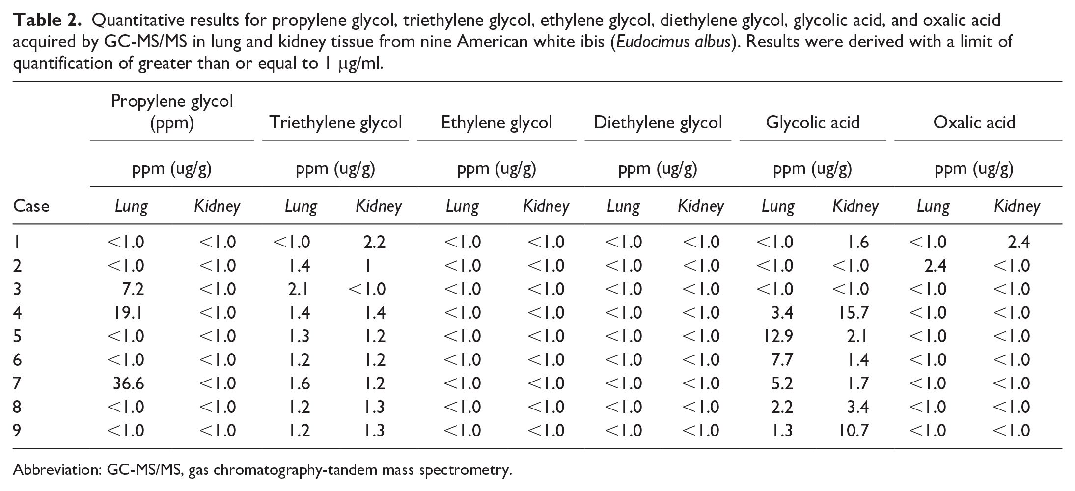
Abbreviation: GC-MS/MS, gas chromatography-tandem mass spectrometry.
Discussion
Detection of TEG and its metabolites in the lungs and kidneys strongly suggests inhalation exposure to TEG, which is known to be one of the primary ingredients, along with PG, in commercial fogging agents used by the submitting zoological institution surrounding this acute mortality event. TEG is suspected to be the primary compound responsible for toxicosis in this case, as it was above the limit of quantification in the lungs of all but case 1, and the kidneys of all but case 3. The presence of traces of ethylene glycol metabolites including oxalic acid in in the kidney of case 1 and lung of case 2 (early dying birds) suggests a pattern of metabolism of TEG ending with the well-known ethylene glycol metabolic route. This is further supported by the near universal presence of the intermediate compound glycolic acid, which was detected in lungs of ibis (cases 4–9) and kidney of 7 ibis (cases 1 and 4–9).
Algae and certain aquatic plants can form the basis of parts of the ibis diet. Oxalic acid is known to be deposited within algae of the Dasycladales and Spirogyra orders, for example. 18 Glycolic acid is also known to be produced by algae such as Chlorella. 26 Therefore, although the pattern of metabolites in the intoxicated ibis suggest a possible relationship to ethylene glycol metabolism (ethylene glycol → glycoaldehyde → glycolic acid → glyoxylic acid → oxalic acid), it is difficult to make this attribution definitively, particularly in light of the lack of further investigation of other ethylene glycol metabolites such as glycoaldehyde or glyoxylic acid, 8 and more importantly, the lack of euthanized unexposed ibis from the same population for metabolite quantitation.
PG was only detected in the lungs of birds that were euthanized by administration of intravenous Euthasol (cases 3, 4, and 7), which contains 18% PG. The lack of PG in case 2 (euthanized) can be explained statistically. PG is metabolized to lactic acid principally from the simple dicarboxylic acid pyruvate in various species. Values found for PG in euthanized animals vary over a wide range of 7–37 µg/g in lung tissue, giving an average of 21.0 ± 14.8 µg/g. If we surmise that case 2 is right at 1 µg/g, the average and standard deviation shift relatively slightly to 15.9 ± 15.7 µg/g. Given the wide range and high resultant standard deviation, it is not inconceivable that one bird in a population fell below the limit of quantitation, particularly given high rates of formation of lactate, an energy-providing compound, in avian species. 11 High turnover of PG to lactate likely explains the lack of PG in non-euthanized animals. The lack of TEG/PG-unexposed euthanized controls was a limitation in this study and confounds PG’s presence in lung tissue; however, histology from 10 ibis that died from non-respiratory-related causes between 2011 and 2024 showed no evidence of pulmonary edema, granulomatous inflammation, perivascular infiltrates, increased mucus production, or epithelial degeneration within the respiratory tract.
In glycol-exposed ibis, the degenerative changes to the epithelial lining of the upper and lower airways, pulmonary edema, perivascular heterophilic infiltrate, and mucus accumulation within airways are indicative of an acute respiratory insult and impairment of the muco-ciliary escalator. Interestingly, previous studies in rodents and lagomorphs have demonstrated toxicity from glycol inhalation to be of minimal concern; however, prolonged exposure to PG has resulted in goblet cell hyperplasia and degenerative changes to the upper respiratory tract, including similar apical cytoplasmic blebbing of epithelial cells, destruction of cilia, and increased mucus production.13,15,25 Lesions associated with acute inhalation toxicity in birds exposed to other aerosolized toxicants, such as polytetrafluoroethylene, include severe pulmonary edema, hemorrhage, and epithelial necrosis with loss of cilia.16,30 Avian influenza was considered as a possible cause of this acute mortality event; however, all ibis tested negative by PCR and did not have specific histologic lesions consistent with avian influenza.
The spectrum of lesions in the birds in this case series varied. Interestingly, four birds with a longer interval between exposure and death (cases 5, 6, 8, and 9) had multifocal granulomas within the lungs surrounding crystalline material, heterophilic cuffing around pulmonary vasculature, and rare necrosis of bronchial epithelium, characterized by cellular debris within bronchial. Multifocally, granulomas surrounded fragmented refractile green crystalline material similar in morphology to calcium oxalate crystals seen in the kidneys with ethylene glycol toxicity. The authors speculate these to be calcium oxalate crystals; however, the material in this case is not birefringent under polarized light and no crystalline material was observed in the kidneys.
While goblet cell hyperplasia and granulomas suggest a more chronic response, the authors suspect this may be due to repeated exposure of the glycol fog, as fogging machines had been used for approximately 6 events in the month prior to this event taking place. Conversely, the granulomas may be unrelated, and the crystalline material they surround may be another substance all together, such as diatomaceous earth which is used on the zoo grounds for pest control.
Because of their small size and the demands of flight, birds have elevated metabolic and respiratory rates, resulting in ventilation of a greater relative volume of air and higher doses of airborne contaminants on a body weight basis than mammals.10,21,30 As a result, birds have historically been used as indicators of air quality. For example, canaries were brought into coal mines to signal when concentrations of toxic gasses like carbon monoxide reached unsafe levels leading to the phrase “canary in the coal mine.” 20 Sensitivity to theatrical fog is suspected to be high as it is comprised of very small particles in a respirable range, that is, below 2.4 µm (small) and often below 1 µm (ultrafine) as defined by the Environmental Protection Agency, that can reach deep into the lungs and be absorbed or cause local effects.14,19
In humans, the most common use of theatrical fog is in the entertainment industry, where workers have the highest risk of work-related asthma.17,19,28 Performers exposed to theatrical fog experience a range of symptoms from mucous membrane irritation to upper airway/voice symptoms to central nervous system depression and lethargy.5,9,17,29 In one case report, multiple fire fighter recruits became ill following exposure to diethylene glycol fog (particle size ranged from 0.5 to 0.7 µm) to simulate fire conditions, and one was hospitalized for acute lipid pneumonia. 6
Because of the low vapor pressure of glycols, whole body aerosol exposure may result in wetting of the skin and feathers, and thus there is concern that in birds, preening may lead to combined inhalation and peroral exposure. 3 This has been studied with ethylene glycol in rats, where it was determined that exposure to an aerosol concentration of 2,500 mg/kg resulted in ingestion by preening of an additional dose of around 1,000 mg/kg.3,27 As such, it is important to consider the potential for absorbed combined dosages from both inhalation and peroral routes.3,27
Based on the findings in this study, careful consideration is warranted with regard to the use of glycol-containing theatrical fog in close proximity to avian species.
Supplemental Material
sj-pdf-1-vet-10.1177_03009858251338853 – Supplemental material for Acute inhalation toxicity in nine white ibis (Eudocimus albus) exposed to theatrical fog containing triethylene glycol and propylene glycol
Supplemental material, sj-pdf-1-vet-10.1177_03009858251338853 for Acute inhalation toxicity in nine white ibis (Eudocimus albus) exposed to theatrical fog containing triethylene glycol and propylene glycol by Jayne S. Ellis, Richard M. Fulton, Andreas F. Lehner and John P. Buchweitz in Veterinary Pathology
Footnotes
Acknowledgements
The authors would like to acknowledge the submitting zoological institution for supplying documentation and photographs of gross findings and histopathology slides. The authors would also like to thank Kurt Williams, DVM, PhD, DACVP for review of the gross and histopathologic findings.
Author Contributions
JSE and RMF reviewed and interpreted the gross and histopathologic findings. JSE captured and edited the photomicrographs and wrote the manuscript with input from RMF, AFL, and JPB. AFL and JPB developed the methodology and performed the toxicologic analysis.
Declaration of Conflicting Interests
The author(s) declared no potential conflicts of interest with respect to the research, authorship, and/or publication of this article.
Funding
The author(s) received no financial support for the research, authorship, and/or publication of this article.
Supplemental material for this article is available online.
References
Supplementary Material
Please find the following supplemental material available below.
For Open Access articles published under a Creative Commons License, all supplemental material carries the same license as the article it is associated with.
For non-Open Access articles published, all supplemental material carries a non-exclusive license, and permission requests for re-use of supplemental material or any part of supplemental material shall be sent directly to the copyright owner as specified in the copyright notice associated with the article.