
Abstract

C. Myocardial adenomatoid tumor.
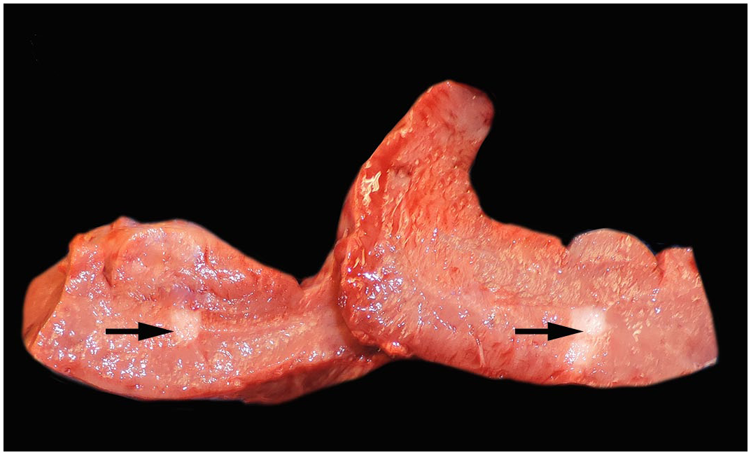
Bovine myocardial adenomatoid tumors are benign and usually incidental findings characterized by cuboidal epithelial cells arranged in tubules, acini, and channels within a dense fibrous stroma in the myocardium. Grossly, these lesions appear as tan-white, firm, plaque-like foci within the left ventricular wall (Image 1, arrows). Cells are immunoreactive for cytokeratin, vimentin, CD30, calretinin, and WT1. The cells are thought to be of mesothelial origin based on immunohistochemistry and ultrastructure analysis. Angiomatosis consists of proliferation of blood vessels. Thyroid adenoma would have more regular acini and luminal colloid. Cardiac mesothelial hyperplasia is characterized by frond-like projections of mesothelium.
Additional reading: Sosa E, Giannitti F, Macías-Rioseco M, et al. Congenital neoplasms in cattle: a literature review and multi-institutional case series. J Vet Diagn Invest. 2025;37(4):559–573.
Contributor: Julie Bedwani, University of Illinois Urbana-Champaign
B. Ureaplasma diversum .
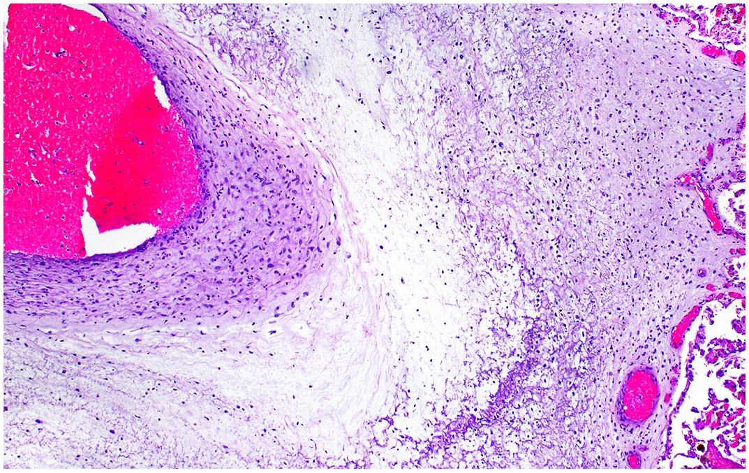
Bovine abortions caused by Ureaplasma diversum typically occur during the third trimester. Histology commonly reveals necrotizing placentitis with moderate to marked expansion of the chorioallantoic stroma by edema, fibrin, few mixed inflammatory cells (neutrophils, lymphocytes, macrophages), and necrotizing arteritis (Image 2), as well as diffuse non-suppurative interstitial pneumonia with marked bronchus-associated lymphoid tissue hyperplasia. Additional findings include arthritis and erosive non-suppurative conjunctivitis. Lesions in other organs have not been described. A diagnosis is supported by positive PCR and/or culture in conjunction with compatible histological lesions. Brucella, Leptospira, and Listeria are not known to cause conjunctivitis.
Additional reading: Santos Junior MN, Macêdo Neres NSD, Campos GB, et al. A review of Ureaplasma diversum: a representative of the mollicute class associated with reproductive and respiratory disorders in cattle. Front Vet Sci. 2021;8:572171. doi:10.3389/fvets.2021.572171.
Contributor: Alvaro Wehrle-Martinez, Massey University, New Zealand
C. Pyrrolizidine alkaloid toxicity.
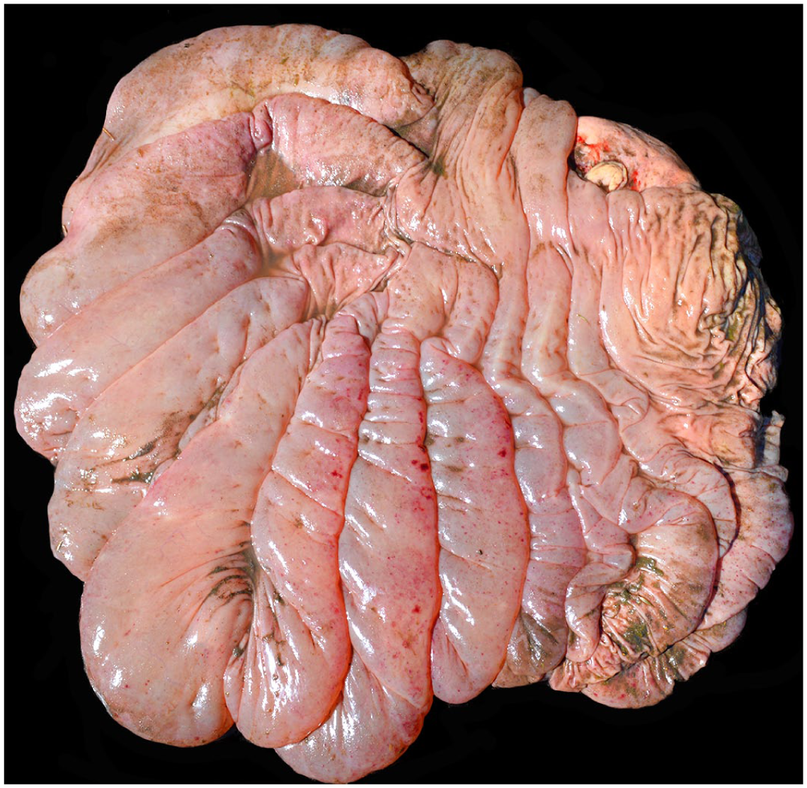
Chronic pyrrolizidine alkaloid toxicity results in chronic hepatopathy characterized by hepatocellular loss and nodular regeneration, with megalocytosis as a characteristic histologic feature, accompanied by periportal to bridging fibrosis and bile duct proliferation. These changes lead to portal hypertension and hypoproteinemia, resulting in edema and ascites. Abomasal edema (Image 3) is common in animals suffering from chronic intoxication with plants containing pyrrolizidine alkaloids (such as Senecio spp). Ruminal acidosis may cause chemical rumenitis but does not result in abomasal edema. Bovine leukosis virus can cause abomasal lymphoma. Clostridium perfringens type D produces enterotoxemia with intestinal, pulmonary, or central nervous system, but not abomasal lesions.
Additional reading: Cullen JM, Stalker MJ. Liver and biliary system. In: Maxie MG, ed. Jubb, Kennedy & Palmer’s Pathology of Domestic Animals. Vol. 2. 6th ed. Amsterdam: Elsevier; 2016:336–338.
Contributor: Ignacio Alvarez, Alvarez Diagnostic Laboratory, Bahia Blanca, Argentina
B. Hardware disease.
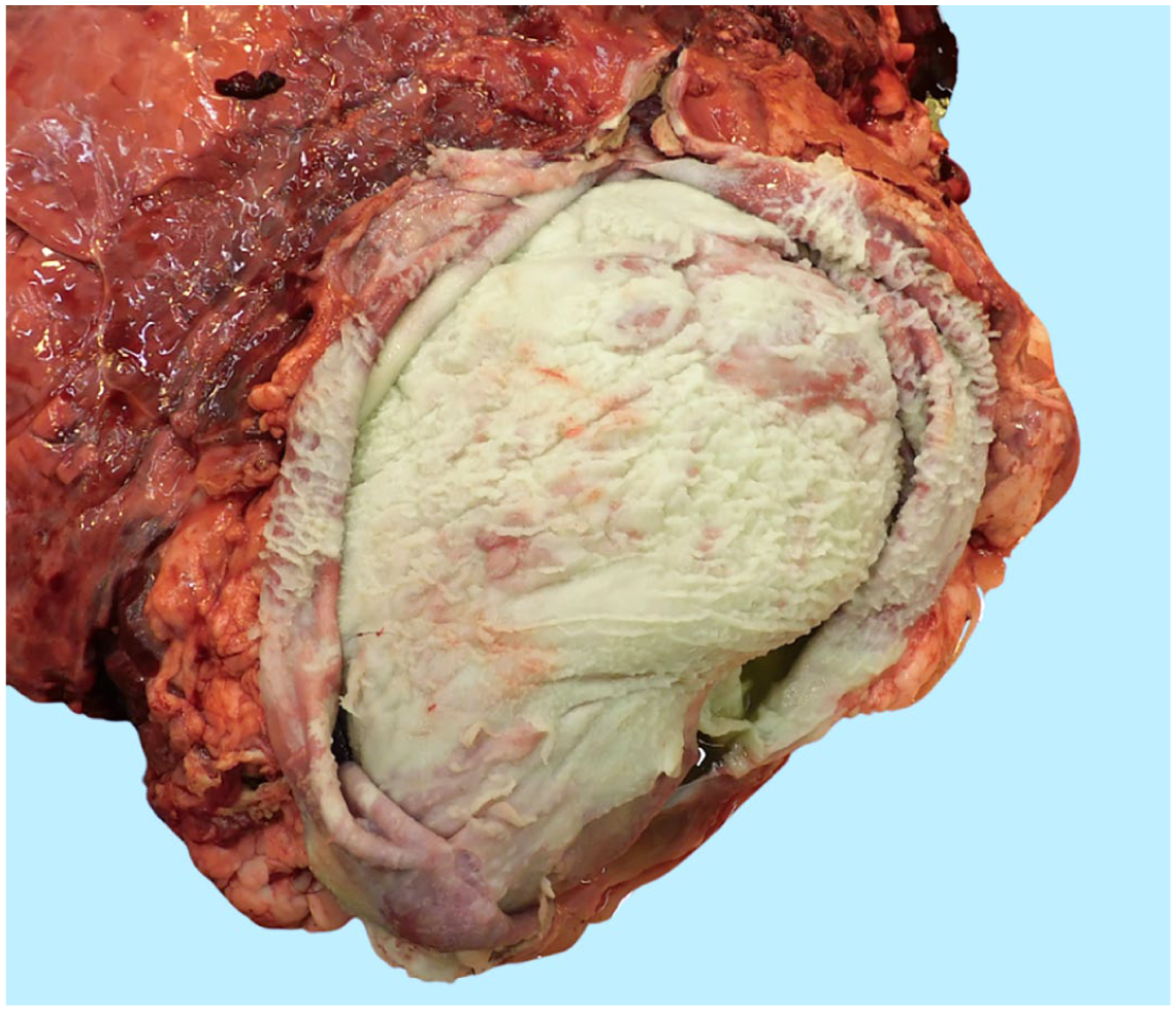
In cattle, hardware disease (traumatic reticuloperitonitis) occurs when a sharp and thin foreign body, often a metal wire or nail, penetrates the reticular wall, usually in the cranioventral direction. When the foreign body advances further cranially and extends through the diaphragm, it may perforate the pericardium, resulting in traumatic pericarditis. The pericardial reaction tends to present as copious and fibrinopurulent pericardial material (Image 4). Brisket and heartwater disease present with hydropericardium without fibrinous pericarditis. Mulberry heart disease is a term classically applied to pigs with vitamin E/selenium deficiency and presents as suffusive hemorrhages on the epicardium.
Additional reading: Uzal FA, Plattner BL, Hostetter JM. Alimentary system. In: Maxie MG, ed. Jubb, Kennedy and Palmer’s Pathology of Domestic Animals. Vol. 2. 6th ed. Amsterdam: Elsevier; 2016:38–39.
Contributor: Cassandra M. Powers, California Animal Health and Food Safety Lab
Veterinary Pathology invites submission of exceptional gross or microscopic images for consideration as an Image Challenge, along with a multiple-choice question and answer. For details, see the Instructions to Authors on the journal website.