
Abstract
Clinical History and Gross Findings
Between July 2024 and September 2025, veterinarians reported deaths in feedlot cattle from 20 commercial operations across 15 municipalities in Mato Grosso do Sul, Brazil. Most affected animals were Nelore or crossbred steers and heifers aged 10 to 24 months, although one outbreak involved 4-month-old calves.
All herds received diets containing cottonseed and/or cottonseed meal, with an increase of at least 30% of these components in the diet compared to previous years. Clinical illness typically emerged 8 to 30 days after feedlot entry. Herd size ranged from 50 to 22,000 cattle, and morbidity and mortality varied widely (0.3%–30% and 0.15%–18.3%, respectively).
The disease progressed acutely, with most animals dying within 1 to 7 days after clinical signs onset. Clinical signs included apathy, reduced feed intake, depression, reluctance to walk, difficulty rising, and delayed responses. Some cattle also had diarrhea, regurgitation, dehydration, and dyspnea. Therapeutic attempts varied among operations and included antimicrobials, anti-inflammatory drugs, and supportive therapy. Reported medications included tilmicosin, penicillin, tetracycline, tulathromycin, oxytetracycline, ceftiofur, flunixin meglumine, dexamethasone, and dipyrone. In several outbreaks, treatments were not recorded.
Clinical pathology results were obtained from 36 cattle across 7 feedlots. Aspartate aminotransferase ranged from 37.5 to 690.2 UI/L (reference: 54–135 UI/L), creatine kinase from 137.6 to 27,125.4 UI/L (88–292 UI/L), creatinine from 1.0 to 30.5 mg/dL (0.4–0.9 mg/dL), phosphorus from 3.4 to 47.6 mg/dL (4.1–7.3 mg/dL), and urea from 26.9 to 519.3 mg/dL (7–19 mg/dL).
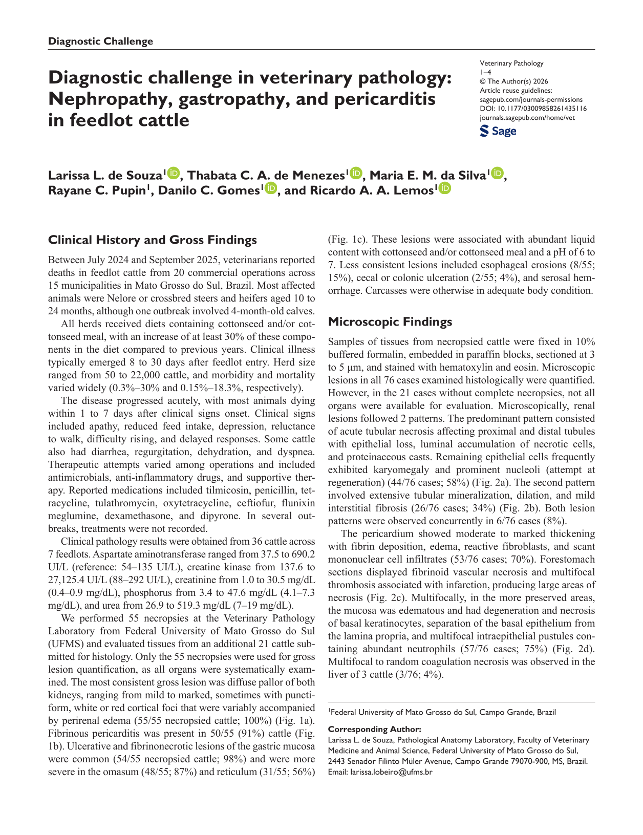
We performed 55 necropsies at the Veterinary Pathology Laboratory from Federal University of Mato Grosso do Sul (UFMS) and evaluated tissues from an additional 21 cattle submitted for histology. Only the 55 necropsies were used for gross lesion quantification, as all organs were systematically examined. The most consistent gross lesion was diffuse pallor of both kidneys, ranging from mild to marked, sometimes with punctiform, white or red cortical foci that were variably accompanied by perirenal edema (55/55 necropsied cattle; 100%) (Fig. 1a). Fibrinous pericarditis was present in 50/55 (91%) cattle (Fig. 1b). Ulcerative and fibrinonecrotic lesions of the gastric mucosa were common (54/55 necropsied cattle; 98%) and were more severe in the omasum (48/55; 87%) and reticulum (31/55; 56%) (Fig. 1c). These lesions were associated with abundant liquid content with cottonseed and/or cottonseed meal and a pH of 6 to 7. Less consistent lesions included esophageal erosions (8/55; 15%), cecal or colonic ulceration (2/55; 4%), and serosal hemorrhage. Carcasses were otherwise in adequate body condition.
Most frequent macroscopic findings in mortality outbreaks in feedlot cattle. (a) Diffusely pale kidneys. (b) Heart. Pronounced fibrin adhered to the surface of the pericardium. (c) Omasum. A focally extensive area composed of irregular and ruptured folds, covered by a thick, whitish-brown material (necrosis).
Microscopic Findings
Samples of tissues from necropsied cattle were fixed in 10% buffered formalin, embedded in paraffin blocks, sectioned at 3 to 5 µm, and stained with hematoxylin and eosin. Microscopic lesions in all 76 cases examined histologically were quantified. However, in the 21 cases without complete necropsies, not all organs were available for evaluation. Microscopically, renal lesions followed 2 patterns. The predominant pattern consisted of acute tubular necrosis affecting proximal and distal tubules with epithelial loss, luminal accumulation of necrotic cells, and proteinaceous casts. Remaining epithelial cells frequently exhibited karyomegaly and prominent nucleoli (attempt at regeneration) (44/76 cases; 58%) (Fig. 2a). The second pattern involved extensive tubular mineralization, dilation, and mild interstitial fibrosis (26/76 cases; 34%) (Fig. 2b). Both lesion patterns were observed concurrently in 6/76 cases (8%).

Histological features of nephropathy and gastropathy in feedlot cattle. Hematoxylin and eosin. (a) Kidney. Numerous necrotic tubules (*) containing amorphous eosinophilic material, cellular debris, and residual tubular epithelial cells with karyomegaly and prominent nucleoli. Necrotic cells and debris completely occlude the tubular lumen (inset). (b) Kidney. Tubules with marked epithelial loss, scattered regenerating cells, or mineralization (arrows), interspersed with mild fibrous connective tissue. (c) Omasum. The lining epithelium is diffusely replaced by fibrin, cellular debris, and clusters of bacteria (arrows). Inset: a blood vessel in the lamina propria has fibrinoid necrosis and infiltration by degenerate neutrophils and mononuclear cells. (d) Rumen. Intraepithelial pustules composed of abundant neutrophils.
The pericardium showed moderate to marked thickening with fibrin deposition, edema, reactive fibroblasts, and scant mononuclear cell infiltrates (53/76 cases; 70%). Forestomach sections displayed fibrinoid vascular necrosis and multifocal thrombosis associated with infarction, producing large areas of necrosis (Fig. 2c). Multifocally, in the more preserved areas, the mucosa was edematous and had degeneration and necrosis of basal keratinocytes, separation of the basal epithelium from the lamina propria, and multifocal intraepithelial pustules containing abundant neutrophils (57/76 cases; 75%) (Fig. 2d). Multifocal to random coagulation necrosis was observed in the liver of 3 cattle (3/76; 4%).
Differential Diagnoses
The consistent epidemiologic pattern of multiple unrelated operations feeding cottonseed and/or cottonseed meal and reporting similar clinical and pathological features indicated foodborne toxicosis as the primary suspicion. Several nephrotoxicants can induce acute tubular necrosis, including heavy metals, mycotoxins, antimicrobial drugs, and toxic plants. 2
Heavy metals such as arsenic, lead, cadmium, and mercury cause tubular necrosis and, in some cases, forestomach injury.2,7,9 Heavy metal contamination is often linked to soil, water, pesticides, or industrial byproducts. 9 Although arsenic was initially suspected due to its ability to cause renal and forestomach necrosis, 7 analytical results revealed only trace levels in all tissues evaluated.
Mycotoxins such as citrinin or ochratoxin A can damage renal tubules, but ochratoxin A is primarily degraded in the rumen, reducing its relevance in cattle. 2 Mycotoxin concentrations in samples of all dietary components measured during the outbreak remained below European regulatory limits, 3 making this differential unlikely.
Among plant toxicities, oak poisoning produces both acute tubular necrosis and gastrointestinal ulceration, 6 but exposure was improbable because affected feedlots were geographically distant and used different feed suppliers. Tannins are present in high concentrations in this plant and are responsible for the toxic effects, but their concentration in cottonseed and/or cottonseed meal from one feedlot was less than 1%.
Fluoride poisoning, another cause of renal and gastrointestinal injury,1,4 was discounted because fluoride concentrations in tissues and drinking water were far below toxic thresholds.
Gossypol, a well-recognized toxicant in cotton byproducts, mainly causes cardiac and reproductive effects and is particularly harmful to monogastrics and calves. Adult ruminants exhibit partial resistance via ruminal binding to dietary proteins.5,10 Importantly, myocardial necrosis, the primary lesion of gossypol toxicosis, was absent in all outbreaks.
Further Investigations and Diagnosis
We conducted toxicologic investigations across 6 feedlot operations, evaluating blood, urine, liver, kidney, rumen contents, hair, water, and feedstuffs, including cottonseed and/or cottonseed meal. Inductively coupled plasma mass spectrometry (ICP–MS) quantified heavy metals, enzyme immunoassays (ELISA) were used to detect aflatoxins, fumonisins, and ochratoxin A, and fluoride concentrations were measured using an ion-specific electrode following hexamethyldisiloxane (HMDS)-facilitated diffusion. Although identical samples were not formally reanalyzed in a second laboratory, toxicologic testing was distributed across multiple independent laboratories according to sample type.
Heavy metal concentrations were uniformly low. Maximum detected values included arsenic at 1.48 µg/L in blood and 6.1 µg/kg in urine, mercury at 21 µg/kg in urine and liver, lead at 1.3 µg/dL in blood and 1.887 mg/kg in liver/kidney, and cadmium at 5 µg/kg in urine and liver. Chromium reached 0.8 µg/L in blood. Rumen, liver, kidney, and hair samples contained only trace arsenic (≤0.032 mg/kg; hair, 215 µg/kg), insufficient for acute or chronic intoxication.
Feed analysis of one feedlot revealed aflatoxins up to 3.63 ppb, fumonisins up to 990 ppb, and ochratoxin A up to 2.37 ppb, far below toxic limits for ruminants. Tannins in cottonseed and cottonseed meal of one feedlot reached only 0.4%. Water samples of 2 feedlots contained minimal contaminants (arsenic, 0.001 mg/L; lead, 0.008 mg/L; cadmium, 0.001 mg/L; mercury, 0.0001 mg/L; chromium, 0.01 mg/L; fluoride, 0.5 mg/L). Fluoride in the liver and kidney of 9 cattle from different feedlots remained extremely low (≤0.0017 µg/g).
Discussion
The epidemiologic, clinical, and pathological findings indicate a toxic syndrome characterized by acute nephropathy with prominent extrarenal lesions consistent with uremia. Acute renal failure allows accumulation of uremic toxins, which increase vascular permeability, impair endothelial function, and induce mucosal necrosis. 8 These mechanisms explain the fibrinous pericarditis and extensive fibrinonecrotic gastrointestinal lesions. Although chronic uremia typically produces more pronounced extrarenal lesions, the widespread vascular necrosis seen here suggests a particularly aggressive toxic insult.
We systematically considered multiple nephrotoxins. Heavy metals can induce tubular necrosis and gastrointestinal injury,2,7,9 but concentrations in all matrices were far below toxic thresholds. Mycotoxins were present at negligible levels and are unlikely to cause disease in ruminants due to ruminal degradation. 2 Antimicrobials and several toxic plants were plausible but lacked epidemiologic support. Oak toxicosis produces the closest lesion pattern, 6 yet exposure was implausible. Fluoride intoxication was ruled out by consistently low tissue concentrations.1,4 Gossypol toxicity was excluded based on the absence of myocardial necrosis and the relative resistance of adult ruminants.5,10
Despite extensive investigation, no recognized toxic agent accounted for the renal and gastrointestinal lesions. However, the epidemiologic link to cottonseed-based feeds remained compelling. All affected operations incorporated higher concentrations of cottonseed and/or cottonseed meal into finishing diets, and disease onset occurred during the first month on feed. The outbreaks occurred in geographically distant operations supplied by products of varying origin, with no consistent changes in feed appearance or odor. Although manufacturing processes could not be directly evaluated and a specific contaminant was not identified, the association between disease occurrence and cottonseed-based feeds was remarkably consistent. To our knowledge, no comparable syndrome combining severe renal tubular necrosis, fibrinous pericarditis, and forestomach infarction has been reported in association with cottonseed and/or cottonseed meal.
These findings have substantial practical importance, as morbidity and mortality reached levels capable of causing major economic losses in feedlot systems. The inability to detect a known agent underscores the limitations of targeted toxicologic assays and highlights the need for broader analytical approaches to detect emerging toxicants in plant-derived feeds.
Collectively, these outbreaks represent acute toxic nephropathy and gastropathy strongly associated with ingestion of cottonseed and/or cottonseed meal, although the causal agent remains undetermined.
Footnotes
Declaration of Conflicting Interests
The authors declared no potential conflicts of interest with respect to the research, authorship, and/or publication of this article.
Funding
The authors disclosed receipt of the following financial support for the research, authorship, and/or publication of this article: Financial support was supplied by Universidade Federal de Mato Grosso do Sul (UFMS/MEC) and Coordenação de Aperfeiçoamento de Pessoal de Nível Superior (CAPES, Brazil)—Finance Code 001. One of the authors (Ricardo A. A. Lemos) had a fellowship from Conselho Nacional de Desenvolvimento Científico e Tecnológico (CNPq), Brazil.