
Abstract
Clostridium chauvoei is an anaerobic bacterium, widespread in the environment, and one of the causes of gas gangrene in animals. We describe an outbreak of gas gangrene in 1- to 3-day-old suckling piglets in a commercial farrowing farm caused by C. chauvoei. Twenty-five litters were affected, resulting in 30 deaths and a ~1% increase in preweaning mortality. Affected piglets had lethargy; fever; anorexia; and purple discoloration of the abdomen, head, and limbs. Without intervention, the disease progressed to death or required euthanasia within 24 hours. Gross and microscopic findings included deep ulcers over the carpal and tarsal joints, sole erosions, muscle necrosis, and muscle and subcutaneous edema and emphysema with intralesional gram-positive rods. Polymerase chain reaction confirmed C. chauvoei infection. All cases occurred in piglets housed in pens with abrasive, solid concrete floors, and skin lesions caused by contact with those floors were presumed to be the port of entry for C. chauvoei.
Clostridia are anaerobic bacteria that can cause enteric, histotoxic, and neurotoxic infections. Gas gangrene, previously called malignant edema, which is a term no longer recommended to be used in veterinary medicine,12,15,16,18 is a histotoxic clostridial infection usually predisposed by traumatic injuries that facilitate the entry and growth of these bacteria in the tissues. As all gas gangrene etiological agents (Clostridium septicum, Clostridium chauvoei, Clostridium novyi type A, Clostridium perfringens type A, and Paraclostridium sordellii) are ubiquitous in the environment, tissue damage permits their entry into tissues and promotes localized anaerobiosis, thereby enabling bacterial proliferation.12,13,16,17,20 In swine, gas gangrene is rare, but when it occurs, it can have high lethality. 14 Most reported cases of gas gangrene in swine are attributed to C. septicum, as is also described in ruminants.13,16,17 However, any of the clostridial species mentioned above can cause gas gangrene, acting alone or in combination.12,13,16 Infections caused by C. chauvoei in pigs are poorly described and are mainly associated with contaminated injection sites when they occur. 17 Here, we describe the epidemiologic, clinic, and pathologic aspects of gas gangrene caused by C. chauvoei in suckling piglets.
The cases occurred in a commercial pig farm housing 150 lactating sows, with an average of 14.1 piglets born alive per sow, 12.6 piglets weaned per sow, a weaning age of 21 days, and 2.4 litters per sow a year. The average preweaning mortality was 10.5%. Half of the lactating sows (75 sows) were housed in pens with a plastic-suspended floor and the other half in 2.2 m × 0.6 m pens with a solid, deteriorated, and rough concrete floor, which allowed for the accumulation of organic matter. Sows were vaccinated against swine neonatal diarrhea (associated with Clostridium perfringens C, Escherichia coli, and rotavirus A) and reproductive failure (associated with porcine parvovirus, porcine circovirus-2, and Erysipelothrix rhusiopathiae). Piglets received an intramuscular iron injection at 3 days of age and were vaccinated against porcine circovirus-2 and Mycoplasma hyopneumoniae infections at 21 days of age.
Twenty-five litters of 1- to 3-day-old piglets were affected in 2 months, resulting in the death of 30 piglets, and an approximately 1% of increase in mortality compared with the period preceding the outbreak. No intramuscular injections were given to the piglets before the onset of the clinical signs. Clinical cases were observed exclusively in litters housed in the pens with solid concrete floors, with a significantly higher incidence in piglets born to lactating sows with high body condition scores, mainly scores of 4 or 5 (1, emaciated; 2, thin; 3, ideal; 4, fat; and 5, overly fat). Because of the high body condition score, these sows had difficulty adopting an appropriate posture while nursing, resulting in the piglets having to kneel on the rough concrete floor for suckling.
During a herd visit by the veterinarian in charge of the farm, a total of 63 piglets had clinical signs, 8 of which were selected for clinical evaluation. The 8 piglets had deep skin ulcers over the carpal joints (Fig. 1a), and 5 of those had similar lesions in the skin over the tarsal joints. They all had marked lameness, apathy, anorexia, and fever. Extensive dark purple discoloration involving the skin of forelimbs, hindlimbs, and ventral abdomen, and the hooves, was observed in 2 of the piglets with skin ulcers (Fig. 1b). Death or terminal disease necessitating euthanasia occurred in less than 24 hours if the animals were not treated. Piglets treated with intramuscular penicillin (25,000 IU) and dihydrostreptomycin sulfate (25 mg/kg) (Penfort PPU; Ourofino, Brazil), soon after the onset of clinical signs, frequently recovered. Two piglets that had severe clinical signs and that had been recently treated with penicillin and dihydrostreptomycin sulfate, as described above, were euthanized in extremis and immediately autopsied on the farm. Pigs were euthanized using an intravenous overdose of ketamine and xylazine, followed by the intravenous administration of a hypersaturated potassium chloride solution.
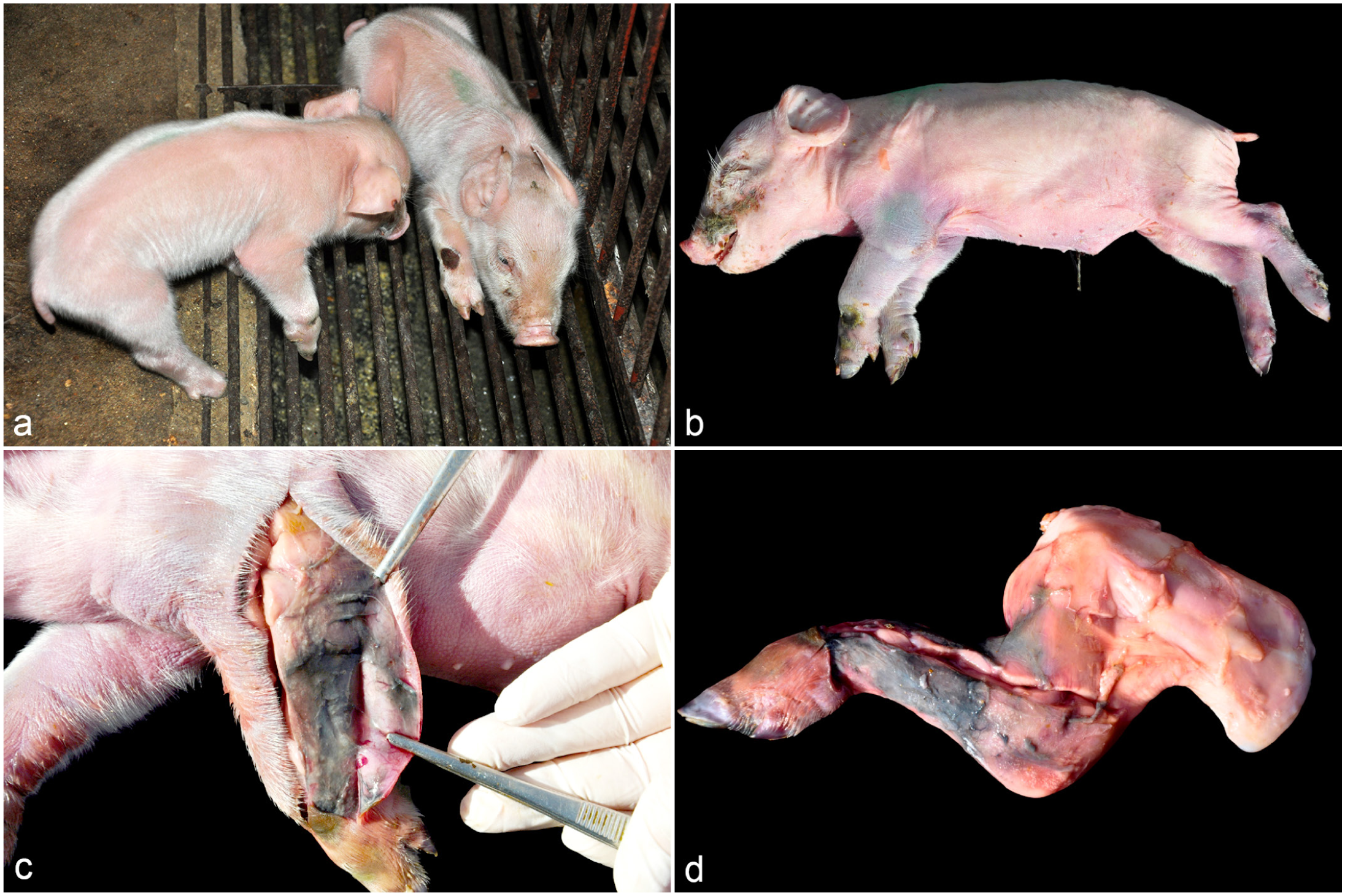
Clinical and gross findings of gas gangrene due to C. chauvoei infection in suckling piglets. (a) Affected piglets with lameness; note the ulcerated skin lesions over the carpal joint region. (b) Both forelimbs and hindlimbs are swollen, with focally extensive areas of red to purple skin discoloration. (c) The surface of the skeletal muscle shows a focally extensive blackened area with mild deposition of filamentous, friable material (necrotizing fasciitis). In addition, the subcutaneous tissue contains yellow gelatinous material consistent with edema. (d) Forelimb removed with necrotizing fasciitis extending up to the scapula region.
Grossly, the skin over the carpal and tarsal joints revealed approximately 1- to 2-cm-diameter ulcers. There was mild to moderate subcutaneous edema and marked black discoloration of the adjacent skeletal muscle under areas of purple skin discoloration (Fig. 1c, d). The soles of hindlimbs and forelimbs were markedly swollen and had areas of mild bruising and erosion. There were also moderate splenomegaly and noncollapsed lungs with mild interlobular edema. No other significant gross abnormalities were observed. Samples of skin, skeletal muscle, hooves, lungs, trachea, heart, esophagus, lymph nodes, liver, spleen, kidneys, small and large intestines, brain, and urinary bladder were collected, fixed in 10% neutral-buffered formalin, and processed routinely for the production of 3-µm-thick hematoxylin and eosin and Gram-stained sections.
Histologically, both piglets had deep skin ulcers with marked edema and mild neutrophilic infiltrate in the epidermis, dermis, subcutaneous tissue, and adjacent skeletal musculature of the limbs. Multifocally, the myocytes had moderate to marked hypercontraction bands with a lack of cross-striations, loss of cytoplasm, or floccular to granular cytoplasm. In the skeletal muscle perimysium and fascia, there were mild, multifocal neutrophilic infiltrates, fibrin deposition, gas bubbles, hemorrhage, and gram-positive rods with round edges that were single or in clusters, many of which had subterminal spores (Fig. 2a, b). Frequently, rods were observed inside blood vessels. A few blood vessels within the skeletal muscle had fibrinoid necrosis characterized by deposition of amorphous eosinophilic material in their walls (Fig. 2c, d). Thrombosis in vessels with fibrinoid necrosis and mild multifocal hemorrhage was observed in the sole of the hooves. Moderate splenic congestion and interlobular pulmonary edema were noted. No other significant microscopic lesions were observed in the other tissues from both piglets examined.
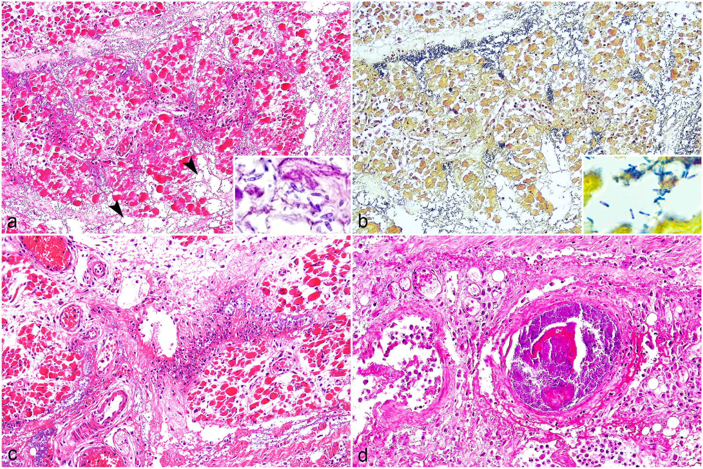
Microscopic lesions of gas gangrene due to C. chauvoei infection in suckling piglets. (a) Skeletal muscle. Surrounding the myocytes, there is edema, muscle necrosis, mild gas bubbles (arrowheads), and numerous intralesional aggregates of bacterial bacilli. Inset: Bacteria are rod-shaped with rounded edges and spores. Hematoxylin and eosin (HE). (b) Sequential section of (a) showing an abundant rod-shaped, gram-positive bacteria. Inset: Higher magnification of the gram-positive bacteria with spores. Gram stain. (c) Skeletal muscle. In some areas, there are mild neutrophil infiltrates and occasional fibrinoid vascular necrosis. HE. (d) Higher magnification of fibrinoid vascular necrosis, intravascular bacteria, and adjacent polymorphonuclear inflammatory infiltrates. HE.
Samples of skin, subcutaneous tissue, and skeletal muscle from the forelimbs and hindlimbs were aseptically collected. Tissue homogenates were inoculated into pre-reduced, reinforced clostridial medium (Merck), and cooked meat medium (Millipore) for anaerobic enrichment and incubated at 35°C–37°C for 24–48 hours under strict anaerobic conditions using GasPak systems. Cultures were streaked onto 5% sheep blood agar plates and incubated anaerobically at 35°C–37°C, with plates examined at 48 and 72 hours. For aerobic cultures, 5% sheep blood agar was used under aerobic conditions at 35°C–37°C for 48 hours. No bacterial growth was observed in any of the samples.
Species-specific polymerase chain reaction (PCR) assays were used for detection of C. chauvoei, 5 C. septicum, 5 C. perfringens, 19 and P. sordellii 11 on samples of skeletal muscle of both piglets. Total DNA was extracted from tissue homogenates using the MagMAX CORE Nucleic Acid Purification Kit (ThermoFisher Scientific, Carlsbad) according to the manufacturer’s instructions. DNA concentration and quality were assessed using fluorometry (FluoroQuant) before molecular analysis. Amplification reactions were performed in a final volume of 20 µL using GoTaq qPCR Master Mix (Promega), species-specific primers and probes, nuclease-free water, and template DNA. Reactions were carried out in a QuantStudio 3 Real-Time PCR System (ThermoFisher Scientific). Appropriate positive controls (American Type Culture Collection—ATCC strains) (Table 1) and no-template controls were included in each run. The real-time PCR assay was positive for C. chauvoei in skeletal muscle samples from both piglets (piglet 1: cycle threshold = 22.7; piglet 2: cycle threshold = 35.9) and negative for the other clostridia tested.
Primers, probes, and positive controls used for PCR detection of histotoxic clostridia.
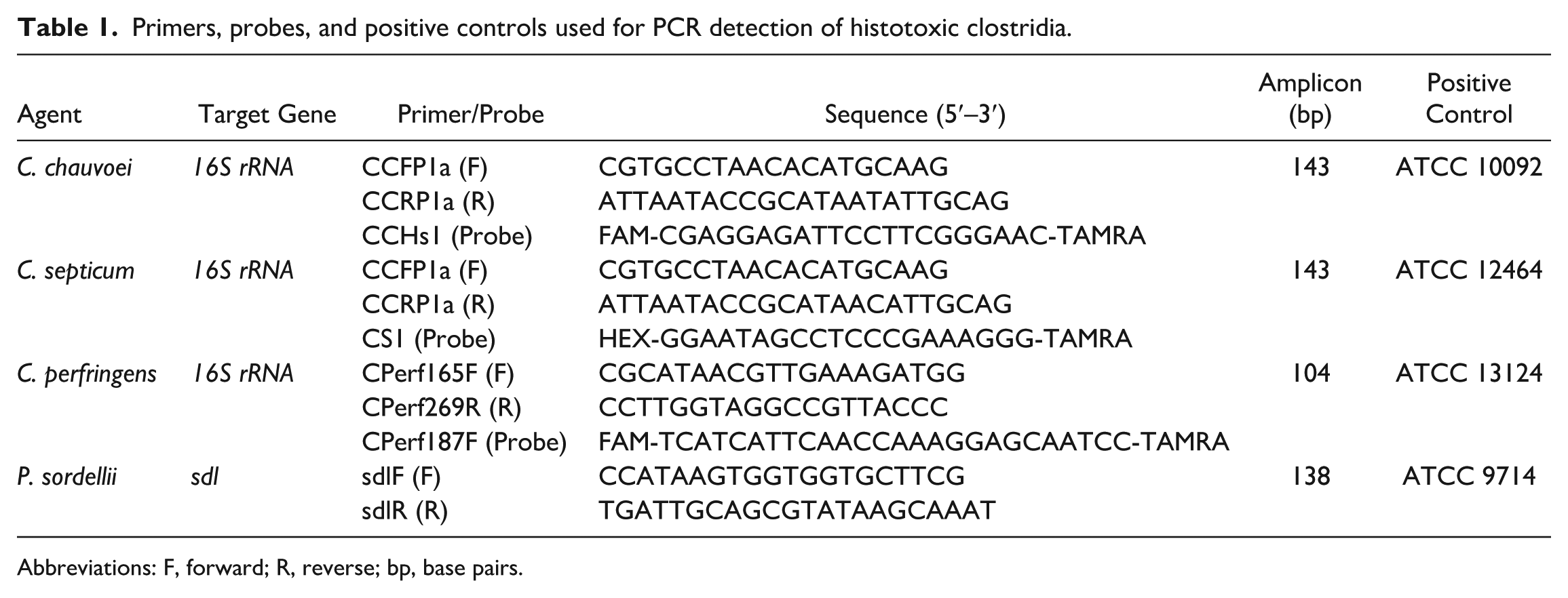
Abbreviations: F, forward; R, reverse; bp, base pairs.
A presumptive diagnosis of gas gangrene was established based on epidemiologic data, clinical signs, and gross and microscopic lesions, and was confirmed by detection of C. chauvoei by real-time PCR in tissues of the affected piglets. The latter was vital to determine the etiology, as several other clostridia (eg, C. septicum, P. sordelli, C. perfringens) can also cause gas gangrene and must be ruled out to confirm the etiology.12,13,16,17
It is speculated that the port of entry of C. chauvoei was the skin wounds, which supports a diagnosis of gas gangrene with exogenous pathogenesis. This is different from the endogenous pathogenesis of bovine blackleg for which no skin or mucosal wounds are required. After bacterial colonization of the wounds, the infection can spread leading to muscular necrosis, edema, and emphysema. The hemorrhage and free hemoglobin contribute to the dark discoloration of affected tissues. 2 A lower number of neutrophils has been described in several histotoxic clostridia infections, including those produced by C. chauvoei.1,9,12,16 Although this is a characteristic of alpha-toxin of C. perfringens–associated gas gangrene, it has been suggested that other clostridial toxins have a similar leukocytoclastic effect. 12 Clostridium chauvoei toxin A (CctA), a pore-forming toxin, is the main hemolytic and cytotoxic toxin produced by this microorganism. 3 Other virulence factors, including DNase, protease, neuraminidase, hyaluronidase, collagenase, and hemolysin, have also been described as contributors to its pathogenicity; however, their precise role in disease development has not yet been fully elucidated.3,16 Most toxins produced by histotoxic clostridia act initially at the local level, inducing tissue necrosis. This necrotic environment subsequently provides optimal conditions for bacterial proliferation and further toxin production. Toxins might gain access to the systemic circulation, leading to toxemia, shock, and death. 16 Nonspecific lesions seen in other organs, such as splenomegaly and alveolar edema, are likely the consequence of toxemia.8,16,17
No aerobic or anaerobic bacteria were isolated from either piglet. Both animals had been treated with antibiotics, possibly suppressing bacterial growth. In these 2 cases, the antibiotic treatment was administered when the animals were in advanced states of disease, which likely explains the lack of successful treatment. Although the lesions were typical of gas gangrene, antemortem antibiotic treatment may have additionally influenced the observed pathological findings, as gas production was not marked in our cases. Molecular methods, such as the PCR used in this study, are very sensitive and can detect the DNA of dead microorganisms, which may explain why the PCR was positive, but culture was negative.
The ulcerative lesions over the carpal and tarsal regions were attributed to repeated friction of the piglets’ skin with the concrete floor. The floor type has been associated with the development of skin, foot, and joint lesions in piglets, with lesions reported to be more severe in indoor-housed animals, particularly on abrasive, solid concrete surfaces, compared with outdoor systems.4,6,10 Lesions such as sole bruising and skin abrasions are described as more prevalent in suckling piglets during the first week of life and decrease progressively until 4 weeks of age. 6 This pattern suggests that the 1-week-old suckling piglets might be at the highest risk for gas gangrene under these conditions.
A higher prevalence and larger size of sole erosion on the hind feet have been reported in pigs housed on solid concrete floors, and it is speculated that this is because the piglets push forward with their hind limbs when suckling. 6 This mechanism may also account for the higher number of cases observed in litters from overweight sows in the present study, as increased difficulty in accessing the teats may intensify friction of the hind legs against the concrete floor, exacerbate skin abrasions, and thereby facilitate entry of C. chauvoei. In addition, impaired nursing postures in overweight sows may have reduced colostrum intake in piglets, leading to lower passive maternal immunity. This may have increased the susceptibility to C. chauvoei infection and represents a potential risk factor. Recently, the isolation of Clostridium spp. also had a high prevalence in ulcerative lesions associated with porcine ear necrosis in field conditions, accounting for 53% of infections. 7 In addition, a case of gas gangrene in a piglet highlighted traumatic skin injuries as a significant entry point for clostridial infections, primarily involving C. septicum and, more rarely, C. chauvoei. 14
After diagnosis, it was suggested to continue the early treatment with penicillin and streptomycin in suckling piglets with marked lameness due to the ulcerated skin lesions in the joints area. In addition, management interventions were implemented, primarily focusing on replacement of the deteriorated floor, adjustment of body condition scores within the herd, and enhancement of disinfection protocols. No further clinical cases were observed after 6 months following the initial outbreak.
Footnotes
Author Contributions
JCOM, FFP, and DLB performed necropsies and made epidemiological investigations. JCOM, FFP, MMP, AHG, and DD performed histological evaluations. CR and AM performed the bacteriological and molecular analysis. The manuscript was written by JCOM, FFP, and FAU with contributions from the other authors.
Declaration of Conflicting Interests
The authors declared no potential conflicts of interest with respect to the research, authorship, and/or publication of this article.
Funding
The authors disclosed receipt of the following financial support for the research, authorship, and/or publication of this article: Financial support for the development of this work was supplied by Conselho Nacional de Desenvolvimento Científico e Tecnológico (CNPq—Number 404897/2023-1) and Coordenação de Aperfeiçoamento de Pessoal de Nível Superior (CAPES)- Finance Code 001.