
Abstract

In baseball players, elbow injuries are the second leading cause of time lost from participation; shoulder injuries are the first. 10 Biomechanical studies have demonstrated that high valgus stress to the medial elbow structures peaks just after the cocking phase of throwing.4,7,13–16,18 This stress creates a valgus overload syndrome, which has been well described.6,11,13,16,18 Lesions attributed to this mechanism include medial collateral ligament sprains, flexor and pronator muscle strain and tendinitis, ulnar nerve neuritis, and avulsion fracture of the medial epicondyle.3,6,12,13,16 In this case report, we report a stress lesion of the proximal, medial ulna in a baseball pitcher.
Case Report
A 14-year-old, right-handed baseball player, who reported right medial elbow pain of 3 months’ duration, was seen at our hospital. For his age, he was a very high-caliber pitcher and was a member of several traveling teams and frequently attended baseball camps. He played only baseball and did no weight lifting. He first noticed insidious onset of the elbow pain during the first 5 minutes of a throwing session with his father. He had no history of injuries to his elbow and had no previous shoulder or elbow problems. He did not hear or feel a pop or snap in his elbow. He did note some vague bruising on the medial side of his elbow that resolved over several days. He reported no paresthesia and no loss of strength in his upper extremity. However, the pain in the right elbow increased during a throwing session a few weeks later. The patient reported a loss of velocity in his pitches over this period. He did not have any locking or catching in the elbow. He had not tried any medication and had not had any physical therapy. His medical history was unremarkable, with no systemic arthritis or problems in any other joints.
On physical examination, the patient was well developed and in no acute distress. His shoulder examination was normal with full motion, normal strength, and no atrophy. Examination of his elbow revealed hyperextension of 5° bilaterally, with equal flexion in both elbows of 145°. Pronation and supination movements were normal. He had no ecchymosis or swelling about the elbow. He did have tenderness on deep palpation 2 to 3 cm distal to his medial epicondyle, in the area of the medial collateral ligament insertion. Manual muscle testing was normal, and testing of his wrist flexor and pronator muscles did not cause any pain. Stress testing with valgus stress revealed no laxity at 30° of flexion or full extension, and it did not reproduce his pain. The neurovascular status was normal, with normal sensation and reflexes in both upper extremities.
Plain radiographs of the elbow showed some mild periosteal reaction on the medial aspect of the proximal ulna (Fig. 1). Three-phase bone scanning substantiated the presence of a lesion on the proximal ulna in the region of the medial collateral ligament insertion (Fig. 2). Magnetic resonance imaging showed no other lesions in the elbow and also revealed edema in the bone at the medial aspect of the proximal ulna, consistent with a fracture or stress reaction (Fig. 3).
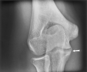
Plain AP radiograph of the right elbow showing periosteal reaction of the proximal ulna (arrow).
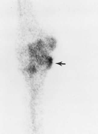
Three-phase bone scans showing stress reaction of the proximal ulna (arrow).
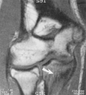
Magnetic resonance imaging scan of the right elbow showing edema of the proximal ulna consistent with stress reaction or fracture (arrow).
The patient was instructed to refrain from throwing for 4 to 6 weeks, but he was allowed to participate in some light weight-lifting. After that period, his tenderness was gone and he followed a progressive throwing program. He was able to return to pitching with no limitations 6 weeks later. After 1 year he had no recurrence of his symptoms, his elbow examination was normal, and he was pitching with no symptoms.
Discussion
Stress lesions of the upper extremity have been described in a variety of sports.5,6,8,9,17 A wide variety of lesions causing medial elbow pain in throwing athletes have been described.3,6,12,13,16 Overhand or throwing athletes generate tremendous forces in the shoulder and elbow. In the elbow these forces take the form of valgus and extension overload.6,16,18 For the elbow, the significant phases of the pitching motion are the late cocking, acceleration, and deceleration phases. The muscle contraction intensity and timing are selectively coordinated to control the external forces in a manner that provides optimal elbow positioning. 15 During the late cocking phase of the throwing motion, the shoulder is abducted approximately 90°, the elbow is flexed approximately 90° to 120°, and the humerus is in maximal external rotation. The acceleration phase is marked by internal humeral rotation and rapid extension of the elbow, which generate tremendous valgus forces about the medial aspect of the elbow joint and valgus extension forces posteriorly.4,7,13,14 These stresses are transmitted primarily to the medial collateral ligamentous complex, flexor carpi ulnaris, and, to a lesser extent, to the the medial flexor-pronator muscle.
Repetitive stress is presumably the cause of the proximal ulnar stress lesions seen in this case. The athlete reported here was not involved in any sports or activities other than baseball. The throwing motion can generate forces high enough to create a tensile stress failure in this region. Traction spurs from the medial side of the coronoid process have been described in a tennis player and in a baseball pitcher.6,12,13 Neither of these patients had fractures of the ulna, and the exact location of the ulnar spurs were not specified.
In the growing athlete, medial elbow problems are most frequently related to stress of the growth plate of the medial epicondyle. The medial epicondylar physis does not completely fuse in girls until age 14 to 15 and in boys until age 17 to 18. Chronic medial epicondylar physeal changes are manifested as a widening of the physis. 13 Acute avulsion of the medial epicondyle due to valgus stress has been reported.6,13 Valgus overload syndrome in the adolescent can include lateral-side lesions such as osteochondritis dissecans and Panner's disease.6,12,13,16 Extension overload of the growing elbow can manifest as a widening of the olecranon physis due to repetitive stress.6,8,13
In throwing athletes with medial elbow pain, plain radiographs typically are normal, although injured or degenerative medial collateral ligaments may show calcifications.6,11–13 Stress radiographs can be performed but may not always demonstrate instability.4,11 Magnetic resonance imaging findings associated with acute medial collateral ligament injuries include edema around the ligament with disruption of the medial collateral ligament fibers. Magnetic resonance imaging findings associated with chronic medial collateral ligament injury include attenuation of the ligament with loss of homogeneity.9,11 In the case presented here, MRI showed edema but no frank rupture of the medial collateral ligament. Magnetic resonance imaging is also useful when evaluating lesions where infection or tumor is suspected. Even though isolated tumors of the ulna are unusual, MRI and three-phase bone scanning were used to rule out any tumors in the young patient in this case report.
Stress lesions of the medial elbow physis can often be treated successfully with just rest. Healing is usually complete within 6 weeks. Stress fractures in adults typically heal within 6 to 8 weeks, depending on the degree of fracture.1,2,4,7 In the case of the adolescent presented here, the symptoms resolved in 6 weeks.
Although stress injuries to the medial elbow of throwing athletes are well documented, lesions to the proximal ulna are uncommon. In adults, these may take the form of a stress fracture or traction spur of the medial aspect of the proximal ulna at the coronoid. In adolescents, stress injury to the same location can occur. Both of these lesions should heal with rest, but continued pain may indicate that other lesions are present.