
Abstract
Bone tunnel enlargement has been reported after anterior cruciate ligament (ACL) reconstruction surgery. Although the long-term outcome of this phenomenon is not yet known, tunnel lysis or expansion may be clinically significant in revision surgery because the enlarged tunnels may complicate graft placement and fixation. There any many proposed theories for tunnel lysis. The most accurate statement is that this condition has a multifactorial etiology. Mechanical and biological causes have been reported, and both contribute to enlarged graft tunnels. This article describes the multiple causes of bone tunnel enlargement after ACL surgery. Future techniques and advances in primary ACL surgery must seek to eliminate this phenomenon.
From 1991 to 1999, the number of ACL reconstructions performed each year rose from 63,000 to more than 100,000, and this number continues to rise. 7 It has been reported that as many as 10% of these patients may experience graft failure and recurrent instability. 52 Therefore, as the number of primary ACL reconstructions rises, so too will the number of revision surgeries.
There are many reasons for ACL reconstruction to fail. The proper cause of each failure must be evaluated prior to revision. The preoperative plan of revision surgery must consider the placement and size of the previous bone tunnels. Tunnel lysis or enlargement often complicates revision surgery. Tunnel enlargement is determined by measuring the widths of the femoral and tibial tunnels on both the lateral and anteroposterior radiographs. The measurements are corrected for magnification and compared to the diameters drilled at surgery. Different tunnel shapes have been described as cone, cavity, cystic, or linear (Figure 1). 32 The etiology of the lysis most likely determines the specific tunnel morphology. Tunnel enlargement in the revision setting presents a difficult technical challenge with possible compromise of graft placement, fixation, and ultimately graft healing within the tunnels themselves. These factors have a significant effect on the final outcome of the revision surgery.
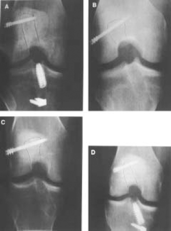
Types of various tunnel morphologies seen. A, linear type; B, cavitary type; C, cystic type; D, cone type. Reprinted with permission from Klein et al: The incidence and significance of femoral tunnel widening after quadrupled hamstring anterior cruciate ligament reconstruction using femoral cross pin fixation. Arthoscopy 79:470-476. Copyright 2003 by Arthroscope Association of North America.
Bone tunnel lysis or enlargement after ACL reconstruction is well documented in the literature (Figures 1, 2, and 3). Although many studies have described the presence of tunnel enlargement, none has shown this to be clinically significant in the short term with respect to laxity of the graft or increased failure rates.1,8,13–16,21,22,24,29,34,38,39,42,48,51,54 Despite this, the presence of large tunnels often severely complicates revision ACL surgery. This may include the need for a staged reconstruction and additional operative procedure with significant cost. The first procedure would involve implant/hardware removal, graft-tunnel debridement, and bone grafting of the tunnels. The revision ACL reconstruction would be performed after the bone graft has incorporated. 18 Authors have reported the need to bone graft tunnels and to delay revision ligament reconstruction. 18 Bone tunnel grafting may be necessary to provide proper placement and fixation of the revision graft.
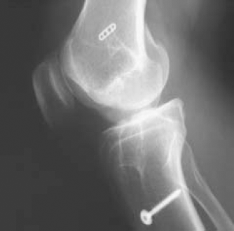
This lateral radiograph of a failed ACL reconstruction 4 years after surgery depicted tunnel enlargement of both the femoral and tibia tunnels. This patient had a staged revision with bone grafting of both tunnels followed by a ligament reconstruction 3 months later.
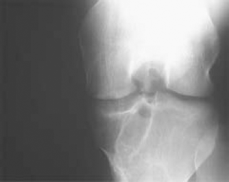
AP radiograph demonstrating enlargement of the tibial tunnel.
The exact etiology of tunnel enlargement is unknown. 22 However, many postulated factors have been de-scribed.13,15,22,24,42 These can be divided into 2 broad categories: mechanical and biological. The different pathoetiologies explain the different shapes of the tunnels with variables including graft type, fixation, and location.13,22,38
Mechanical factors include motion of the graft within the tunnel, fixation methods/devices, stress shielding of the graft, improper graft placement, and accelerated rehabilitation.1,8,13,15,16,22–25,35,38,42,46,48,51Graft swelling, the use of allograft tissue, synovial fluid propagation within bony tunnels, and increased cytokine levels within the knee are all biologic modes of inducing osteolysis and eventual radiographic evidence of tunnel enlargement.10,11,13,14,16,24,28,33,34,39,41,54,55 Much of the literature has supported the mechanical theory; however, the biologic theory clearly is a factor, similar to what has been reported in the joint arthroplasty literature. 41 In this article, discussion will focus on both mechanical and biologic factors, the clinical results, and the long-term implications associated with bone tunnel enlargement after ACL reconstruction. The cause and prevention of tunnel enlargement must be understood by the surgeon to improve surgical techniques and graft alternatives that may prevent the need for tunnel bone grafting if revision surgery is required.
Mechanical Factors
Graft-Tunnel Interface Micromotion
The predominant theory for tunnel enlargement after ACL reconstruction has been motion at the graft-tunnel interface. Current graft substitutes and fixation techniques have been unable to recreate the normal attachment sites of the ACL. As a result, tunnel enlargement continues to be seen with all types of grafts and fixation methods.1,13,15,16,22,24,29,38,42,48,49,51 However, the proposed mechanism in the attainment of large tunnels may be quite different for each graft source and/or fixation method/device.
Morgan et al and others have shown that aperture fixation of the femoral and tibial tunnels may prevent tunnel lysis in some studies. 36 The autologous bone-patellar tendon-bone (BPTB) graft complex provides a stiff construct with the addition of interference screw fixation and rapid biologic bone-to-bone fixation/healing. If properly performed, it may allow for aperture fixation within the femur itself without allowing any graft-tunnel motion. In contrast, typically the tibial bone plug is fixed at a point distal to the joint line at a nonanatomic site, allowing increased graft motion within the proximal tibial tunnel.1,16,22,24,25 Moreover, this graft is rectangular in shape with a thickness of approximately 4 mm. This creates a “dead space” within the tibial tunnel proximal to the bone plug.16,22 Studies have shown that synovial fluid may track along the graft and the bone tunnel within the dead space described above.16,22,24 Because there is a mismatch between the graft and tunnel, motion in the sagittal plane is theoretically allowed to occur during flexion and extension in the early perioperative period. Insalata et al termed this motion the “windshield wiper effect.” 24 Many studies in the literature demonstrate tunnel enlargement primarily within the tibial tunnel proximal to the fixation site and a minimal such effect within the femoral tunnel.1,16,22,24 Morgan et al described a technique using a retrograde bio-interference screw for aperture fixation of the tibial tunnel. The authors reported an absence of postoperative tunnel expansion with this technique. 36
Hamstring autografts in many previous studies have exhibited higher rates of tunnel enlargement than BPTB autografts have.13,24,51 All of these studies have 1 thing in common. The fixation of the hamstring tendons was performed by using a low stiffness construct, that is, fixation point distant from the joint line.22,50 This allows more graft elongation to occur with flexion and extension of the knee, and this has been referred to as the “bungee effect” in the literature.22,23,33 This longitudinal motion occurs at the graft-tunnel interface. It is frequently seen with fixation methods such as polyester tape (Endo-Button, Smith & Nephew, Andover, Massachusetts) (Figure 2). Subsequently, the femur has been the primary location of the tunnel widening secondary to its long tunnel length and subsequent pistoning of the graft within the tunnel.37,43,51
Clatworthy et al performed a prospective study comparing the incidence of tunnel widening between patellar tendon and hamstring grafts. 13 They used a Mitek anchor for femoral fixation, and tibia fixation consisted of staples for the hamstrings and interference screws for the patellar grafts. They showed a significant increase in tunnel widening both in the femur and tibia in the hamstring group at a minimum of 12 months after surgery. This study suggests that the bone-to-bone interface may play a role in decreasing micromotion of the graft once incorporation occurs. Insalata et al performed another comparison study using suspensory fixation for the hamstrings (Endobutton femur, post for tibia) and interference screw fixation for the patellar grafts. 24 Again, there was a statistically significant difference with respect to tunnel enlargement in both the femur and tibia. They concluded that the distant points of fixation for the hamstring grafts created a larger force moment during graft cycling, potentially leading to tunnel widening. In a recent prospective randomized study, Webster et al noted similar results as those reported above. 51 They noted that tunnel widening stabilized after the first few months, possibly indicating graft-tunnel incorporation. The longer time required for graft incorporation resulted in more time available for graft-tunnel micromotion. Therefore, the fixation of the soft tissue graft was important.
Although one study reported hamstring autograft fixation augmented with biodegradable interference screws decreased tibial tunnel diameter, a very recent prospective randomized study, fixing the hamstring autograft close to the joint line with interference screws, did not decrease tunnel enlargement.8,49 Buelow et al 9 suggested that the insertion of large interference screws not only compresses the graft in the bone tunnel but also significantly enlarges the bone tunnel itself and, therefore, is an iatrogenic process that occurs at time zero. Further expansion is possible.
The maturation process of the graft substitute in the joint has been extensively studied in animal models. The ligamentization process was similar among graft ligament substitutes and among species. The ligament maturation within the tunnel progresses similarly to the articular portion of the graft but at a slower rate. 26 The granulation interface tissue between the tendon and the tunnel wall is replaced by a thin fibrous tissue in the dead space. By 12 weeks, the fibrous tissue becomes dense, and the tendinous portion of the graft is completely adherent to the tunnel wall.13,26,53 With time, remodeling of the fibrous tissue to bone likely occurs. The graft-to-tunnel fixation therefore seems to have an initial critical period prior to complete adherence. Factors affecting the lack of graft ingrowth in the tunnel include provocation of graft movement within the tunnel (weak fixation), too early movement of knee (aggressive rehabilitation or noncompliant patient), and mismatch of the graft to the tunnel size (rectangular graft in round tunnel). This may be why tunnel widening is most commonly observed within the first 3 months after reconstruction and then subsequently stabilizes over time.
Accelerated Rehabilitation
There has been a dramatic change in protocol concerning rehabilitation activities after ACL reconstruction in past years. Shelbourne and Nitz described their “accelerated rehabilitation” protocol in 1990, and it now constitutes the backbone of most postoperative therapy protocols across the country. 46 It entails full weightbearing, immediate unlimited range of motion, strengthening exercises emphasizing full extension of the knee, and an early return to athletic activity (4-6 months). This has significantly decreased the incidence of arthrofibrosis, loss of motion (especially extension), and anterior knee pain of postsurgical patients in the past few years. It has significantly lowered surgical morbidity and improved functional outcome.
However, this accelerated rehabilitation protocol may have a disadvantage, especially in knee ligament constructs with low stiffness or weak/compromised fixation as seen in hamstring grafts and tibia bone. Accelerated rehabilitation will compromise any graft-tunnel interface that is not united, and micromotion appears. Graft-to-tunnel healing could possibly be compromised from motion occurring between this interface during cycling of the knee. This may be especially true in the hamstring ACL group fixed with low stiffness constructs. 5 An animal study has demonstrated tendon-to-bone healing occurring anywhere from 8 to 12 weeks postreconstruction. 40 As a result, graft-tunnel motion (ie, windshield wiper effect or bungee effect) would impede or delay this process from taking place.
Many studies have documented that tunnel enlargement is an early phenomenon occurring within 3 months after ACL reconstruction prior to the tibial tunnel filling with dense fibrous tissue.8,16,22,38,51 This may in part be due to the increased stresses placed on the graft during the accelerated rehabilitation process before graft-tunnel incorporation. However, there are no prospective studies identifying this as a possible causative or contributing factor to increased incidence of bone tunnel enlargement.
Fixation
Tunnel enlargement may effectively occur at time zero. The compressive stiffness of the screw and graft is often greater than the cancellous bone of the proximal tibial metaphysis leading to tunnel enlargement at time zero. Buelow et al reported that the immediately postoperative bone tunnel area was 75% larger in hamstring graft fixation with bioabsorbable interference screws. 9 The insertion of an interference screw apparently not only compresses the graft in the tunnel but also leads to an enlargement of the bone tunnel itself. Compressive stiffness of the fixation device graft construct versus cancellous bone is an additional variable.
Improper Graft Placement
There has been an association in the literature made between improperly placed tibia tunnels in ACL reconstruction and the incidence of tunnel enlargement.30,38,54 Zijl et al noted a trend with more anteriorly placed tibial tunnels having more tunnel enlargement radiographically. 54 There was a significant correlation between malposition of the tunnel and clinical outcome in their study. Previous studies indicate that anteriorly placed grafts may become impinged along the intercondylar roof resulting in increased graft forces, delayed incorporation, and subsequent failure. These increased graft forces may become distributed at the tunnel entrances, resulting in tunnel lysis. Improper tibia tunnel placement and the angle of that tunnel determine femoral tunnel placement and angle if done endoscopically. Segawa et al concluded that 1 of the main factors associated with tunnel enlargement is the location of tunnels and the angle of the femoral tunnel. 43 An acute femoral tunnel angle may increase the mechanical stress on the anterior margin of the femoral tunnel. The windshield wiper motion of the graft may be enhanced by the changing tension in the graft due to tunnel malposition.
Biological Factors
Allografts
Tunnel enlargement has been associated with the use of allograft tissue for ACL reconstruction. The proposed mechanism may involve immunologic rejection (cellular or humoral) of the graft by the host. Earlier reports in the literature have demonstrated decreased long-term clinical results using allograft tissue.14,15,28,39,42 The sterilization process used in the past was the primary cause of the inferior results seen with these grafts.28,39,55 Jackson et al reported on 7 patients who underwent allograft ACL reconstructions. The allografts were sterilized using ethylene oxide. They all experienced persistent synovitis with severe cellular responses and eventually required removal of the allograft. 28
Fresh-frozen and cryopreserved grafts without ethylene oxide sterilization have been used with more favorable clinical results.14,20,34 However, a recent retrospective study has shown an increased incidence of tunnel lysis with allo-grafts compared to autogenous tissue. Fahey et al performed a radiographic study comparing patellar tendon autografts and patellar tendon allografts 1 year postoperatively. 15 All grafts were fixed either by press fitting the femoral side or using a screw when necessary and interference screw fixation on the tibia. They showed a statistical significant increased incidence of tunnel enlargement in the allograft group as compared to the autograft group. They postulated a subclinical immune response responsible for this difference. Clinical outcomes were not adversely affected. Linn et al reported on fresh-frozen Achilles tendons without a calcaneal bone block fixed using screws and washers on both the femur and tibia. They reported that most patients had evidence of tunnel enlargement; however, the grafts were clinically stable. 34
In contrast, some studies have shown no difference with respect to the incidence of tunnel lysis between autografts and allografts.42,54 Schulte et al in a retrospective study reported no difference in tibial tunnel enlargement in allo-graft and autograft patellar tendon grafts. 42 In this study, however, all patients did exhibit evidence of tibial tunnel widening, but no statistical difference was noted. In another retrospective study, tunnel enlargement was no different between the same 2 groups. 54 Several recent animal studies have shown no evidence of immune reaction or inflammation using fresh-frozen allografts.4,22,47 Based on the current literature, it is difficult to conclude that there is an increased risk of tunnel lysis with allograft tissue as compared with autograft.
Cytokines and Synovial Fluid
Altered cytokine levels in the synovial fluid of patients with ACL injuries have been identified in several studies.10,11,22 Cytokines are small proteins that act as local intracellular messengers and are synthesized during inflammatory or antigenic stimulation. Macrophages have the ability to release cytokines and other inflammatory mediators. High levels of these cytokines may stimulate osteoclastic activity, leading to bone resorption. This has especially been noted in the total joint literature where high levels of macrophages and cytokines are noted around loose implants.3,17,19,31,41,45,55 The cytokines most frequently mentioned include interleukin 1, interleukin 6, interleukin 8, tumor necrosis factor alpha, and prostaglandin E2. Interleukin 1 receptor antagonist protein and transforming growth factor beta are 2 cytokines with protective properties against the catabolic effects of other cytokines. This inflammatory process may be a significant contributor to bone osteolysis and subsequent widening after ACL reconstruction. Along with the acute trauma of ACL injury, graft necrosis and bone necrosis from drilling have also been implicated in the release of inflammatory cytokines creating a hostile environment within the knee joint.2,12,15
In normal synovial fluid, a high concentration of interleukin 1 receptor antagonist protein exists while low levels of the other cytokines are present.10,11,22 Thus, a protective environment exists. After ligament rupture, large increases of the interleukins and tumor necrosis factor occur acutely. Associated injuries such as bone bruises and meniscus tears may affect the cytokine profile. These cytokines may decrease and stabilize over time or continue to be elevated.10,11,22 Moreover, interleukin 1 receptor antagonist protein apparently becomes depleted over time and is somewhat undetectable in chronic-ACL situations. Consequently, the cartilage protective function of this cytokine is lost.
Several MRI studies have shown synovial fluid tracking between the graft-bone tunnel interface8,13 (Figure 4). The bone tunnel is subsequently exposed to the increased levels of cytokines within the synovial fluid possibly inducing osteolysis and eventual radiographic evidence of tunnel enlargement. Some authors support this theory as to why there is an increase in tibial tunnel lysis in patellar tendon grafts.16,22,24
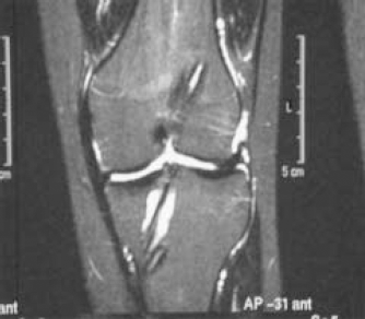
MRI demonstrating fluid in the tibia tunnel.
Graft Swelling and Pressure Effect
Graft swelling may contribute to tunnel enlargement. Jackson et al in a goat model reported an increase in cross-sectional area of 50% in autografts and allografts following ACL reconstruction, which was still evident for 6 months after the reconstruction. 27 A human MRI study also showed graft swelling but to a lesser degree. An average of a 12% increase in size was noted.8,13 The resulting pressure effect of the graft against the bone tunnel may cause an area of necrosis stimulating cytokine production and subsequent bone resorption. However, no study has correlated swelling of the graft with tunnel widening.
Also, the use of metal or bioabsorbable interference screws in the graft incorporation area may cause a local inflammatory response resulting in cell necrosis and associated bone tunnel enlargement. 22 Similarly, no data correlating the immediate enlargement of the tunnel by interference screws and progressive tunnel widening are available. There is not much known about the biological resorption of biodegradable screws.
Discussion
In general, the exact etiology of bone tunnel enlargement after ACL reconstruction is unknown. Mechanical and biological theories have been proposed to explain tunnel widening. This process is most likely multifactorial, arising both from mechanical and biological etiologies.
There have been several observations regarding the timing and progression of tunnel widening. The tunnel diameter increase is significantly higher within the first 6 weeks following surgery. There is minimal change between 3 months and 2 years, and finally a decrease in tunnel diameter has been seen at 3 years after surgery for autografts and allografts.13,16,38 Therefore, this is a relatively acute phenomenon occurring within the first 3 months postreconstruction. However, in a patient with persistent lack of healing of the graft in the tunnel, the tunnel widening may continue to persist.
Different shapes of the tunnels have been shown radiographically (cone, cavity, cystic, or linear) at different follow-up intervals. The distance of the fixation site from the intra-articular attachments may be a relevant factor in the shape of the tunnel and amount of tunnel enlargement. The wide variability in the incidence of tunnel widening and the shape modification of the tunnels is undoubtedly a multifactorial phenomenon.
Possible mechanical explanations for tunnel enlargement are bone resorption due to improper tunnel placement, stress shielding, and motion at the graft-tunnel interface. There is believed to be a windshield wiper effect as the graft oscillates from its fixation point as the knee is put through a range of motion. 22 This ongoing motion is thought to disrupt tendon-to-bone healing. Reducing the graft length to its intra-articular portion increases graft stiffness and graft isometry, which minimizes the potential for creep failure within the tunnel.6,25,51 Anatomic fixation of the grafts (aperture fixation) should be able to minimize adverse mechanical effects as well as to limit the exposure of bone tunnels to synovial fluid.6,36,44,49
There are many potential biologic explanations for tunnel enlargement. Cytokines are capable of directly or indirectly affecting bone resorption.33,41 It is proposed that cytokines released at the time of ACL injury or surgery may lead to osteolysis. The cytokines may also be involved with an immune response in allografts, graft swelling, and an inflammatory response by the synovium within the tunnel. Studies with MRI have shown synovial fluid tracking between the graft-bone interface.8,13 Therefore, cytokines within the synovial fluid have the access to induce osteolysis in the tunnel.
The etiology of tunnel widening remains unknown but is most likely multifactorial. Although tunnel enlargement does not appear to adversely affect clinical outcome in the short term, the long-term relationship with potential knee laxity or increased traumatic failure rate is unknown. Certainly, tunnel enlargement may severely complicate revision surgery, and the results of revision surgery show increased laxity compared to primary surgery. Large tunnels create problems with graft fixation and graft placement, thus compromising results of revision surgery. Patients with significant tunnel lysis may need a staged reconstruction with bone grafting of the tunnels followed by delayed ligament reconstruction. Not only does this require a second surgery, but the potential for additional giving-way episodes further risks injury to the menisci and articular cartilage. Therefore, it should be important to avoid tunnel enlargement. The surgeon must understand possible causes of tunnel widening and attempt to tailor the ACL reconstruction to minimize these risks. Studies have shown that aperture fixation in the femoral and tibial tunnels may decrease tunnel lysis. Autografts may cause less tunnel lysis than allografts because of a proposed cellular reaction to allograft tissue. Proper tunnel placement has been clearly shown to improve results, and postoperative rehabilitation methods need to allow for sufficient graft incorporation. Therefore, techniques that optimize graft biology, fixation, placement, and rehabilitation need to be identified and practiced. Future research should be directed at preventing this phenomenon and evaluating the relationship of tunnel enlargement with knee laxity and the outcome of revision ligament surgery.