





Abstract
Background: Double-bundle anterior cruciate ligament reconstruction replicates the 2 functional bundles of the native ligament, the posterolateral and the anteromedial, to control anteroposterior and rotational laxity.
Hypothesis: Double-bundle anterior cruciate ligament reconstruction laxity should be affected by the way grafts are tensioned.
Study Design: Controlled laboratory study.
Methods: Fourteen intact cadaveric knees were instrumented in a 6 degree of freedom rig, and kinematics throughout flexion-extension were recorded with an electromagnetic system under a 90-N anterior force or a 5-N·m internal rotation torque. Anteromedial and posterolateral bundle bovine extensor tendon grafts were fixed to load cells on the tibia, and tension was adjusted to match the intact knee anteroposterior laxity with 3 different protocols: (1) anteromedial bundle first and then posterolateral bundle at 90° and 20° of flexion, respectively; (2) posterolateral bundle first and then anteromedial bundle at 20° and 90° of flexion, respectively; and (3) both bundles together at 20° of flexion. Finally, a single-bundle graft positioned at 10 o'clock was tensioned at 20° of flexion.
Results: Lower graft tensions were required to match intact knee laxity in double-bundle anterior cruciate ligament reconstruction. Tension patterns with knee flexion were independent from the tensioning protocol. Protocols 1 and 2 overconstrained anteroposterior laxity, whereas protocol 3 matched intact knee anteroposterior laxity throughout the range of motion. The single-bundle reconstructions had excess anteroposterior laxity in flexion. Rotations were better restored with double-bundle protocols 2 and 3.
Conclusion: Knee laxity after double-bundle anterior cruciate ligament reconstruction is affected by the sequence in which the grafts are tensioned.
Clinical Relevance: Double-bundle anterior cruciate ligament reconstruction ensures better laxity restoration than does single bundle when both bundles are fixed together.
Double-bundle (DB) ACL reconstruction is being introduced to clinical use, and this is supported by scientific evidence that the replication of both the anteromedial bundle (AMB) and the posterolateral bundle (PLB) of the native ligament ensures better control of anteroposterior and rotational laxity than do traditional single-bundle (SB) procedures. 33 36 The first descriptions of DB techniques did not aim at replicating the real native ACL attachment.1,21,24,27,29,32,43 On the contrary, more recently, anatomical DB procedures have been described and validated.2,11,28,39,40 Furthermore, although early clinical experiences 1 29 did not show a clear advantage with DB techniques, more recent series have demonstrated some advantage, at least at short-term follow-up.2,28,40
Despite recent anatomical studies on native bundle attachments,12,18,26,34,42 which should improve tunnel positioning, we are not aware of studies on how best to tension DB grafts. Proper graft tensioning is critical for restoring normal anteroposterior laxity in SB reconstructions, 4 and nowadays it is accepted that excessive tension in the graft is not required and that tensioning toward extension is advisable.4,30,35,41 The way the grafts are tensioned (force applied and knee flexion angle) should influence DB procedures as well. With 2 independent intra-articular bundles replicating the native ACL, load sharing should be expected. However, stress shielding of one bundle might occur if the other is too tight. Similarly, the combined effect of 2 tight bundles with possible subsequent overconstraint of the joint might also be anticipated.
Up until now, in the developmental phase of DB techniques, different tensioning strategies were employed at surgery. In general, some surgeons tensioned both grafts at the same flexion angle, 39 40 whereas others 2 11 tensioned them at different flexion angles based on the rationale that the 2 native ACL bundles show different tensional behavior through flexion-extension. 3 This investigation tests the effects of different DB graft-tensioning strategies on knee laxity and bundle tension. The aim is to identify the best protocol, among the tested ones, for restoring normal patterns of knee laxity with anatomical DB ACL reconstruction. We hypothesized that the sequence of tensioning the grafts at different angles of knee flexion should affect both knee laxity and graft tension.
Materials and Methods
Seventeen fresh-frozen cadaveric knees from consented donations (mean age, 64.5 years; range, 48-74 years) were obtained from a tissue bank (IIAM, Jessup, Pa) in conformity with a local research ethics committee permit and related legal requirements. They were thawed overnight before use. Approximately 200 mm of thigh and shank were available in each specimen. The skin and the muscles were not removed. The fibula was secured to the tibia in its anatomical position with cortical screws, and a 400-mm-long aluminum rod was fixed with bone cement in the tibial medullary canal and secured with additional transverse screws. The femoral end was cemented in a tubular steel pot aligned coaxial with the tibial intramedullary rod while the knee was held at 0° of extension with the tibial rod vertical. This minimized the varus-valgus moment during passive flexion-extension. The steel pot was removed from the bone cement cylinder after the cement had set.
The femoral cement cylinder was secured in a nonferromagnetic rig that allowed manual passive knee flexion-extension by moving the femur with the unconstrained tibia hanging vertically (Figure 1). A Steinmann pin was drilled mediolaterally across the proximal tibia perpendicular to the shaft, and 2 semicircular hoops were mounted on this, 1 anterior and 1 posterior. These could be connected to weights via pulleys and strings, resulting in an anterior or posterior tibial drawer force without imposing a rotation torque or inhibiting natural coupled tibial rotation. A polyethylene disc (200 mm) was secured to the distal end of the tibial rod. Weights connected via pulleys and strings to opposite poles of the disc resulted in an internal or external rotation torque (Figure 1).

Specimen position in the rig was adjusted to approximately match knee and rig flexion-extension axes. Manual passive flexion-extension movements were applied to the femur; the motion of the hanging tibia was otherwise unconstrained. The anterior or posterior force was applied with weights connected to the proximal tibia. Internal or external rotation torque was applied with weights connected to both sides of a polyethylene disc secured at the end of the tibial rod.
Six degree of freedom knee motion was measured with an electromagnetic system (Nest of Birds, Ascension Technology, Burlington, Vt) whose accuracy had been validated previously 9 and found to be ±0.06 mm in a 30-mm translation and ±0.4° in a 20° rotation. The system consisted of a magnetic field transmitter and 3 sensors. Two sensors were press fitted into custom-made mounting blocks that had previously been fixed rigidly to the femoral and tibial shafts with screws and bone cement. The third sensor was instrumented with a stylus, calibrated, and used to digitize bony landmarks. All the equipment employed in the study was tested to ensure that it was nonferromagnetic and that it did not affect the electromagnetic measuring system.
After exposing the medial femoral epicondyle through a standard medial parapatellar approach, its center was digitized. The apex of the lateral epicondyle was digitized after exposing it through a limited lateral approach to the distal femur. The center of the proximal femoral medullary canal was digitized to calculate the femoral anatomical axis. The bony landmarks described above were digitized with the knee in extension, and the relative position of the tibia to the femur at this moment was assumed to be the zero. The position of 0° of extension was defined when the tibial intramedullary rod was parallel to the potted shaft of the femur in the sagittal plane. The 6 degree of freedom kinematics were described using the 3-cylinder open-chain model of Grood and Suntay. 20 This article presents flexion-extension rotation about the transepicondylar axis, internal-external rotation about the long axis of the tibia, and anteroposterior translations in the directions perpendicular to these axes. The 6 degree of freedom data on the relative position of the tibia with respect to the femur were recorded with no external loads applied while the knee was intact. Thus, the only loading was the weight of the tibia, plus the intramedullary rod and pulley, which hung vertically below the distal femur. The femur was flexed-extended in the test rig above the hanging tibia, which was free to float in the other 5 degrees of freedom such as anteroposterior translation and internal-external rotation. These tibiofemoral relative motions were not inhibited or altered by the test rig; this test configuration has been used previously. 19 This data set was used as the “intact knee” datum, and changes from this were recorded as a result of adding other loads or performing a ligamentous procedure, and those changes are presented in the “Results” section.
The limits of tibial anteroposterior translation and internal-external rotation through a flexion cycle were recorded under the application of a 90-N anterior and then posterior drawer force and again with a 5-N·m internal and then external rotation torque.
After intact knee laxity was recorded, the AMB and PLB were isolated and separated with the knee at 90° of flexion under the application of a 90-N tibial anterior drawer force, which tightened the AMB and left the PLB relaxed. Under this loading condition, the 2 bundles can be easily separated by blunt dissection. We had much experience separating the ACL into its functional fiber bundles, 3 18 in addition to knowing the work of others.12,26,34,42 The bundles were cut in the midsubstance, their tibial and femoral attachments demarcated, and ACL-deficient limits of tibial motion recorded. During all the experiments, the capsule was left open as it has been demonstrated not to affect knee anteroposterior laxity. 44 The effect of a medial parapatellar incision on rotational laxity is not known; by leaving the incision open, we avoided variability caused by repeated suturing and reopening the joint.
Two 6-mm outside-in tunnels were drilled in the femur and in the tibia in the center of each bundle attachment with a commercially available drill guide (Arthrex, Naples, Fla). Size-specific, double-stranded, pretensioned bovine extensor tendon grafts were lubricated with synthetic grease (Castrol Limited, Swindon, United Kingdom) to reduce friction and passed through the tibial and femoral tunnels to reconstruct both bundles. Bovine extensor grafts have previously been shown to have mechanical properties that are not significantly different to those of human hamstrings tendons. 14 For the proximal fixation, the grafts were looped over a 3-mm K-wire fixed rigidly to the femoral cortex across the tunnel entrances. The free ends of the grafts exited the tibial tunnels and were connected with stiff No. 2 Ultrabraid sutures (Smith & Nephew, Inc, Andover, Mass), which had been sewn to the grafts in a Roman sandal fashion, to 2 load cells (Entran Sensors & Electronics, Fairfield, NJ), which had been secured previously to the tibial intramedullary rod via 2 perpendicular threaded bars that allowed the load cells to be adjusted coaxial with the tibial tunnels (Figure 2).
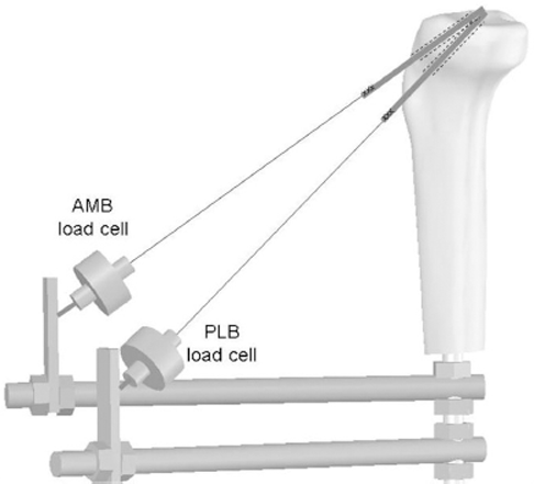
Load cells were positioned in line with the tibial tunnels using an adjustable mounting and then were connected to the distal ends of the grafts. AMB, anteromedial bundle; PLB, posterolateral bundle.
Under a 40-N tibial posterior drawer force, which resembled the action of the weight of the leg at the moment of fixation during surgery, tension in the free end of each graft was increased in 5-N increments and the resulting anterior translation laxity measured with the 90-N drawer force. If it did not match the laxity of the intact knee, the tension was adjusted while the knee again had 40-N posterior drawer and the anterior laxity remeasured until it matched the intact state within ±0.5 mm. On average, 5 tension settings per bundle were tested before matching the intact knee laxity. The second graft was tensioned using the same protocol, at its specific angle of knee flexion, again until the anterior laxity matched that of the intact knee within ±0.5 mm. Anteroposterior and rotational laxity resulting from the loads described above, as well as tension changes in the load cells, was then recorded across the range of knee flexion-extension after 3 tensioning strategies: (1) AMB graft tensioned first at 90° of flexion and then PLB at 20° to match intact knee anterior laxity, (2) PLB tensioned first at 20° and then AMB at 90° to match intact knee anterior laxity, and (3) both bundles tensioned together at 20° with the same tension. The rationale was therefore to tension the AMB, dominant in flexion, at 90° and the PLB, acting mainly in extension, at 20°, when a sequential tensioning protocol was used. Simultaneous tensioning in slight flexion (20°) was tested in consideration of its clinical use by some surgeons 39 40 because it is known that both fiber bundles are tight when the knee is close to extension. 3 6
After completing the recordings, the 4 tunnels were filled with polyester resin. After the resin became hard, single tibial and femoral 8-mm tunnels were drilled in the centers of the native ACL attachments at the junction between the 2 bundles, which had been identified previously. An 8-mm pretensioned double-stranded bovine extensor tendon graft was passed into the joint. It was fixed and tensioned in the same fashion as that of the DB grafts to match the anterior laxity of the intact knee at 20° of knee flexion. The envelopes of laxity were measured again, as described above.
Continuous kinematics and tension data were interpolated to single degree of flexion increments and analyzed using SPSS 13.0 for Windows (1989-2004, SPSS Science Inc, Chicago, Ill). Tibial rotations and translations from each loading condition were normalized to the anteroposterior and rotational position of the intact knee in the neutral condition without external loads at each degree of knee flexion. Mean differences and 95% confidence intervals of the differences in translations and rotations between the different knee conditions and intact knee envelope of laxity were calculated at single degree of flexion increments and plotted. It was assumed, according to the sign test, that when the confidence interval of a difference did not change sign, then the difference was statistically significant (P < .05).
Results
Anterior cruciate ligament transection resulted in a significant tibial anterior translation throughout the range of motion and slight but significant tibial internal rotation (Figure 3). The limits of posterior translation and external rotation were not affected measurably by the section of the ACL or by any of the ACL reconstruction procedures, so those data are not presented.

A, mean limit of intact knee anterior laxity in response to 90-N anterior drawer force (continuous line) +95% CI band of the tibial position after cutting the ACL (gray area). B, mean limit of intact knee internal rotation laxity in response to 5-N·m internal rotation torque (continuous line) +95% CI band of the tibial position after cutting the ACL (gray area). CI, confidence interval.
The highest tension to match intact knee anterior laxity was required when the PLB was tensioned first: 43 ± 29 N in the PLB graft and 60 N total tension (Table 1). The other reconstructions all required a similar total tension to match the intact knee laxity: 37 N for AMB graft tensed first, 34 N for both DB grafts tensed together, and 38 N for the SB graft. Table 1 also shows large SDs on the mean PLB first and SB graft tensions, so some knees required large graft tensions to restore anterior laxity to the intact values with those protocols.
Graft Tension Required With a 40-N Tibial Posterior Force to Match Intact Knee Laxity With 90-N Anterior Drawer Force Applied a

Data are means ± SD (N = 14). AMB, anteromedial bundle; DB, double bundle; PLB, posterolateral bundle; SB, single bundle.
Significantly different from DB AMB first and DB together (P < .05 after Bonferroni adjustment).
Tension patterns in the flexing knee with an anterior drawer force were not affected by the tensioning strategy: the load cell connected to the PLB grafts always showed the largest variation in tension with peak values at full extension (Table 2 and Figure 4). The tension in the load cell connected to the AMB graft changed less across the range of motion, with a peak around 30°. Overall tension values, on the contrary, were affected by the tensioning strategy. In DB reconstructions, the load cell connected to the graft that was tensioned first, particularly the PLB graft, recorded the highest tension values. The lowest tension under an anterior force was recorded when both bundles were fixed together (Table 2 and Figure 4). With an internal rotation torque, the tension tended to be less than under an anterior drawer force, particularly in the distal AMB graft load cell (Figure 5).
Maximum Graft Tension and Angle of Knee Flexion at Which It Was Recorded With a 90-N Anterior Drawer Force Applied a
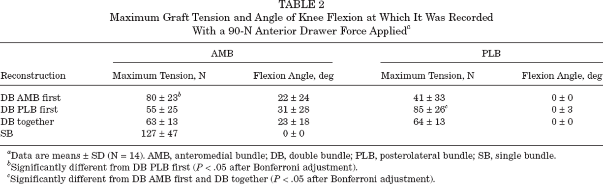
Data are means ± SD (N = 14). AMB, anteromedial bundle; DB, double bundle; PLB, posterolateral bundle; SB, single bundle.
Significantly different from DB PLB first (P < .05 after Bonferroni adjustment).
Significantly different from DB AMB first and DB together (P < .05 after Bonferroni adjustment).
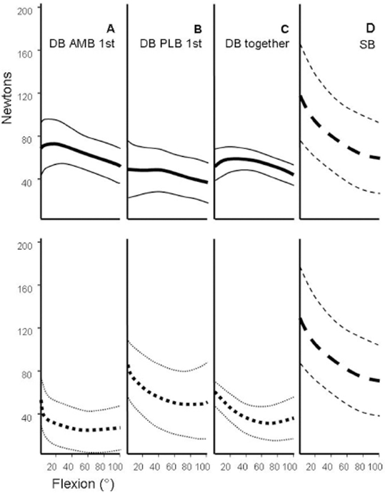
A-C, tension in the AMB graft (continuous line, upper row) and PLB graft (dotted line, lower row) under a 90-N anterior force versus knee flexion (mean ± SD, N = 14). D, mean tension in SB graft (mean ± SD, N = 14) under a 90-N anterior force. A, the AMB graft in a DB reconstruction was tensed first. B, the PLB graft in a DB reconstruction was tensed first. C, both grafts in a DB reconstruction were tensed together. AMB, anteromedial bundle; DB, double bundle; PLB, posterolateral bundle; SB, single bundle.
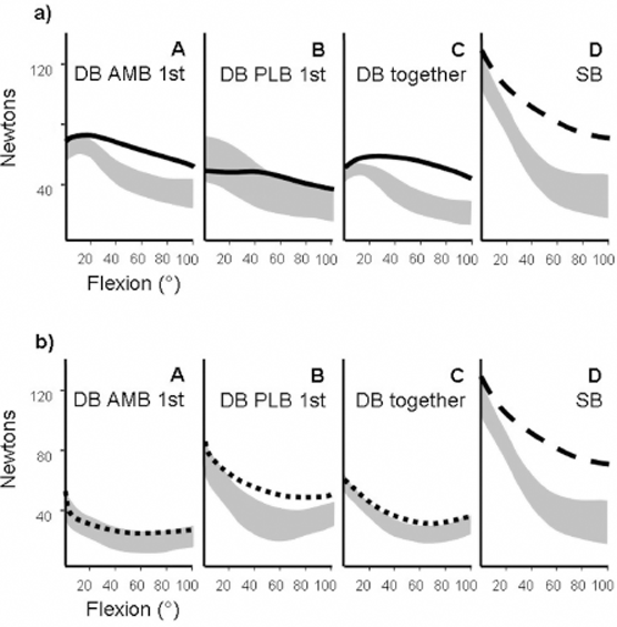
(a) A-C, mean tension (continuous line) in the AMB graft with a 90-N anterior drawer force and ±95% CI of the tension with an isolated 5-N·m internal rotation torque (gray area). D, tension in an SB graft with an anterior drawer force and 95% CI of the tension under an isolated 5-N·m internal rotation torque. (b) A-C, mean tension (interrupted line) in the PLB graft with a 90-N anterior drawer force and ±95% CI of the tension with an isolated 5-N·m internal rotation torque (gray area). D, tension in an SB graft with an anterior drawer force and 95% CI of the tension under an isolated 5-N·m internal rotation torque. AMB, anteromedial bundle; CI, confidence interval; DB, double bundle; PLB, posterolateral bundle; SB, single bundle.
Although all the ACL reconstruction procedures were quite effective in controlling tibial anterior laxity, there were differences in their ability to restore intact knee laxity patterns (Figure 6). Inconsistent matching of intact knee anterior laxity in flexion was recorded when the AMB was tensioned first and in SB reconstruction. The 95% confidence interval in Figure 6 shows that some knees had been overconstrained or underconstrained. Significant overconstraint of the anterior translation limit was recorded in both flexion and extension when the PLB graft was tensioned first. The most consistent match to intact knee laxity across the range of motion was obtained when both bundles were fixed together.
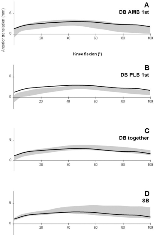
A-D, 95% CI band of the anterior translation limit after the different ACL procedures (gray area). Continuous lines represent intact knee anterior laxity limit under a 90-N anterior force. There was significant overconstraint in both flexion and extension after the PLB graft was tensioned first (B). The larger CI in A and D indicates less consistent control of knee laxity. AMB, anteromedial bundle; CI, confidence interval; DB, double bundle; PLB, posterolateral bundle; SB, single bundle.
Intact knee rotational laxity was controlled most consistently when both bundles were fixed together and when the PLB graft was fixed first, although some small but significant differences from normal persisted in the flexed knee. Inconsistent control of knee rotations, indicated by the large confidence intervals, was recorded when the AMB graft was tensioned first and by the SB reconstruction (Figure 7).
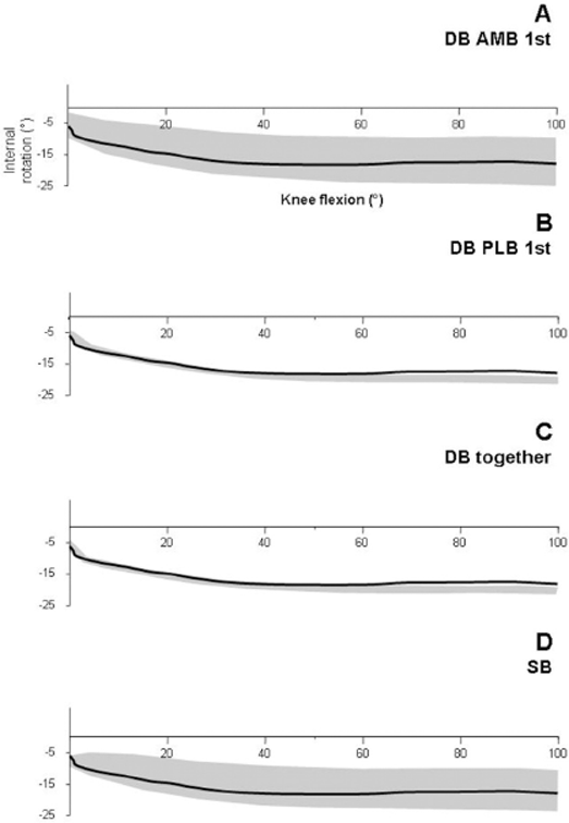
A-D, 95% CI band of the internal rotation limit after the different ACL procedures (gray area). Continuous lines represent intact knee internal rotation limit under a 5-N·m internal torque. Although statistically significant increases of internal rotation laxity were seen in protocols B and C, the small CI indicates more consistent control of tibial rotation than with protocols A and D. AMB, anteromedial bundle; CI, confidence interval; DB, double bundle; PLB, posterolateral bundle; SB, single bundle.
Discussion
The main finding of this investigation was that knee laxity and graft tension after anatomical DB ACL reconstruction were affected by the way the 2 grafts were tensioned. If the 2 bundles were fixed simultaneously at 20° of flexion and relatively low tension applied (17 N for each bundle), intact knee laxity restoration was obtained consistently with a low tension in the grafts throughout the range of motion. This was not possible with sequential bundle tensioning, which resulted in overconstraint of anterior laxity if the PLB graft was fixed first or in unpredictable anteroposterior and rotational laxity if the AMB graft was fixed first.
The tension patterns in the grafts when the knees were flexing were not affected by the tensioning strategy. In both load cells, peak values were always recorded toward knee extension. In the PLB graft load cell, tension decreased rapidly when the knee was flexed, whereas in the AMB graft load cells, a small tension decrease was recorded. This reflects the behavior of the native ACL bundles, which has been reported previously with different methodologies.3,5,16 The rationale was therefore to tension the AMB, dominant in flexion, at 90° of knee flexion and the PLB, acting mainly in extension, at 20° of knee flexion, when a sequential tensioning protocol was used.
Sequential graft bundle tensioning required more tension than did simultaneous tensioning to match the intact knee laxity with a DB graft, especially when the PLB was tensioned first. This is a consequence of controlling the whole of the anterior laxity increase with 1 bundle only before tensioning the second bundle. When the AMB was tensioned first, a low tension (19 N) was needed because the AMB is the dominant bundle in flexion, whereas the PLB is relaxed. Conversely, if the PLB was tensioned first at 20°, it required a high tension (43 N) because control of anterior laxity in extension is shared with the AMB.
Mechanical interactions between the graft bundles were shown clearly by sequential bundle tensioning. When the AMB was tensioned first to match the intact knee laxity at 90° of flexion, the subsequent tensioning of the PLB at 20° resulted in a tendency toward overconstraint in flexion, likely because of the extra effect of the PLB (Figure 5). Similarly, with the PLB tensioned first, overconstraint near extension can be explained by the additional stabilization provided by the AMB fixed last in flexion and tightening in extension.
The importance of DB graft tensioning has been emphasized also by Miura et al, 25 who compared simultaneous and sequential graft tensioning. Different from our experimental setup, they did not explore the effects of graft tensioning sequence (AMB or PLB first) and used a fixed amount of tension without adjusting it to match intact knee laxity. As a result, with simultaneous graft tensioning, they observed graft overload and some anterior laxity overconstraint.
The SB reconstructions were tensed at 20° knee flexion to match the intact knee laxity, and that resulted in less consistent control of anterior translation and internal rotation than did DB procedures in the flexed knee. As expected, higher tension was required in an SB graft. The inferiority of SB to DB reconstructions has been documented previously for controlling tibial anterior translation, 33 internal rotation, 36 and the pivot shift. 23 The inability of SB procedures to restore intact knee laxity has been demonstrated also in vivo. 17 A more lateral SB femoral tunnel location has been advocated by some to improve the rotational control of the reconstruction. 37 In this study, the SB tunnels were positioned at the centers of the native ACL attachments, which resulted in lower femoral positioning than did conventional SB techniques, which locate the femoral tunnel high in the intercondylar notch close to the roof. Despite its more anatomical rotation, the SB graft was not sufficient to restore intact knee limits of rotation consistently, and this has been related to its inability to eliminate the rotational component of the pivot shift in vitro. 23
The role of the PLB of the natural ACL remains controversial; it has been suggested that it acts more as a rotatory than a translation restraint. 42 It is known that the natural PLB tightens when the knee extends and is then a restraint to anterior translation 3 and that the PLB of a DB reconstruction behaves in the same way. 36 Observations in this study support this concept. A high graft tension is required to control anterior translation if the graft structure is only a secondary restraint. In this study, the total tension required to fix simultaneously both bundles at 20° was less than that applied to the PLB only and to an 8-mm SB graft at the same angle, suggesting that the PLB was less efficient at controlling anterior translation. In addition, the PLB tension was still high with an internal rotation torque, whereas AMB tension decreased. Thus, the different bundle function hypothesis, that the AMB controls anterior translation and the PLB controls rotation, respectively, is supported by these experiments.
These experiments found that transection of the ACL resulted in only a few degrees’ increase in rotational laxity, which is likely to have only minimal clinical significance. This has been reported previously. 15 On the other hand, the ACL controls the pivot-shift phenomenon, which is a combination of abnormal rotations and translations 8 and is the best predictor of patients’ subjective instability. 22 Further understanding of the pivot-shift phenomenon with regard to the limits of rotations and translations is needed to relate the effectiveness of laboratory procedures on the ACL to clinical instability. The present study has found that when both grafts of a DB reconstruction were tensed simultaneously at 20° of knee flexion, the knees had much more consistent control of both anterior translation and internal rotation than did SB reconstructions.
This experiment inevitably suffered the drawbacks associated with work on elderly specimens in vitro. Furthermore, the conclusions reached refer to time zero conditions and may be influenced subsequently by the effects of chronic graft relaxation and graft remodeling, which have been demonstrated to affect the fate of ACL reconstruction.5,7,31,38 The above limitations have been offset by the ability to make a series of comparative intraspecimen tests on a range of ACL reconstruction protocols. Further, the test setup allowed carefully controlled loads to be applied and full 6 degree of freedom kinematic data to be collected by motion sensors attached directly to the bones. It has been shown that anteroposterior laxity measurements in vitro 10 duplicate clinical laxity tests of the passive ligament restraints, 13 but there is a lack of knowledge about clinical rotational laxity.
Conclusion
This study has confirmed in vitro that DB ACL reconstruction is superior to an SB reconstruction in replicating intact knee laxity in both anterior translation and internal rotation. It has also demonstrated that the graft tensioning procedure affects knee translations and rotations. Among the test protocols used, simultaneous tensioning of both bundles of a DB reconstruction at 20° of knee flexion best restored intact knee laxity. Level 1 clinical evidence that a DB ACL reconstruction gives a significant advantage over an SB reconstruction is still missing, but this study will contribute to improving DB surgical technique.
Clinical Relevance
In anatomical DB ACL reconstruction, in which the 2 grafts are placed in tunnels at the centers of the anatomical ACL bundle attachments in the femur and tibia, tensioning of both AMB and PLB grafts simultaneously in slight knee flexion (20°) and with low tensions (20 N) gave the best results for returning translational and rotational knee laxity to normal, among the tensioning protocols examined.