





Abstract

Dear Editor:
I read with interest the article by Ferretti et al titled “Double-Bundle Anterior Cruciate Ligament Reconstruction: A Computer-Assisted Orthopaedic Surgery Study” (36[4], pages 760–766). The authors should be praised for their effort in investigating the effect of double-bundle reconstruction on knee biomechanics in an in vivo model. Much further work is needed in this arena; they should be congratulated for their efforts.
In this article, the authors performed single- and double-bundle ACL reconstruction using a computer navigation system. They concluded that double-bundle surgery did not show significant differences in anterior tibial displacement and internal and external rotation when compared with single-bundle ACL reconstruction. In their Figure 3 showing the passed grafts, the tibial anteromedial (AM) and posterolateral (PL) grafts are oriented in a medial-lateral relationship. Based on research from the University of Pittsburgh and the recent article of Edwards et al, 1 the size and shape of the AM and PL bundle insertion sites may vary. Most commonly, they are oriented in an anterior-posterior instead of a medial-lateral relationship. This has been shown quite nicely in multiple anatomical publications over the past year.
The native ACL and PCL do not impinge. The PCL triangle described by Lawhorn and Howell 2 can be observed in native knees. When the ACL is reconstructed in an anatomical fashion, this triangle should be reproduced. It was stated that their femoral AM tunnel was drilled at the 11-o'clock position, which is hard to interpret as the knee is a 3-dimensional structure, and the clock position varies with different flexion angles and position of the arthroscope.
The “o'clock position” clearly means different things to different people. It should be replaced by a system that is clearly specific, anatomically based, and objectively quantified. Unfortunately, the authors’ CT scan did not render more information on their femoral tunnel placement. The tunnel placement may have been nonanatomical and vertical because the PCL triangle appears closed.
I would also recommend a more “objective” evaluation system for placement of the graft on the tibia. I would ask the authors to specify which bony and soft tissue landmarks they used for tunnel placement. The article by Ferretti et al (Figure 3, p. 763) shows a posteriorly positioned AM bundle. The authors failed to specify exact anatomical location points of their 2 tibial entry locations in the tibia at the level of native ACL anatomical site. They stated the “guide wire was placed 7 mm anterior to the PCL insertion site.” Cadaveric studies have shown that fibers of the native ACL attach much further anteriorly than just 7 mm. Tibial placement of both groups may have been relatively nonanatomical in this study.
It cannot be emphasized enough that double-bundle ACL reconstruction or single-bundle reconstruction does not necessarily mean that the surgery was done in an anatomical fashion. To compare the difference between single- and double-bundle ACL surgery, it is imperative to perform the surgery anatomically. As Zantop et al 3 demonstrated in this same issue of AJSM, a nonanatomical double-bundle ACL reconstruction provides a totally different biomechanical effect than does an anatomical double-bundle ACL surgery. As would be expected, this fails to show any clinical superiority to single-bundle reconstruction. I hope that these questions will help educate our readership to better interpret the results of this study while contributing to a better understanding of ACL insertion site anatomy and reconstruction. I congratulate the authors for stimulating us all to learn more about ACL reconstruction.
Coming to the aspects pointed out by the reader, we can reply as follows:
The relationship between the 2 bundles is strongly influenced by the position of the scope and by degrees of flexion of the knee; therefore, no significant data can be assumed by a single image as Figure 3 of our original article. In our study, tunnel placement was done following suggestions of Freddie Fu, who personally instructed 2 of the authors who spent a visiting fellowship in Pittsburgh just to learn more about the original anatomical reconstruction. Figure 1, a postoperative CT scan showing the tibial tunnels’ position in an anteroposterior relationship rather than in an anteromedial (AM) relationship as presumed by the endoscopic image, should clarify this aspect.
Postoperative CT scan at a level just below the tibial articular surface showing the relationship between anteromedial and posterolateral tibial tunnels.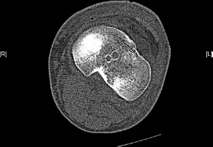
The position of the AM bundle was performed following a well-standardized surgical procedure made much easier and more reproducible by the outside-in technique used to drill the femoral tunnel. A special guide is used to position the entry of the tunnel just 5 mm in front of the “over-the-top” position as suggested by Edwards et al. 1 The effectiveness of the reconstruction of the AM bundle is confirmed by its biomechanical effect in significantly reducing both anterior tibial translation and internal rotation. The position of the tibial tunnel was identified according to the excellent study of Morgan et al 3 who recognized, in a cadaveric study on 50 specimens, the PCL as the more reliable and reproducible landmark for positioning the tibial tunnel entry.
We agree with the reader about the importance of an anatomical tunnel placement; however, our study demonstrates that even if all efforts are made to anatomically reconstruct the ACL, the addition of the posterolateral bundle to the AM bundle does not reduce significantly the internal rotation of the tibia at 30° of knee flexion, as already demonstrated in 1976 by Furman et al 2 on cadaveric knees.