






Abstract
Various lesions can occur after anterior dislocation of the shoulder joint. 16 Additional damages to the soft tissue, cartilage, humeral head, rotator cuff, and glenoidal cavity can occur if the dislocation is repeated. Moreover, the severity of the accompanying lesions after primary dislocation can be increased by repeated dislocation and subluxation of the joint.
Many authors have reported the presence of intra-articular lesions after primary dislocation of the shoulder joint, but there is little direct research comparing prevalence of the accompanying lesions between primary and recurrent dislocation of the shoulder joint and the relationship between accompanying lesions and recurrent dislocation. 2
The purpose of this study is to investigate the prevalence of accompanying lesions, including types of anteroinferior labrum injuries, using diagnostic arthroscopy and magnetic resonance arthrography (MRA), in patients with primary or recurrent anterior dislocation of shoulder joint.
Materials and Methods
Study Participants
All patients with traumatic anterior shoulder dislocation were recommended for MRA and diagnostic arthroscopy of the shoulder joint. From May 2004 to May 2008, a total of 168 patients were admitted; among them, 144 underwent diagnostic arthroscopy and MRA after informed consent. Thirty-three patients had primary shoulder dislocation and 111 had recurrent dislocation. There were no patients with a bilateral dislocation. We defined primary dislocation as a 1-time occurrence of traumatic anterior shoulder dislocation and recurrent dislocation as 2 or more occurrences of traumatic anterior shoulder dislocation. Inclusion in the primary dislocation group was based on patients having no history of anterior dislocation or other symptoms of shoulder joint injury prior to dislocation. All patients who were included in this study reported a history of a traumatic event that had caused either anterior dislocation or recurrent episodes of anterior instability. We defined dislocation as displacement of the humeral head into a locked position anterior to the glenoid, necessitating manual reduction.
The occurrence of dislocation was verified radiographically in all patients. For the recurrent dislocation group, radiographs were taken at least twice to confirm the dislocation.
If there was evidence of multidirectional instability of the ipsilateral and contralateral sides, the patients were excluded from the study. Multidirectional instability still lacks a uniform definition that is accepted by many orthopaedic surgeons. Laxity implies an ability to subluxate or dislocate, whereas instability refers to symptomatic laxity. Multidirectional instability is then identified as a clinical syndrome that occurs when shoulder laxity produces symptoms. 1,7,14,15 Symptomatic multidirectional instability was diagnosed based on the following criteria: (1) current complaints of shoulder discomfort and symptoms of instability and (2) positive sulcus sign, either positive load-and-shift and/or positive apprehension test.
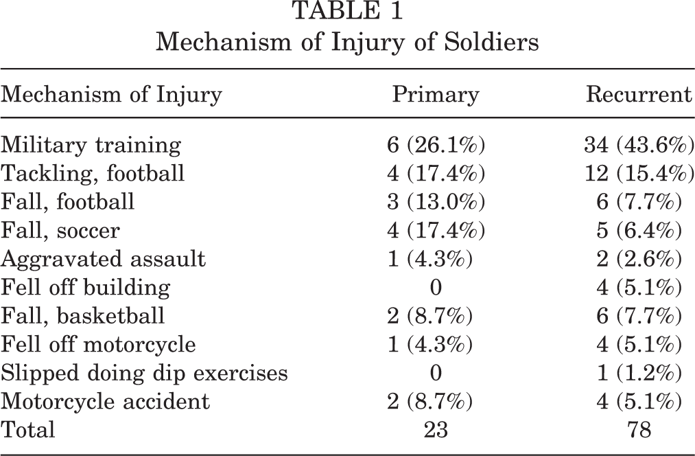
The average age was 23.4 ± 8.8 years (range, 17-33 years) in the primary dislocation group and 25.2 ± 11.7 years (range, 18-38 years) in the recurrent dislocation group. There were 32 males (96.9%) and 1 female (3%) in the primary dislocation group and 105 males (94.5%) and 6 females (5.4%) in the recurrent dislocation group. Nineteen patients (57.5%) had involvement of the dominant shoulder and 14 (42.4%) had involvement of the nondominant shoulder in the primary dislocation group. Sixty-eight patients (61.2%) had involvement of the dominant shoulder and 43 patients (38.7%) had involvement of the nondominant shoulder in the recurrent dislocation group. With regard to the vocations of the patients, in the primary group there were 23 soldiers (69.6%), 6 athletes (18.1%), 3 laborers (9%), and 1 homemaker (3%). In the recurrent dislocation group, 78 were soldiers (70.2%), 11 were athletes (9.9%), 13 were students (11.7%), 7 were laborers (6.3%), and 2 were homemakers (1.8%). Among the 6 athletes in the primary group, 2 (33.3%) were collision athletes. In the recurrent dislocation group, 4 (36.3%) were collision athletes. The mechanisms of injury in the soldiers are described in Table 1.
Mechanism of Injury of Soldiers
The time from dislocation to operation was an average of 14 ± 11.3 days (range, 2-32 days) in the primary dislocation group. The time from last dislocation to operation was an average of 18 ± 4.8 months (range, 10-32 months) in the recurrent dislocation group. The average age was 20.4 ± 5.8 years (range, 13-35 years) at the time of first dislocation, and time from the earliest dislocation to operation was an average of 3.4 ± 6.9 years (range, 2 months-14 years). The number of dislocations reported by patients was an average of 7.8 ± 9.2 (range, 2-30); 2 to 5 dislocations were reported by 34 patients (30.6%), 6 to 10 in 40 patients (36%), 11 to 20 in 22 patients (19.8%), and more than 20 in 15 patients (13.5%). Surrounding events that led to the initial dislocation were remembered by 132 of 144 patients; 83 (62.8%) reported a sports injury, 31 (23.4%) reported falling down, and 18 (13.6%) indicated a traffic accident.
This study was approved by our institute’s ethical review board and informed consent was obtained from all patients.
Evaluation Methods
All findings were recorded prospectively using a special evaluation form. At the time of diagnostic arthroscopy, the surgeon only checked the MRA itself without seeing the official reading of the radiologist. A radiologist who specializes in the musculoskeletal system provided readings of the MRA results. The radiologist was also blinded to the diagnostic arthroscopy findings. Once the MRA and arthroscopy were complete, we compared the diagnoses. To investigate correlation between the preoperative MRA and postoperative arthroscopic diagnosis, we diagnosed lesions, such as labrum lesions and superior labrum anterior and posterior (SLAP) lesions, using the intraoperative arthroscopic findings as a standard.
MRA Protocol
Intra-articular contrast injection was performed under ultrasound guidance using a posterior approach under aseptic conditions. An average of 15 mL (range, 10–20 mL) of diluted gadolinium (0.1 mL of 0.5mmol/mL gadolinium [Dotarem, Guerbet, France] in 20mL of normal saline) was injected. The magnetic resonance images were acquired with either of the two 1.5-T whole-body scanners (Magnetom Sonata, Siemens Medical Systems, Erlangen, Germany, or Intera-NT, Philips Medical Systems, Best, Netherlands), using a dedicated receive-only shoulder coil. After acquisition of scout images in axial, coronal, and sagittal planes, pulse sequences employed using the Siemens system were fat-saturated T1-weighted (repetition time in milliseconds/echo time in milliseconds = 443/14) images in the axial and oblique coronal planes, T1-weighted images in the sagittal plane (518/14), fat-saturated turbo spin-echo T2-weighted (3690/74) images in the sagittal and oblique coronal planes, and proton density (3500/43) images in the axial plane. Pulse sequence parameters for the Philips system were fat-saturated T1-weighted (425/15) images in the axial and oblique coronal planes, T1-weighted images in the sagittal plane (529/15), fat-saturated turbo spin-echo T2-weighted (3269/70) images in the sagittal plane, and oblique coronal plane and proton density (2281/15) images in the axial plane. Imaging parameters common to both scanners were field of view, 16 cm; matrix, 256 × 160; slice thickness, 3.0 mm; interslice gap, 10%.
Examination Under Anesthesia
Instability of the shoulder joint was measured under general anesthesia. If the humeral head was not completely dislocated from the glenoid rim, it was classified as grade 1, dislocation of the joint toward the anterior glenoid rim and patients with spontaneous reduction after dislocation were classified as grade 2, and patients without spontaneous reduction were classified as grade 3. 5,9
Arthroscopic Procedure
With the patient in the lateral decubitus position, the arthroscope was inserted through a posterior portal, and anterosuperior and anteroinferior portals were made when necessary. Anteroinferior labrum injuries and accompanying lesions were investigated first. Then, after observation through the posterior portal, accompanying lesions were examined through the anteroinferior portal and then the anterosuperior portal. In particular, the shape of glenoid and the range of abnormal glenoidal labrum lesion were studied through the anterosuperior portal. After the operation, the findings were recorded and sorted into 5 categories, including anteroinferior labral lesions, capsular lesions, SLAP lesions, bony structure lesions, and rotator cuff lesions. Abnormal manifestations of the anteroinferior labrum were divided into Bankart lesions, anterior labrum periosteal sleeve avulsion (ALPSA) lesions, bony Bankart lesions, and glenoid articular rim disruption (GLAD) lesions. The ALPSA lesions were classified into either free or adherent ALPSA lesions, depending on whether the lesion adhered to the glenoid neck. The extent of each glenoidal labrum lesion was recorded using a clock-face system with 12 o’clock representing the bicipital anchor. An extensive glenoidal labrum lesion was considered to be present if it extended above the 3-o’clock position or below the 5-o’clock position.
A judgment of capsular laxity was based on the presence of a “drive-through” sign and redundancy of the capsule and glenohumeral ligaments observed during diagnostic arthroscopy of the shoulder joint. Other criteria were the presence of external rotation greater than 100° and at least 50% anterior humeral head translation during arthroscopy. 16
Through the posterior portal, a calibrated arthroscopic probe was used to measure and quantify degree of anterior glenoid bone loss, if any, with reference to the central bare spot of the glenoid. The bare spot of the glenoid was located at the geometric center of the inferior glenoid, allowing accurate measurement of anatomy of the bony glenoid even in the cases of bone loss. An inverted pear-shaped glenoid has been reliably shown to represent a bony deficiency of the anteroinferior glenoid of 20% to 25%. 8,10
We used the χ2 test for frequency comparisons between the 2 groups and the unpaired t test for continuous variables (age, number of dislocations). The level of significance was P < .05.
Results
Five patients (15.1%) had grade 1 instability, 14 (42.4%) had grade 2 instability, and 14 (42.4%) had grade 3 instability in the primary dislocation group. Thirty-four patients (30.6%) had grade 2 instability and 77 (69.3%) had grade 3 instability in the recurrent dislocation group. All the patients who had grade 1 instability were in the primary dislocation group. Intra-articular findings revealed 4 capsular tears and 2 humeral avulsions of the inferior glenohumeral ligament. The size of capsular tear with grade 1 instability was small, less than 1 cm in size. There was a statistically significant difference between the 2 groups (P = .048).
Hemarthrosis was evident in 32 patients (96.9%) in the primary dislocation group and in 12 patients (10.8%) in the recurrent dislocation group (P = .024). There were 12 patients with hemarthrosis in the recurrent dislocation group, and they underwent surgery within 14 days following the most recent dislocation.
Lesions of Anteroinferior Labrum
In the primary dislocation group, there were 8 (24.2%) Bankart lesions, 9 (27.2%) free ALPSA lesions, 4 (12.1%) bony Bankart lesions, and 1 adhesive ALPSA lesion. In the recurrent dislocation group, there were 68 (61.2%) Bankart lesions, 11 (9.9%) free ALPSA lesions, 13 (11.7%) bony Bankart lesions, 16 (14.4%) adhesive ALPSA lesions, and 1 GLAD lesion (0.9%) (Table 2). In our study, the category of bony Bankart lesions did not include cases of inverted pear-shaped glenoid, but rather only bony defects of less than 20% of the glenoid with anterior glenoid bone loss <5 mm. Inverted-pear glenoids were included in a separate category.
Prevalence of Anteroinferior Labral Abnormalities and Capsular Abnormalities a
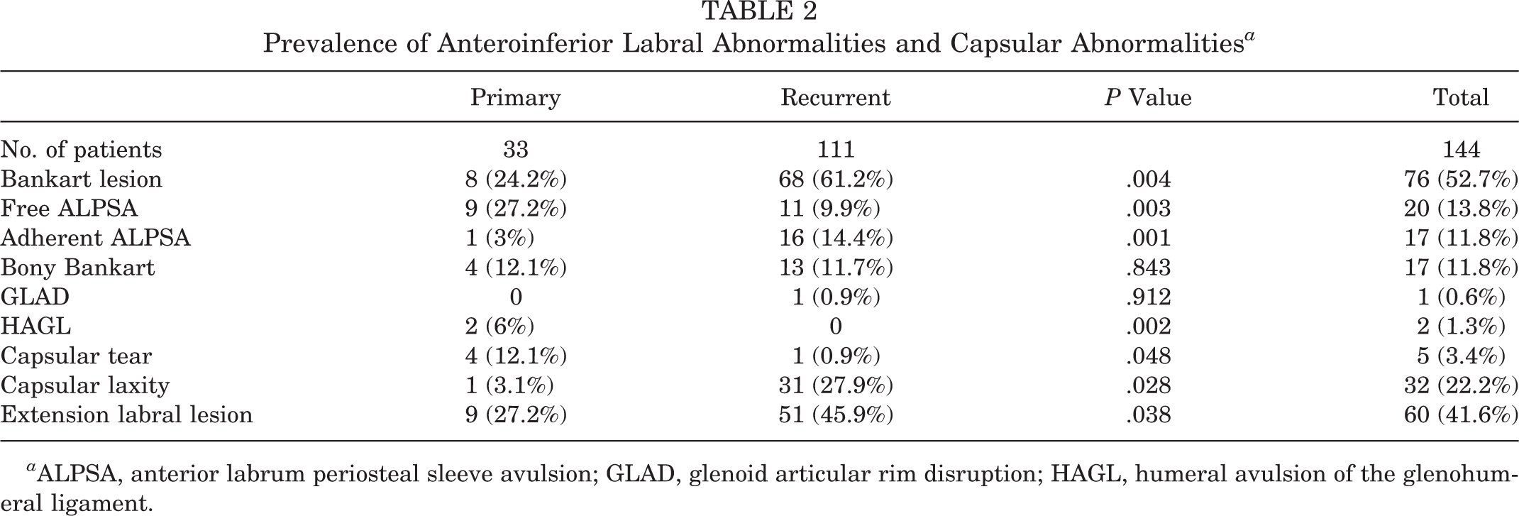
ALPSA, anterior labrum periosteal sleeve avulsion; GLAD, glenoid articular rim disruption; HAGL, humeral avulsion of the glenohumeral ligament.
Within the capsulolabral complex, 22 (66.6%) anteroinferior labral lesions that were limited to the anterior glenoid rim not extended to the capsular portion were found in the primary dislocation group, and 109 (97.1%) patients in the recurrent dislocation group were noted to have the same lesions as the primary dislocation group. There was a statistically significant difference between the 2 groups (P = .002). There were 9 (27.2%) and 51 (45.9%) patients with extensive labral lesions in the primary and recurrent dislocation groups, respectively. This difference was statistically significant (P = .038).
Capsular Lesions
In the primary dislocation group, humeral avulsion of the glenohumeral ligament (HAGL) lesions were found in 2 patients (6%), capsular tears in 4 (12.1%), and capsular laxity in 1 (3%). In the recurrent dislocation group, there was 1 patient (0.9%) with a capsular tear and 31 (27.9%) with capsular laxity (Table 2). No HAGL lesions were noted. The difference in capsular laxity was statistically significant between the 2 groups (P = .028).
SLAP Lesions
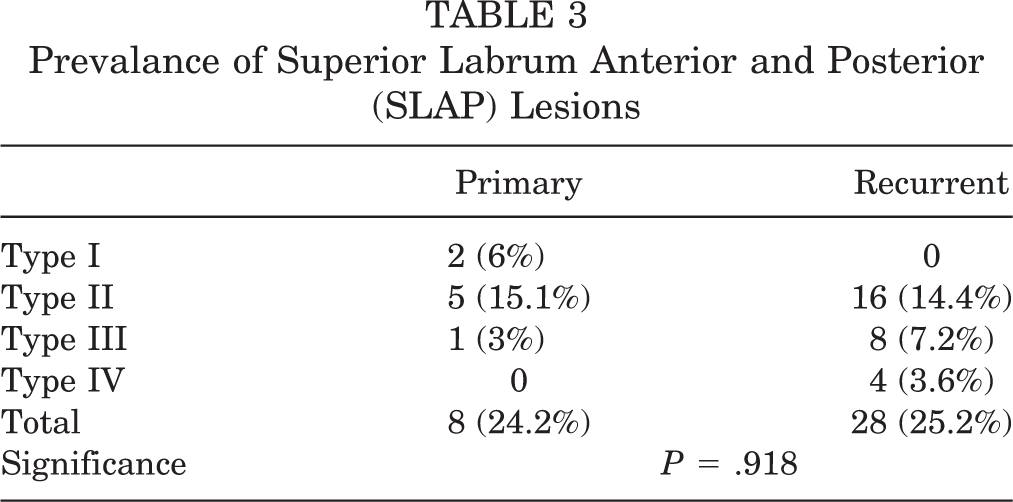
In the primary dislocation group, there were a total of 8 SLAP lesions (24.2%). Type I was found in 2 (6%) patients, type II in 5 (15.1%) patients, and type III in 1 patient (3%). Of the 28 SLAP lesions found in the recurrent dislocation group, 16 (14.4%) were type II, 8 (7.2) were type III (7.2%), and 4 (3.6%) were type IV (Table 3). There was no statistically significant difference between the 2 groups (P = .918).
Prevalance of Superior Labrum Anterior and Posterior (SLAP) Lesions
Bony Lesions
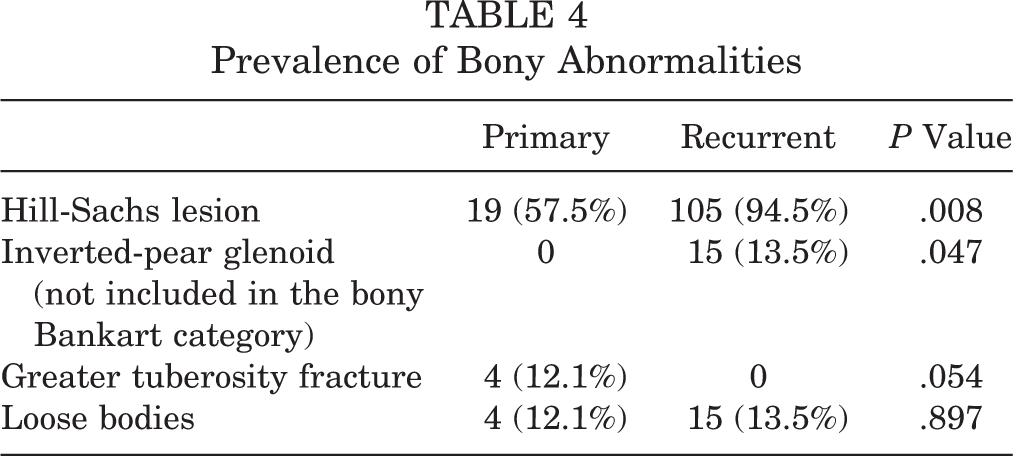
In the primary dislocation group, 19 (57.5%) Hill-Sachs lesions and 4 (12.1%) humerus greater tuberosity fractures were noted. In the recurrent dislocation group, 105 (94.5%) Hill-Sachs lesions and 15 patients (13.5%) with an inverted pear-shaped glenoid with attritional glenoid bone loss without bony fragments were found (Table 4). For Hill-Sachs lesions and inverted pear-shaped glenoids, there were significant differences between the 2 groups (P = .008/P = .047). In particular, the inverted pear-shaped glenoid was observed only in the recurrent group. In our study, all the cases with the inverted pear-shaped glenoid showed attritional glenoid bone loss without bony fragment.
Prevalence of Bony Abnormalities
In the primary dislocation group, an intra-articular loose body was noted in 4 patients (12.1%), and in 15 patients (13.5%) in the recurrent dislocation group. The loose body was removed during the arthroscopic procedure. The intra-articular loose body was either cartilage or osteochondral bone. It was generated in the humeral head or glenoid rim by the impact of the dislocation. Particularly, 3 patients (75%) with a loose body in the primary dislocation group were also found to have greater tuberosity fractures. Two of these patients also had a bony Bankart lesion. The loose bodies were bony fragments that originated from the bony Bankart lesion. A loose body from the anatomical neck portion of the humerus by fracture of the greater tuberosity was observed in the other patient.
Lesions of the Rotator Cuff
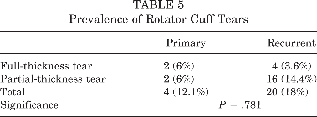
Partial rupture of the rotator cuff was observed in 18 patients (12.5%). Among them, 2 (6%) were in the primary dislocation group and 16 (14.4%) were in the recurrent dislocation group. Also, complete rupture was observed in 6 patients—2 in the primary dislocation group and 4 in the recurrent dislocation group (Table 5). All the partial ruptures of the rotator cuff were on the articular side, involving less than 25% of the rotator cuff.
Prevalence of Rotator Cuff Tears
Surgical Correlation
Preoperative MRA in the primary dislocation group did not discover type II SLAP lesions in 2 patients and a Hill-Sachs lesion in 1 patient. Two additional lesions were detected on arthroscopy, but missed by MRA in the recurrent dislocation group. These were a type II SLAP lesion and a lesion with a loose body. However, with the exception of these 5 cases, surgical findings in all patients coincided with MRA findings (96.5%).
Discussion
Generally, the Bankart lesion has been thought to be the most common lesion associated with anterior shoulder joint dislocation. However, the prevalence of Bankart lesions in the primary dislocation group in our study was only 24.2%, which is much lower than the data reported in previous studies. It is difficult to compare our results directly with those of other studies, as previous studies did not make detailed classifications of anteroinferior labrum lesions, such as Bankart and ALPSA lesions, as we did. Even if we gathered all of the anteroinferior labrum lesions in the primary dislocation group, a labral lesion was identified in only 22 (66.6%) patients.
Free ALPSA lesions were found in 27.2% of the primary dislocation group compared with 9.9% in the recurrent dislocation group. However, the prevalence of adhesive ALPSA lesions increased significantly in the recurrent dislocation group. We believe that the degree of displacement of free ALPSA lesions increased as time passed and as the dislocation was repeated, and the lesion became adhered to and fixed by granulation tissue of capsule membrane and coagulated blood on the inner side of inferior glenoid neck and converted to the adhesive ALPSA lesion. Extensive labral lesions and capsular laxity also had a higher frequency in the recurrent dislocation group. Additional damage in the glenoid labrum and joint capsule may be induced as dislocation of the joint and minute, incomplete subluxations are repeated and the anteroinferior labrum lesion is extended upward and downward; capsular laxity is seen as the chronic form of capsular tear in the recurrent dislocation group.
In the case of SLAP lesions, there was no statistical difference seen between the 2 groups, but 4 patients with type IV lesions were observed in the recurrent dislocation group. All the patients who had Bankart lesions also had accompanying extended anterosuperior tears. The extension may progress to the long head of biceps, when the Bankart lesion expands in the anterosuperior direction.
In our study, HAGL lesions and capsular tears were more prevalent in the primary dislocation group than in the recurrent dislocation group. Two patients with HAGL lesions and 4 with capsular tears were found in the primary dislocation group; all were treated with nonoperative management without repair during diagnostic arthroscopy, which resulted in excellent clinical outcomes. Bottoni et al 4 performed arthroscopy on 10 shoulders and found capsular tears that were less than 1 cm long in 2 patients. They thought that because all of these capsular tears were small, most may have healed without becoming a recurrent dislocation. We also found that capsular tears in the primary dislocation group were less than 1 cm in length. Spontaneous healing was achieved without recurrent dislocation during the 2 years of follow-up. In addition, on instability test under general anesthesia, all demonstrated grade 1.
In contrast, Ogawa and Yoshida 12 described extensive capsular tears in their patients with recurrent anterior dislocations. An isolated capsular tear was found in 5 (1.5%) of 333 shoulders treated with an open repair for the recurrent dislocation. They suggested that an extensive capsular tear cannot be healed spontaneously. We found that, in our recurrent dislocation group, capsular laxity was more prevalent than capsular tears. As dislocation is repeated, we believe the size of the tear is increased, and it is thought that this causes insufficiency of the inferior glenohumeral ligament, becoming a cause of further recurrent anterior dislocations.
In the recurrent dislocation group, there was a significantly higher prevalence of Hill-Sachs lesions and inverted pear-shaped glenoids. Lo et al 8 stated that meaningful cartilage and bone defect is induced by the repetitive collision of the humeral head and anteroinferior glenoid rim in the recurrent dislocation of the shoulder joint. In our series, Hill-Sachs lesions were observed in all of the 15 patients with an inverted pear-shaped glenoid. In their study, Lo et al 8 they suggested that a contact sports participant with an inverted pear-shaped glenoid is likely to have recurrent dislocation. Normal pear-shaped glenoids became inverted pear-shaped glenoids when 27% to 30% of the glenoid cavity was removed. Itoi et al 6 reported that instability was induced by a 21% glenoid defect. In our research, severe grade 3 instability was observed in all of the 15 patients showing the inverted pear-shaped glenoid. Therefore, if a Hill-Sachs lesion is suspected with a large-scale defect and inverted pear-shaped glenoid, a preoperative CT scan is needed to find the extent of the bone defect.
The prevalence of labral lesions in acute shoulder dislocation has been reported in some studies. For example, Norlin 11 reported the prevalence as 100% and Arciero et al 3 reported them as 97% of Bankart lesions. Rotator cuff tears were not reported in these studies. In contrast, a total of 18 patients (12.5%) with partial rupture were found in our investigation. Porcellini et al 13 proposed that there was a strong linkage in the instability because of dislocation and rotator cuff rupture, and the strength of relation was higher in the cases with repetitive dislocations of more than 7 times.
We attempted to identify prevalence and variety of accompanying lesions by comparing the findings of MRA taken before operation to the findings of arthroscopy. Antonio et al 2 noted that, with the high resolution provided by MRA, radiologists are able to further classify these labral avulsions into various lesions in addition to the Perthes/Bankart categorization used in previous studies, and suggested a role for early MRA to assist in treatment triage. The ability to base treatment plans on MRA is limited because there is no definite classification of displacement of the detached capsulolabral complex in MRA yet. However, if the anteroinferior labrum is completely avulsed with no attachment to the glenoid, and lies adjacent to the middle glenohumeral ligament, we consider that complete reduction through nonoperative treatment is difficult. In our study, except for 5 cases that were missed by MRA, surgical findings in all lesions also coincided with MRA findings (96.5%). Thus, we believe that MRA is an accurate method for assessing accompanying lesions.
Conclusion
Various forms of anteroinferior labral lesions were observed in the investigation of prevalence of secondary concomitant lesions. The recurrent dislocation group showed a significantly higher prevalence of labral lesions and bony lesions in comparison with the primary dislocation group, suggesting that they increase in prevalence with repeated recurrences of shoulder dislocation. Also, in our series MRA was an accurate method to assess accompanying lesions in first-time and recurrent anterior dislocation of the shoulder, suggesting that it is a useful tool to guide treatment.
Footnotes
The authors declared that they had no conflicts of interests in their authorship and publication of this contribution.