
Abstract
Proximal hamstring tendinopathy (PHT) was first described by Puranen and Orava 30 in 1988. Symptoms occur in athletes involved in a variety of sports, particularly in sprinting and endurance sports. The characteristic complaint of chronic PHT is ill-defined pain, especially while performing sports activities or when sitting, in the area of the ischial tuberosity that radiates distally to the popliteal fossa. Askling et al 1-3 have found that a combination of extensive hip flexion and knee extension can lead to a specific acute injury in the proximal hamstring tendons for a variety of sport activities. However, in our experience, and in the experience of others, 21,28 the pain typically appears and gradually increases without being triggered by any acute event.
A previous MRI study 10 demonstrated an increased tendon girth and an intrasubstance signal heterogeneity of the hamstring tendons in patients with chronic PHT. The histopathologic findings in a more recent study 21 showed rounding of the tenocyte nuclei, increased ground substance, collagen disintegration, and increased vascular proliferation in the biopsy specimens from the hamstring tendons of patients with chronic PHT.
Taken together, these data indicate that the pathologic changes observed in this condition are similar to those observed in other tendinopathies. 37 In view of this similarity, the rationale for the use of shockwave therapy (SWT) in the treatment of PHT in our study was based on the effectiveness of SWT in the treatment of other tendinopathies reported in previous studies. 5,14-16,31-36,38,41
As with other tendinopathies, the first option is nonoperative, including changes in, or the interruption of, the sports activity, nonsteroidal anti-inflammatory drugs (NSAIDs), physiotherapy, and stretching and strengthening exercises of the hamstring muscles. No controlled studies are available.
To our knowledge, no prospective clinical studies have been performed to assess the effectiveness of SWT compared with traditional conservative treatment (TCT) in patients with chronic PHT.
The purpose of this randomized controlled clinical study was to evaluate the effectiveness and safety of SWT, and to compare the effects of SWT with those of TCT in patients with chronic PHT.
As a null hypothesis, we hypothesized that SWT or TCT are equally effective in patients with chronic PHT. In this study, chronic PHT was defined as recurrent pain and tenderness attributable to degenerative changes in the proximal hamstring tendons persisting for at least 6 months.
Materials and Methods
Between 2004 and 2006, 40 patients with MRI-verified chronic PHT were enrolled in this study. This randomized controlled clinical study was approved by an institutional review board. All patients were informed of the potential risks of treatment as well as of the possibility of being randomly included in either the SWT group or TCT group.
Magnetic resonance images were analyzed by an experienced musculoskeletal radiologist (with 30 years of experience in musculoskeletal radiology) who was blinded to the diagnosis. Signal intensity abnormalities of the proximal hamstring tendon substance (defined as high signal intensity compared with the low signal intensity of normal tendon) on T1- and T2-weighted images were evaluated as qualitative criteria on MRI. In addition, the presence of a longitudinal split of the proximal hamstring tendon before its point of insertion was recorded. The radiologist was asked to render one of the following diagnoses for each proximal hamstring tendon, according to a simple MRI grading system devised by Khan et al 20 for Achilles tendinopathy: grade 1, a normal tendon; grade 2, a thickened tendon with homogeneous signal intensity; or grade 3, intratendinous high signal intensity, which was diagnosed when there was a signal intensity change that was predominantly visible on T1-weighted images, because in these sequences the intensity change was not affected by the “magic-angle” effect. 12
Written informed consent was obtained from all the patients before their participation in the study.
Inclusion criteria were chronic PHT, diagnosed clinically (Puranen-Orava test, 30 fast hamstring-stretch test, and hamstring strength test 43 ) and by means of MRI, and a pain score of ≥4 cm on the visual analog scale (VAS) at the first evaluation.
Briefly, the Puranen-Orava 30 test entails actively stretching the hamstring muscles in the standing position with the hip flexed at approximately 90°, the knee fully extended, and the foot on a support.
For the fast hamstring-stretch test, the patient lies in the supine position with the legs fully extended; the examiner grasps the symptomatic leg behind the heel with 1 hand and at the knee with the other hand, flexes the hip and knee maximally, and then rapidly straightens the knee. This test, which was proposed by the lead author (A.C.), was found to have a high intra- and interobserver reliability, with an intraclass correlation coefficient of .87 and .85, respectively. These tests were considered positive if they evoked or exacerbated the pain typically experienced by the patient within the proximal hamstring tendons.
Hamstring strength was assessed according to Young et al, 42 with the patient placed in the prone position with the hip extended and the knee at 30° of flexion. The examiner pulls down with 1 hand behind the heel while the patient resists and attempts to maintain the position or flex beyond 30°, and the examiner’s other hand is placed on the belly of the hamstring to assess the quality of the muscular contraction. Hamstring weakness was recorded using the Medical Research Council (MRC) power grading system 9 and, according to Young et al, 42 was considered as normal, mild, moderate, or severe (MRC power grading of 5, 4.5, 4, and 3.5, respectively) when compared with the nonaffected side.
The patients to be enrolled in the study had to have at least 2 positive clinical tests, and the MRI had to be positive (grade 2 or 3) in all patients
Exclusion criteria were lumbar sciatic pain, piriformis syndrome, ischial tuberosity avulsion, ischiogluteal bursitis, or hamstring muscles tears; pregnancy; implanted pacemaker; blood coagulation disorders or use of anticoagulant drugs; age <18 years; inflammatory or neoplastic disorders; and any treatments administered in the past 4 weeks.
The patients were interviewed to obtain demographic data as well as data on the onset time of pain, the possible cause of injury, the presence and intensity of pain in the sitting position or when driving a car, previous treatment, former and present state of sports activity, and the sport and/or daily living activities that triggered or exacerbated the pain.
During the physical examination, a differential diagnosis was made between PHT and lumbar sciatic pain, piriformis syndrome, and hamstring muscles tears. A positive Lasègue sign and reduction or impairment of Achilles and patella reflex tests were used to make the differential diagnosis between PHT and lumbar sciatic pain. A positive Lasègue sign and tenderness on deep gluteal palpation over the belly of the piriformis muscle, positive FAIR (flexion, adduction, and internal rotation) test, 40 and a positive Freiberg sign 13 (pain caused by forced internal rotation of the extended thigh) and Pace sign 29 (pain caused by resisted abduction in conjunction with external rotation of the thigh) were used to make the differential diagnosis between PHT and piriformis syndrome. All these findings and signs were considered positive if they recreated the characteristic symptoms that the patient experiences with activities in both the buttock and radicular component. 4
The onset of pain and MRI findings were used to make the differential diagnosis between PHT and hamstring muscle tears.
Pelvic MRI was routinely performed before treatment in all cases. However, if one of the aforesaid conditions was suspected on the basis of the clinical findings, additional radiographs, electroneuromyographic (ENMG) studies, and MRI of the lumbar spine or of hamstring muscles were performed before randomization.
Twelve of the 52 patients who underwent the first evaluation did not satisfy the inclusion criteria (Figure 1). Five patients suspected to have a hamstring muscle tear by the physician underwent thigh MRI. A hamstring muscle tear was confirmed in all 5 patients. The remaining 7 patients, suspected by the physician to have pain originating in the lumbar spine, underwent radiography, lumbar MRI, and ENMG. These examinations confirmed the clinical suspicion of lumbar sciatic pain attributable to an L5-S1 herniation in 5 patients and an L4-L5 spondylolisthesis in the remaining 2 patients.
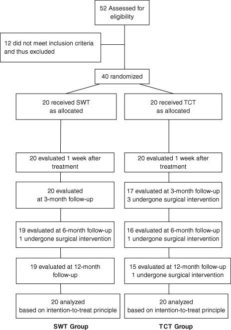
Flowchart of the study.
Therefore, 40 professional athletes (27 men and 13 women) were enrolled in this study. Their demographic and baseline characteristics, as well as their sports activities, are shown in Table 1. The first patient was randomized on February 1, 2004, while the last patient was randomized on September 30, 2006. Patients were randomly assigned to receive either SWT (SWT group) or traditional conservative treatments (TCT group) by means of a computer-based 1:1 randomization scheme and sealed envelopes (Figure 1).
Demographic and Baseline Characteristics of Both Groups a
Values are the mean ± standard deviation. SWT, shockwave therapy; TCT, traditional conservative treatment; CI, confidence interval; NPRS, Nirschl phase rating scale; VAS, visual analog scale.
Shockwave Treatment Group
All 20 patients in the SWT group (Table 1) received 4 SWT sessions, each of which was performed at weekly intervals, with 2500 shocks per session at a pressure of 4 bar (equal to an energy flux density of 0.18 mJ/mm2). The treatment frequency was 10 shocks/s. The total energy flux density of the treatment session was approximately 450 mJ/mm2. Shockwaves were provided by a radial shockwave generator (EMS Swiss Dolorclast, Milano, Italy) consisting of a control unit, a handpiece, and a medical air compressor. The compressor creates pneumatic energy, which is used to accelerate a projectile inside the handpiece. When the projectile strikes the metal applicator, which is 15 mm in diameter, a shockwave, which is distributed radially from the metal applicator to the pain zone, is created. The control unit modulates the number of impulses, the intensity (expressed in bar), and the frequency (expressed in hertz).
Adopting the principle of clinical focusing, the area of maximal tenderness was treated in a circumferential pattern, starting from the area of maximum pain level. The treatment area was prepared with ultrasound coupling gel to minimize the loss of shockwave energy at the interface between the shockwave metal applicator and skin. No analgesic drugs or local anesthetic were administered before, during, or after treatment.
Patients were treated in the supine position with the hip maximally flexed and the knee at 90°; the shockwave metal applicator was positioned perpendicular to the area corresponding to the pain over the proximal hamstring tendons (Figure 2).
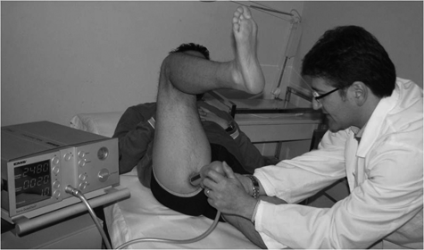
Application of shockwave treatment.
No ambulatory aids, immobilization, or other cointervention were used. Weightbearing and unrestricted range of motion were allowed immediately. Placement of an ice pack over the treated area for 15 to 20 minutes every hour was recommended for 4 hours after each SWT session.
Although daily life activities and training were permitted during the treatment period, patients were instructed to avoid activities and/or exercises that would increase the severity of their symptoms. Competitions were avoided, and the time to return to competitions was made on a case-by-case basis.
Traditional Conservative Treatment Group
Twenty patients were assigned to TCT (Table 1), consisting of rest (in the first week), an NSAID (in the first week), physiotherapy (in the first 2 weeks), and an exercise program (in the last 3 weeks).
Ibuprofen 600 mg twice daily (1200 mg total) was administered as the NSAID for the first week. Physiotherapy included ultrasound 11 in the continuous mode, with an intensity of 1.2 W/cm2, on a daily basis for the first 2 weeks, and transverse friction massage 6 3 days a week for the first 2 weeks. The exercise program included conventional stretching and strengthening exercises for hamstring muscles, performed 3 days a week for the last 3 weeks. Before starting each training session, participants performed a warm-up with 10 minutes of low-intensity stationary biking without resistance. The stretching protocol consisted of the following: sitting hamstring stretch with anterior pelvic tilt 4 × 20 seconds; standing hamstring stretch with anterior pelvic tilt and slow side-to-side rotation during the stretch, 4 × 20 seconds; and contract-relax hamstring stretch in standing with foot on stool, 4 sets of 10-second contraction and 20-second stretch. The strengthening protocol included isotonic exercises, performed using isotonic machines or free weights, in closed or open kinetic chain, were as follows: prone leg curls, 4 × 6 repetitions × 50% of 1 RM (1 repetition maximum); standing leg curls, 3 × 10 repetitions × 30% of 1 RM; standing hip flexion, 3 × 10 repetitions × 30% of 1 RM; standing hip extension, 3 × 10 repetitions × 30% of 1 RM; dead lift, 4 × 6 repetitions × 50% of 1 RM; alternate lunge with opposite trunk rotation and barbell over the shoulder, 3 × 10 repetitions × 30% of 1 RM; half squat, 4 × 6 repetitions × 50% of 1 RM; half-squat jump, 3 × 10 repetitions × 30% of 1 RM; and counter-movement jump, 3 × 10 repetitions × 30% of 1 RM.
Outcome Measures
Patients were assessed before treatment, and 1 week and 3, 6, and 12 months after the end of treatment by an independent treatment-blinded physician. The study procedure itself was conducted by a second physician who was aware of the treatment but who was not in any way involved in assessing the patients before or after the treatment.
No validated disease-specific questionnaires are available for PHT. Therefore, generic outcome measures (pain severity and recovery) were chosen as primary and secondary outcome measures.
Primary Outcome Measures
The first primary end point was a decrease of 3 points in the mean self-rated pain intensity score between baseline score and the score 3 months after the end of treatment. The self-rated pain intensity score was recorded on a 10-cm horizontal VAS with 0 cm labeled “no pain” and 10 cm labeled “worst pain I have ever had.”
The second primary end point was a 2-phase decrease in the mean of the Nirschl phase rating scale (NPRS) 25 between baseline and 3 months after the end of treatment. The NPRS is a 7-phase (1-7) assessment of pain and activity limitations caused by overuse injuries. 25 Phase 1 on this scale indicates mild pain after exercise that resolves within 24 hours. Phase 2 indicates pain after exercise that exceeds 48 hours but resolves with warm-up. Phase 3 indicates pain that accompanies exercise but does not alter activity. Phase 4 indicates pain that accompanies exercise and alters activity. Phase 5 indicates pain caused by heavy daily living activities. Phase 6 indicates intermittent pain at rest that does not disturb sleep or pain caused by light daily living activities. Phase 7 indicates constant pain at rest (dull ache) and pain that disturbs sleep.
Secondary Outcome Measures
The first secondary end point was the number of patients who achieved a reduction of at least 50% in the VAS score from the baseline to both 1 week after the end of treatment and 3 months after the end of treatment.
The second secondary end point was the degree of recovery from the baseline to 3 months after the end of treatment, measured on a 6-point Likert scale (“completely recovered” to “much worse”). Success rates were calculated by dichotomizing responses. Patients who referred to themselves as “completely recovered” or “much improved” were counted as successes, whereas patients who referred to themselves as “somewhat improved,” “same,” “worse” or “much worse” were counted as failures.
Statistical Analysis
The calculation of the number of patients was based on the first primary outcome. To detect a difference of 3 points in the VAS scores with a level of significance of 5% and a power of 80%, the necessary sample size was determined to be 17 patients per group. Assuming a dropout of 15%, 20 patients per group were required. The VAS scores were assumed to be 2 points in the SWT group and 5 points in the TCT group, with a common standard deviation of 3 points.
The primary efficacy end point was prospectively defined as a reduction of 3 points in the VAS score from baseline to 3 months after the end of treatment.
Statistical analyses were performed using SAS version 8.2 (SAS Institute Inc, Cary, North Carolina). All outcome analyses were performed according to the principle of intention-to-treatment. 18
A 2-way analysis of variance, with group as the between-patients factor and time as the within-patients factor, was used to assess the presence of significant differences between the SWT and TCT groups and within each group before treatment and at the scheduled follow-ups for the VAS score and NPRS. A Tukey post hoc comparison was used to assess significant differences between mean values when a significant main effect and interaction were found. The model for all the analyses included the main effects of treatment, time, and the treatment × time interaction. Significance levels for multiple comparisons were adjusted with the Bonferroni procedure. The level of significance was set at P < .05. A 95% confidence interval (CI) was also calculated.
A 2-sided χ2 test was carried out to compare the number of patients who achieved a reduction of at least 50% in the VAS scores in the SWT group with that in the TCT group 1 week and 3 months after the end of treatment; the level of significance was set at 5%.
The Fisher exact test was carried out to compare the percentage of success in Likert rating scale 3 months after the end of treatment; the level of significance was set at 5%.
A 2-sided χ2 test was carried out to compare the number of patients who were able to return to their preinjury level of sports activity; the level of significance was set at 5%.
Results
The patients were followed for a mean of 10.7 months (range, 1-12 months). Six patients were excluded from the further follow-up: 3 were excluded at 3 months; 2, at 6 months; and 1, at 12 months (Figure 1). Five (25%) of these patients were in the TCT group, while 1 (5%) was in the SWT group. All 6 patients were excluded because they had undergone a surgical intervention between the end of treatment and the follow-ups. Nevertheless, based on the intention-to-treat principle, 18 the data for these 6 patients were included in the data analysis.
Primary Outcome Measures
Regarding the first primary end point, 2-way analysis of the VAS score revealed a significant effect of treatment (F = 41.21, P <.001) and a significant treatment-time interaction (F = 48.13, P < .001). Three months after the end of treatment, a significant improvement in the mean VAS score (F = 38.75, P < .001) was observed in the SWT group. No significant difference was observed in the TCT group in the VAS score 3 months after the end of treatment (F = 8.33, P = 0.43).
The mean pain score in the SWT group was 7.1 ± 1.1 points at the baseline and 2.1 ± 1.9 points at 3 months after the end of treatment. The mean score in the TCT treatment group was 7.0 ± 1.9 points at the baseline and 6.8 ± 2.2 points at 3 months. At 3 months after the end of treatment, the between-group difference was 4.7 points (95% CI, 1.3-7.2; P < .001).
The mean changes in the VAS score from the baseline to 1 week and 6 and 12 months after the end of treatment are shown in Table 2.
Mean VAS Score at Baseline, and 1 Week and 6 and 12 Months After the End of Treatment in Both Groups a
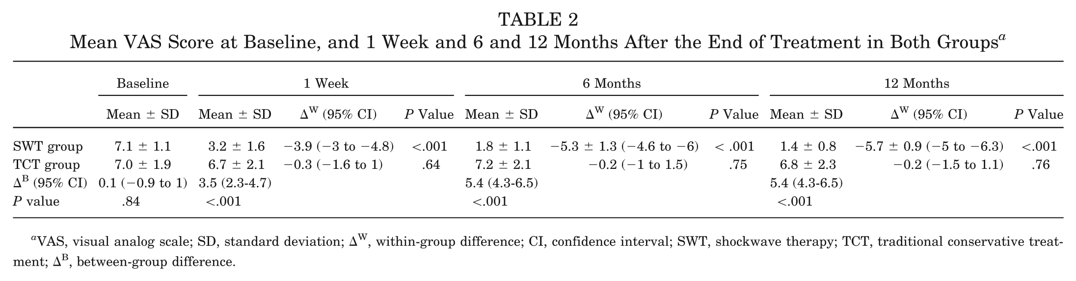
VAS, visual analog scale; SD, standard deviation; ΔW, within-group difference; CI, confidence interval; SWT, shockwave therapy; TCT, traditional conservative treatment; ΔB, between-group difference.
With regard to the second primary end point, a significant improvement in phase was observed on the NPRS (F = 45.21, P < .001) in the SWT group at 3 months after the end of treatment, whereas there was a significant worsening in the TCT group at the same time point (F = 17.26, P = .06). In the SWT group, the mean NPRS score improved from 5.1 ± 0.8 points at the baseline to 1.8 ± 1.0 points at 3 months after the end of treatment. In the TCT group, the mean NPRS score worsened, although not significantly, from 5.3 ± 1.0 points at the baseline to 5.5 ± 1.2 points at 3 months after the end of treatment. At 3 months after the end of treatment, the difference between groups was 3.7 points (95% CI, 1.4-4.8; P < .001).
The mean changes in the NPRS from the baseline to 1 week and 6 and 12 months after the end of treatment are shown in Table 3.
Mean NPRS Score at Baseline, and 1 Week and 6 and 12 Months After the End of Treatment in Both Groups a
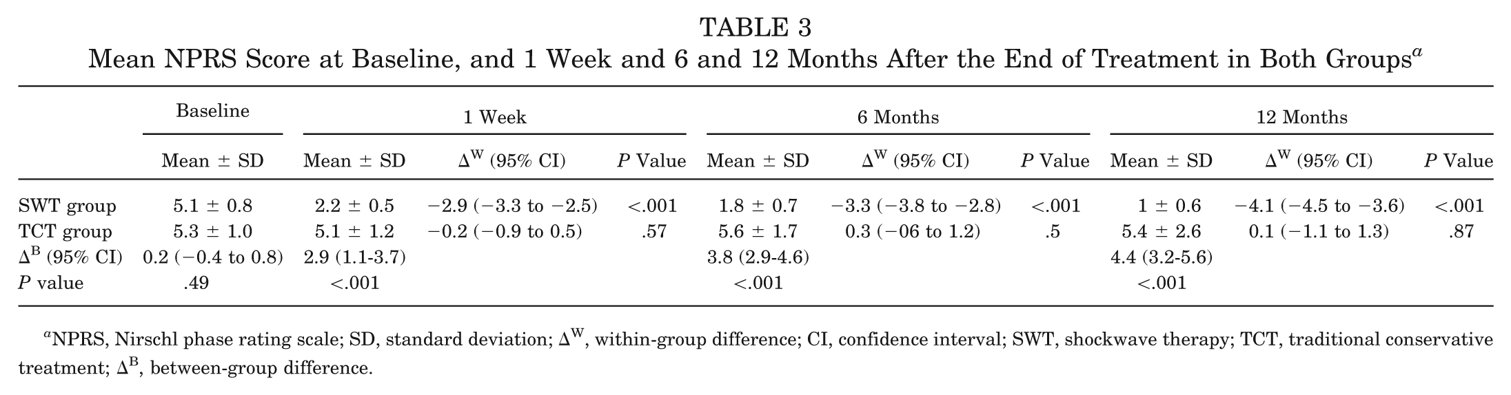
NPRS, Nirschl phase rating scale; SD, standard deviation; ΔW, within-group difference; CI, confidence interval; SWT, shockwave therapy; TCT, traditional conservative treatment; ΔB, between-group difference.
Secondary Outcome Measures
With regard to the first secondary end point, at 1 week after the end of treatment, the treatment was successful in 14 (70%) of the 20 patients in the SWT group compared with 4 of the 20 patients (20%) in the TCT group; significant differences emerged in the rate of successful treatment between the 2 groups (χ2 = 10.23, P < .001). At 3 months after the end of treatment, 17 (85%) of the 20 patients in the SWT group and 2 (10%) of the 20 patients in the TCT group achieved a reduction in pain of at least 50%; the differences in the rate of successful treatment between the 2 groups were significant (χ2 = 11.19, P < .001).
With regard to the second secondary end point, the Fisher exact test revealed that the percentage of patients with Likert scale scores of “1” (completely recovered) or “2” (much improved) (ie, successful results) at 3 months after the end of treatment was significantly higher in the SWT group than in the TCT group (P < .001).
At 3 months after the end of treatment, no patients in the SWT group but 7 patients (35%) in the TCT group reported a worsening in symptoms compared with the pretreatment symptoms.
The 1-week and 6- and 12-month Likert scores after the end of treatment for both groups are shown in Table 4.
Summary of Likert Scores for Both Groups a
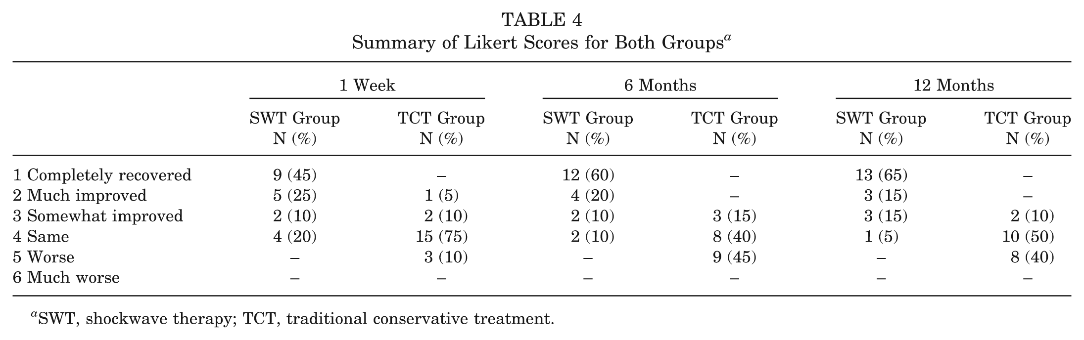
SWT, shockwave therapy; TCT, traditional conservative treatment.
Return to Sports Activity
Three months after the end of treatment, 16 (80%) of the 20 patients in the SWT group were able to return to their preinjury professional level of sports activity. The mean time taken to return to their preinjury professional level of sports activity was 9 weeks (range, 6-15 weeks). None of these patients have had any reinjury during the 12-month follow-up period. By contrast, none of the patients in the TCT group were able to return to their preinjury professional level of sports activity at the same time point (χ2 = 10.36, P < .001).
Discussion
Chronic PHT is an overuse syndrome of unknown origin that is associated with a degenerative process of the hamstring tendons, particularly of the semimembranosus tendon. 21 Clinically, patients with PHT report gradually increasing pain at the level of the ischial tuberosity. Some patients report radiating pain from the ischial tuberosity to the popliteal fossa while sitting for a prolonged time or during sports activities. However, no symptoms distal to the knee have been reported.
Previous studies have reported that results of TCT are somewhat unpredictable, with pain and tenderness frequently recurring. 21,42
The options afforded by surgery, which is indicated for cases that do not respond to nonoperative treatments, include a transverse tenotomy of the thickened semimembranosus tendon 3 to 4 cm distal to the origin, sutured to the biceps femoris tendon to prevent excessive retraction, followed by removal of any adhesions around the sciatic nerve, 21 or simply the release of the thickened fascia between the hamstring tendons, ischial tuberosity, and the sciatic nerve. 42
Unfortunately, as the studies involving surgery were based on retrospective case series, the results of surgical management cannot easily be compared with our results. Good results between 75% and 90% are reported after surgery. 21,42
The results of the current study show that the SWT treatment yielded better results than TCT and comparable results to surgery in the management of patients with chronic PHT. Results of TCT might be better after a longer period of training therapy.
More recently, Askling et al 3 showed that a stretching exercise can determine the onset of proximal hamstring tendon pain. However, previous studies suggest static stretching may help reduce injury rates 8,17,19 and improve recovery from injury 23,24 in the hamstring muscle-tendon complex.
As suggested by a recent in vitro study, low mechanical stretching may be beneficial to tendons by promoting differentiation of tendon stem cells into tenocytes, whereas large mechanical load stretching may be detrimental by promoting differentiation of some tendon stem cells into adipogenic, chondrogenic, and osteogenic lineages, resulting in lipid accumulation, mucoid formation, and tissue calcification, which are typical features of tendinopathy at later stages. 43 These data could explain why low mechanical load stretching and high mechanical load stretching may have contrasting effects on tendon tissue.
In our experience, a controlled and gentle stretching program did not exacerbate or provoke pain, and we do believe this form of treatment has a role in the management of chronic PHT.
The proportion of patients who returned to their preinjury level of sports of activity 3 months after treatment in the SWT group was 80% (20 patients), with a mean time lapse of 9 weeks (range, 6-15 weeks). It is noteworthy that the mean time taken by the SWT-treated patients in our study to return to their preinjury level of sports activity was lower than that taken by patients in a previous study who were treated surgically. 21
In this study, no local or regional anesthesia was used, all the patients tolerated the procedure well (there were no adverse complications), and none of the patients required further treatment.
Although the SWT mechanism of action in tendinopathies has not yet been fully understood, many authors 5,14-16,31-36,38,41 have achieved good results in the treatment of tendinopathies using SWT. The advantages of its use in the clinical setting include the stimulation of tendon tissue healing and the modulation of pain mechanisms. Some studies based on animal experiments have reported that SWT significantly increases the diffusion of cytokines across vessel walls into the pain-generating region, thereby stimulating the tendon healing response, 7 and significantly reduces the nonmyelinated sensory fibers, 27 calcitonin gene-related peptide, 39 and substance P release. 22 Other investigators have demonstrated that SWT acts on the pain system by means of hyperstimulation analgesia, which involves stimulation of a brainstem feedback loop through serotonergic activation via the dorsal horn that exerts descending inhibitory control over pain. 26
This study does have the following limitations: (1) the number of patients enrolled was small, although it did meet the power requirement; (2) the follow-up period was not long enough to determine the long-term effects of the SWT treatment and to assess its effects on the long-term quality of life in our patients; and (3) the lack of specific measurements.
However, the differences in scores between the 2 groups and within the SWT group itself were significant, and the results suggest that the use of SWT is not only safe but also more effective than TCT in the management of patients with chronic PHT.
The results of this study add to the growing number of favorable reports pointing to the efficacy of SWT as a treatment for chronic tendinopathies.
Conclusion
This study demonstrates that SWT is a safe and effective treatment for chronic PHT. Additional studies are needed to confirm these findings.
Footnotes
The authors declared that they had no conflicts of interests in their authorship and publication of this contribution.