
Abstract

Dear Editor:
With great interest we read the article by Pernin et al, “Long-Term Follow-Up of 24.5 Years After Intra-Articular Anterior Cruciate Ligament Reconstruction With Lateral Extra-Articular Augmentation,” in the June 2010 issue. 1 This study assessed the influence of medial compartment and medial meniscus status on the outcome of open anterior cruciate ligament (ACL) reconstruction in combination with a lateral extra-articular augmentation. Total medial meniscectomy and medial articular cartilage damage were shown to be risk factors for development of medial compartment osteoarthritis in this ultra long-term follow-up. We commend the authors on a very nice study that also demonstrates the need for continued long-term follow-up in all surgical patients.
In their surgical technique, the authors describe that the femoral tunnel position was determined by palpation of the over-the-top zone, then moving a few millimeters distally and forward. The tibial tunnels were placed in the posterior third of the ACL insertion site. The description of these tunnel positions may lead to an ACL reconstruction not being placed in an anatomic position, which can potentially increase the risk of degenerative changes following abnormal knee kinematics. 2 Anatomic ACL reconstruction may better restore normal native knee kinematics and therefore is hypothesized to decrease the progression of osteoarthritis. Indeed, from our clinical experience with long-term follow-up after ACL reconstruction, we see that degenerative changes occur to a lesser degree when the tunnels are anatomically placed. As can be appreciated in Figure 1, a more oblique tunnel placement of the femoral tunnel, 35° on AP radiograph, is indicative of a more anatomic position in this long-term follow-up after open ACL reconstruction. It can also be seen that minimal osteoarthritic changes have occurred.
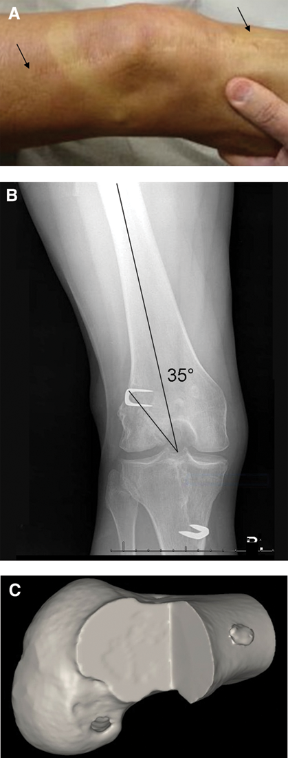
Patient 27 years after open ACL reconstruction of the right knee. A, The scar indicates an open procedure was performed. B, Radiograph shows a tunnel angle of 35° with minimal osteoarthritic changes. C, 3-dimensional computed tomography scan shows an anatomic femoral tunnel position.
We wonder if the authors considered ACL tunnel position as a risk factor for osteoarthritis in their study or in their discussion, and if they performed 3-dimensional computed tomography scanning or other imaging modalities to evaluate tunnel position. This type of imaging would be a valuable addition in determining ACL graft placement.
We recognize the importance of concomitant injuries and their effect after ACL reconstruction, and we would like to commend the authors on their efforts in conducting this meticulous and detailed study and providing us with a better insight in the development of osteoarthritis after ACL reconstruction.
Footnotes
One or more of the authors has declared the following potential conflict of interest or source of funding: Our institution receives research funding from Smith & Nephew for research on ACL reconstruction that is not related to the research presented in this contribution.