






Abstract
Arthroscopy of the hip is considered a safe and effective surgical procedure with a low frequency of complications. 22 Complications associated with the early history of hip arthroscopy focused on technical factors such as traction time, fluid management, and instrument breakage.13,39 With both technical advances and an improved understanding, the breadth of arthroscopic procedures has expanded to address both soft tissue and osseous disorders of the hip. 28 Arthroscopic osteoplasty for proximal femoral and acetabular dysmorphology associated with femoroacetabular impingement (FAI) has been reported with favorable clinical outcomes comparable with the results reported after open debridement and surgical dislocation. 5 However, these technical advances may have correspondingly introduced a new set of complications associated with hip arthroscopy.
While heterotopic ossification (HO) is a well-documented complication after open traumatic and reconstructive hip surgery, the incidence of HO after arthroscopic procedures of the hip remains undefined. 22 A 5% to 90% incidence of HO has been reported after open reduction of acetabular fractures or total hip arthroplasty.8,23 The variable incidence and severity of HO may be multifactorial and reflect differences in surgical technique, predisposing risk factors, and use of nonsteroidal anti-inflammatory drugs (NSAIDs) and radiation therapy for HO prophylaxis. 45 Documented risk factors for HO include male sex, history of HO in the ipsilateral or contralateral hip, ankylosing spondylitis, obesity, old age, and osteoarthritis.2,8
Most cases of postoperative HO are asymptomatic and commonly identified as incidental radiographic findings.8,17,35 In general, Brooker grades I and II HO have minimal symptoms and no significant effect on hip mobility. More severe Brooker grades III and IV HO present with stiffness and discomfort that compromise surgical outcomes.34,41,45 If symptoms are refractory and significantly impair function, a revision procedure to surgically excise focal areas of ectopic bone growth may be necessary. 8 The incidence of HO after open surgical treatment of FAI is not insignificant.14,18 In this regard, differences in the incidence and severity of HO between arthroscopic and open surgical approaches to the hip may be paramount in developing an effective treatment plan that minimizes morbidity and perioperative complications.
An improved characterization of the incidence and severity of HO after arthroscopic surgery of the hip may also help to define the role for routine prophylaxis. The literature includes no established protocols for HO prophylaxis after hip arthroscopy. In hip arthroplasty and acetabular fracture fixation, NSAIDs and radiation therapy are 2 well-established methods of preventing HO postoperatively. ¶ The purpose of this study was to report on the short-term incidence, severity, and location of HO in a consecutive large series of arthroscopies performed to treat both osseous and soft tissue disorders of the hip. We hypothesized that the incidence of HO after arthroscopic surgery is comparable with that after open surgical dislocation of the hip. A secondary purpose was to identify potential differences in the incidence of HO with the addition of indomethacin to an existing naproxen-based postoperative prophylaxis protocol after arthroscopic hip surgery.
Materials and Methods
This study was approved by our Institutional Review Board. From July 2008 to July 2010, 696 hip arthroscopies were performed by the senior author to treat FAI, snapping hip syndrome, or peritrochanteric space disorder (PSD) in patients with pain and/or limited range of motion. These cases were subsequently screened to identify and exclude all patients with significant chondral degeneration (greater than or equal to Tonnis II changes), previous hip surgery, or existing HO at the time of presentation. In addition, patients with diagnosed and surgically treated pigmented villonodular synovitis, synovial chondromatosis, Legg-Calve-Perthes disease, and slipped capital femoral epiphysis were excluded from the study. After the screening process, 616 of the 696 surgical cases met the inclusion criteria. These hips were all diagnosed with and treated for FAI, internal snapping hip syndrome (ie, psoas tendon), and/or PSD (Figure 1).

Selection criteria flow chart. PVNS, pigmented villonodular synovitis; SC, synovial chondromatosis; SCFE, slipped capital femoral epiphysis.
All patients undergoing surgery were entered into a prospective database and managed with a standardized postoperative rehabilitation program. This included immediate range of motion and directed physical therapy aimed at restoration of hip abductor and flexor strength, with continuous passive motion daily for 6 weeks.
Nonsteroidal anti-inflammatory drugs were routinely used for HO prophylaxis. Between July 2008 and July 2009, patients received naproxen (Naprosyn EC, Roche, Nutley, New Jersey) 500 mg twice daily for 30 days (protocol 1) (Figure 2). After July 2009, this protocol was modified to include indomethacin (Indocin SR, Merck, Whitehouse Station, New Jersey) 75 mg daily for 4 days followed by naproxen (Naprosyn EC, Roche) 500 mg twice daily for 30 days (protocol 2). To offset potential gastrointestinal complications due to indomethacin, the patients received omeprazole (Prilosec, AstraZeneca, Wilmington, Delaware) 20 mg daily for the first 4 postoperative days.

Heterotopic ossification prophylaxis protocol.
The change in protocol was implemented to eliminate the infrequent postoperative occurrence of HO after arthroscopic hip surgery that was observed by the senior author. Peer-reviewed trauma literature suggested that the addition of indomethacin may be more effective than other nonsteroidal medications in prophylaxis against HO after acetabular fracture and soft tissue injury.3,11,30 The decision to use the combined approach of a short course of indomethacin and omeprazole followed by naproxen for chemoprophylaxis was based upon an evidence-based review of the literature and discussion with gastroenterologists at our institution. The goal was to maximize the benefits of HO prophylaxis attributable to indomethacin while minimizing any potential significant systemic side effects, most notably ulcer disease and gastrointestinal bleeding.
Statistical analysis was completed to compare the demographics between cohorts treated with HO prophylaxis protocols 1 and 2. Patient age, gender, diagnosis, and surgical procedure were determined for all cases (Table 1). Further analysis of the surgical procedure was completed to define the number of osteoplasties for femoral, acetabular, or combined deformity as well as the ratio of labral debridements to labral refixations among patients diagnosed and surgically treated for FAI. To consolidate data on hips with varying soft tissue injury, these procedures were all categorized as treatments for PSD or snapping hip syndrome and included iliopsoas release, gluteus medius repair, adductor release, piriformis release, trochanteric bursectomy, and iliotibial band lengthening. The preoperative alpha angle was also defined for isolated cam and combined FAI cases to compare the severity of deformity between treatment groups respectively (Table 1).
Patient Demographics a

SD, standard deviation; LD, labral debridement; LR, labral refixation; PSD, peritrochanteric space disorder (iliopsoas release, gluteus medius repair, trochanteric bursectomy, iliotibial band release, adductor release, piriformis release).
Shown as n (%), with % value = N/protocol total × 100.
All patients had follow-up at 10 days, 6 weeks, 3 months, 6 months, 1 year, and 2 years postoperatively. Physical examinations were performed to document range of motion and manual resisted strength testing of the involved and contralateral hips. Radiographic images, including an anterior-posterior and elongated femoral neck view (Dunn lateral) of the hip, were obtained at 6 weeks and 1 year. Patients with radiographic evidence of HO and discomfort in the hip had computed tomography (CT) scans to further characterize the location and severity of heterotopic bone formation. Multidetector helical CT scans were obtained through the hip utilizing 0.625-mm slice thicknesses. Sagittal and coronal reformatted images were obtained. In addition, 3-dimensional (3-D) volume–rendered images and version analysis were obtained using GE Advantage Windows Workstation, Volume Viewer software (Fairfield, Connecticut). Patients with refractory symptoms and failure to respond to nonoperative treatment underwent surgical exploration and resection of HO.
Heterotopic ossification was classified using the Brooker system (Figure 3). 10 Plain radiographs were independently reviewed by 3 of the authors to determine the Brooker grade and approximate the location of HO. The reported grade represents a consensus of the 3 reviewers. Computed tomography scans were examined to gain a better understanding of the specific soft tissue affected (joint capsule, rectus femoris tendon, or abductor musculature).

Brooker classification. 10 (A) Grade I is isolated bony islands. (B) Grade II is bone spurs from the pelvis or proximal end of the femur with more than 1 cm of space between opposing surfaces. (C) Grade III is bone spurs from the pelvis or proximal end of the femur with less than 1 cm of space between opposing surfaces. (D) Grade IV is bridging or ankylosis of the hip. Reproduced with permission of the British Editorial Society of Bone and Joint Surgery. 8
Statistical Analysis
All statistical evaluations were carried out using SAS 9.1.3 (SAS Institute, Cary, North Carolina). Odds ratios were calculated to investigate associations between protocols as well as surgical characteristics and the development of HO, with P < .05 defined as significant. In addition, a multiple logistic regression model was developed to further evaluate predictors of HO formation and compare demographics between the treatment groups.
Results
Patient Demographics
Of 616 arthroscopic surgeries, 29 hips (4.7%) were identified with postoperative HO on plain radiographs. These included 21 male hips (72.4%) and 8 female hips (27.6%). The average age of the patient at the time of surgery was 30.6 years (range, 15-57 years). The mean clinical follow-up time was 13.2 months (range, 2.9-26.5 months) (Table 1).
All cases of HO occurred in patients treated with osteoplasty for symptomatic FAI. Of the 29 hips with postoperative HO, 7 hips (24.1%) had isolated cam impingement treated with a femoral osteoplasty, and 2 hips (6.9%) had isolated rim impingement lesions treated with an acetabular rim resection. Twenty hips (69.0%) had both rim and cam impingement lesions and were treated with a combined femoral osteoplasty and acetabular rim resection. Both of the isolated rim resections and 1 of the 20 combined procedures had an iliopsoas lengthening via a transcapsular approach.
An analysis of demographics was performed to compare the patient populations that received the naproxen-based HO prophylaxis protocol (protocol 1) and the indomethacin-modified protocol (protocol 2) (Table 1). The mean age was 32 ± 12.1 years and 30.7 ± 11.2 years for protocols 1 and 2, respectively, and was not significantly different (P = .16). In addition, no significant differences in gender (χ2 = 0.0012, P = .97) or treated diagnoses of isolated rim, cam, or combined deformity (χ2 = 4.99, P = .17) or soft tissue injury/PSD (χ2 = 3.82, P = .051) were noted between treatment groups. A small but statistically significant difference in the alpha angle was noted between groups, with mean alpha angles of 56.72° ± 12.41° and 58.95° ± 12.59° in group 1 and group 2, respectively (P = .0043). In addition, a significantly greater number of labral repairs were performed in group 2 compared with group 1 (χ2 = 175.23, P < .0001) (Table 1).
HO Classification
Brooker classification of HO at final follow-up revealed 18 hips (62.1%) with grade I HO, 4 hips (13.8%) with grade II HO, 6 hips (20.6%) with grade III HO, and 1 hip (3.5%) with grade IV HO. 10 Fourteen hips (48.3%) had HO located anterior to the joint, and 15 hips (51.7%) had HO located lateral to the joint.
Effect of HO Prophylaxis
Two hundred seventy-seven (45.0%) of the 616 hips received protocol 1 HO prophylaxis. Twenty-three (79.3%) of the 29 hips with HO belonged to the protocol 1 treatment group. There were 13 hips with grade I HO, 4 hips with grade II HO, 5 hips with grade III HO, and 1 hip with grade IV HO (Figure 4). The probability of developing HO with only naproxen for HO prophylaxis was 8.3% (23/277).
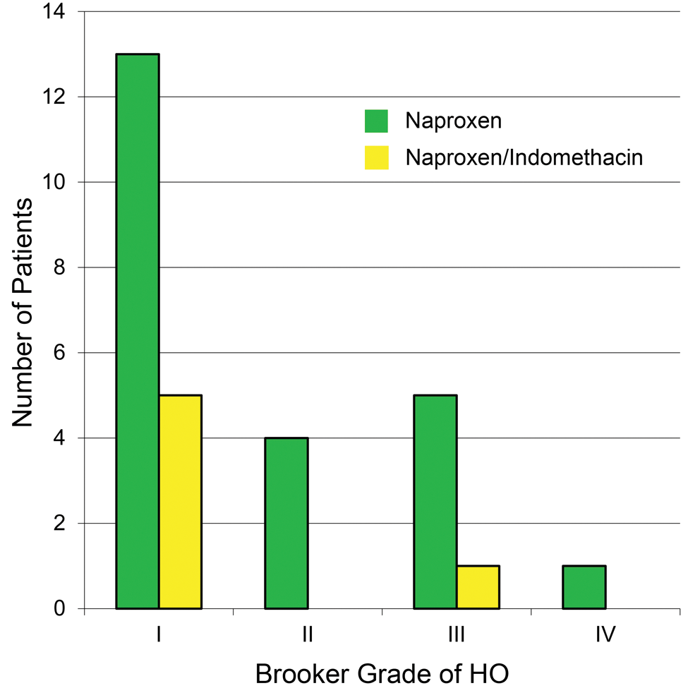
Distribution of heterotopic ossification in both groups according to the classification system of Brooker et al. 10
Three hundred thirty-nine (55.0%) hips received protocol 2 HO prophylaxis. In this group, only 6 hips of the 339 surgeries (1.8%) that included indomethacin along with naproxen for HO prophylaxis developed HO postoperatively. This was 20.7% (6/29) of the total cases of HO and consisted of 5 hips with grade I HO and 1 hip with grade III HO.
Patients who underwent hip arthroscopy in our series and had HO prophylaxis before July 2009 (protocol 1) were 4.36 times more likely (95% confidence interval [CI], 1.72-10.97) to develop HO postoperatively than those who had protocol 2 (P < .05). The odds of developing Brooker grade I were greater for those on protocol 1 (odds ratio [OR], 3.86; 95% CI, 1.36-10.73) than protocol 2 (P < .05). There were no statistically significant differences in the odds of developing more severe (greater than grade I) HO between the 2 protocols; however, this was likely because of the small number of cases available, resulting in limited statistical power.
Surgical Treatment
Among the 29 hips that developed HO postoperatively, 7 underwent revision surgery to excise ectopic bone at a mean duration of 11.6 months (range, 5.2-16.2 months) after the index procedure. All of these hips had ectopic bone located anterior to the joint. Six hips received protocol 1 HO prophylaxis, and 1 hip received protocol 2 HO prophylaxis. These patients included 6 men and 1 woman, with an average age of 33.0 years (range, 23-57 years). The decision to perform revision surgery was made on an individual basis and was based on the severity of pain and restricted range of motion despite an extensive course of nonoperative rehabilitation. There were 2 cases of grade I HO, 1 case of grade II HO, 3 cases of grade III HO, and 1 case of grade IV HO. The 3 hips with either grade I or grade II HO underwent arthroscopic procedures to remove the bone. The other 4 hips with either grade III or grade IV HO required an open excision of bone and a capsulotomy. Immediately after revision surgery, each hip was given a single dose of 700 cGy of radiation therapy as prophylaxis for HO; only 1 of the 7 patients received postoperative indomethacin because of sensitivity or allergy to NSAIDs.
There were no statistically significant associations found between the type of arthroscopic procedure performed and the development of HO. This is likely because of the relatively small number of patients who developed HO postoperatively and the fact that the majority of cases were performed for symptomatic FAI. However, the data clearly demonstrate that the performance of arthroscopic osteoplasty with a capsular cut in male patients represented the majority of cases, and this group is likely at highest risk. Seven cases (~1%) required revision procedures to excise HO. Six of the 7 cases requiring excision of HO were male patients, and all 7 had undergone arthroscopic osteoplasty with a capsular cut. Six of 7 cases were not treated with indomethacin postoperatively because of NSAID sensitivity or allergy. There were no cases of recurrence of HO after excision whether it was performed open or arthroscopically.
Multiple logistic regression was carried out to explore relationships between HO and predictor variables while controlling for interactions. Variables included type of procedure performed (soft tissue, acetabular, femoral, both acetabular and femoral osteoplasty) and prophylaxis protocol. Only prophylaxis protocol 1 was found to be a significant predictor for the development of HO relative to protocol 2 (OR, 4.36; 95% CI, 1.72-10.97).
Discussion
The primary purpose of this study was to define the incidence, severity, and location of HO in a large series of hip arthroscopies performed by the senior author to surgically manage soft tissue and osseous disorders of the hip. We hypothesized that the incidence and severity of HO after hip arthroscopy would be comparable with that reported after open surgical dislocation to treat FAI. In the current series, we determined an overall incidence of 4.7% (29/616) of postoperative HO after arthroscopic hip surgery. The incidence of HO for patients who received only naproxen was 8.3% (23/277) and was significantly reduced to 1.8% (6/339) after implementation of indomethacin into the postoperative prophylaxis protocol (P < .05). The overall incidence of HO after arthroscopic FAI surgery in this series (4.7%) was lower than in most studies reporting the incidence after open surgeries, although the range is wide (2.7%-37%). Patient age, gender, and treated diagnoses were not significantly different between treatment groups. Only 7 cases (~1%) with refractory symptoms required a revision surgery to excise ectopic bone. In all of these revision cases, HO was located anterior to the hip joint. In addition, these cases included 6 in protocol 1. Based on our results, HO is an uncommon but significant complication after hip arthroscopy and is more common in male patients treated with osteoplasty for symptomatic FAI. Revision surgery to excise HO may be required in patients who are experiencing refractory pain and/or restricted motion. The combination of indomethacin and naproxen may be a more effective prophylaxis than naproxen alone for HO after hip arthroscopy.
Before the current study, HO has been a well-documented complication after open traumatic and reconstructive hip surgery. The incidence of HO after hip replacement surgery ranges from approximately 20% to 30%. 38 In a retrospective review of 5122 total hip arthroplasties without HO prophylaxis, Amstutz et al 1 reported 1719 (34%) cases of HO. The incidence of more severe types of HO with pain and stiffness was approximately 14.5%. 1 A 5% to 90% incidence of significant HO has been reported after open reduction of acetabular fractures. Karunakar et al 23 reported 21 patients (17.3%) with Brooker grades III and IV HO after operative treatment of displaced acetabular fractures.
Heterotopic ossification has also been reported as a complication after open surgical procedures to address FAI. In their initial report on the technique of surgical dislocation of the adult hip, Ganz et al 18 reported a 37% rate of HO. This consisted of 68 grade I, 9 grade II, and 2 grade III cases of HO. The most common site of HO in this series was at the tip of the greater trochanter. However, the 2 hips with grade III HO had formation of new bone at the acetabular rim that ultimately required excision. 18 In contrast, Beaulé et al 4 reported 1 case (2.7%) in a series of 37 hips treated with open femoral head-neck osteochondroplasty for FAI. This patient developed grade IV HO that required excision at 10 months postoperatively. 4 Studies on combined arthroscopy and open head-neck osteochondroplasty have also included postoperative complications related to HO. Clohisy et al 14 reported 4 cases (11.4%) of grade I HO in a series of 35 patients who underwent a combined closed and open procedure. None of these patients required additional surgery. Using a similar combined approach, Laude et al 29 reported 1 case (1%) of HO in 100 hips treated for FAI. This patient developed grade II HO and underwent revision surgery at 33 months postoperatively.
The incidence of HO after hip arthroscopic osteoplasty remains poorly defined. Historically, HO is considered a rare occurrence after arthroscopy of any joint. There are only 4 existing reports of HO after elbow arthroscopy.19,21,40,41 In addition, arthroscopic shoulder procedures such as subacromial decompression, distal clavicle resection, and rotator cuff repair carry a small risk of postoperative complications due to symptomatic HO.6,7,9,16,25 However, hip arthroscopy for FAI offers unique technical challenges compared with these other arthroscopic procedures. The osseous resection is often substantial, and the restricted access, distinct compartments, and investing musculature may alter the risk for HO compared with other arthroscopic procedures. 31
In a review of 300 hip arthroscopies for treating FAI, Randelli et al 37 reported 5 cases of HO (1.6%). These cases were part of a control group (n = 15) that did not receive any HO prophylaxis because of either patient noncompliance or contraindications to NSAIDs. The treatment group (n = 285) received a nonstandardized NSAID protocol. 37 Larson and Giveans 27 reported 6 cases (6.3%) of HO in a series of 96 hips treated arthroscopically for FAI. Only one of these patients developed a significant functional limitation, resulting from ossification of the iliopsoas tendon. This motion deficit was nearly completely resolved at 1 year and required no further treatment. Of 207 hips treated arthroscopically for FAI, Byrd and Jones 12 reported 1 case (<1%) of capsular heterotopic bone formation. Although the grade of HO was not reported, this patient achieved a high Harris Hip Score.
The overall rate of HO in the current study (4.7%) is lower than that reported in comparable large series of open surgical dislocation for FAI.14,18 Open surgical dislocation of the hip joint to treat FAI is a fairly extensive procedure that requires a trochanteric osteotomy and mobilization of the abductor musculature. For this reason, arthroscopic treatment of FAI may help reduce the incidence of HO relative to open and combined approaches. Arthroscopic surgery may also benefit from continuous irrigation of the joint and periarticular tissues that evacuates hematoma and bone debris, both widely recognized precursors to HO.2,38 Some learned key points and an algorithm employed by the senior author to minimize the incidence of HO after hip arthroscopy are provided in Figure 5.
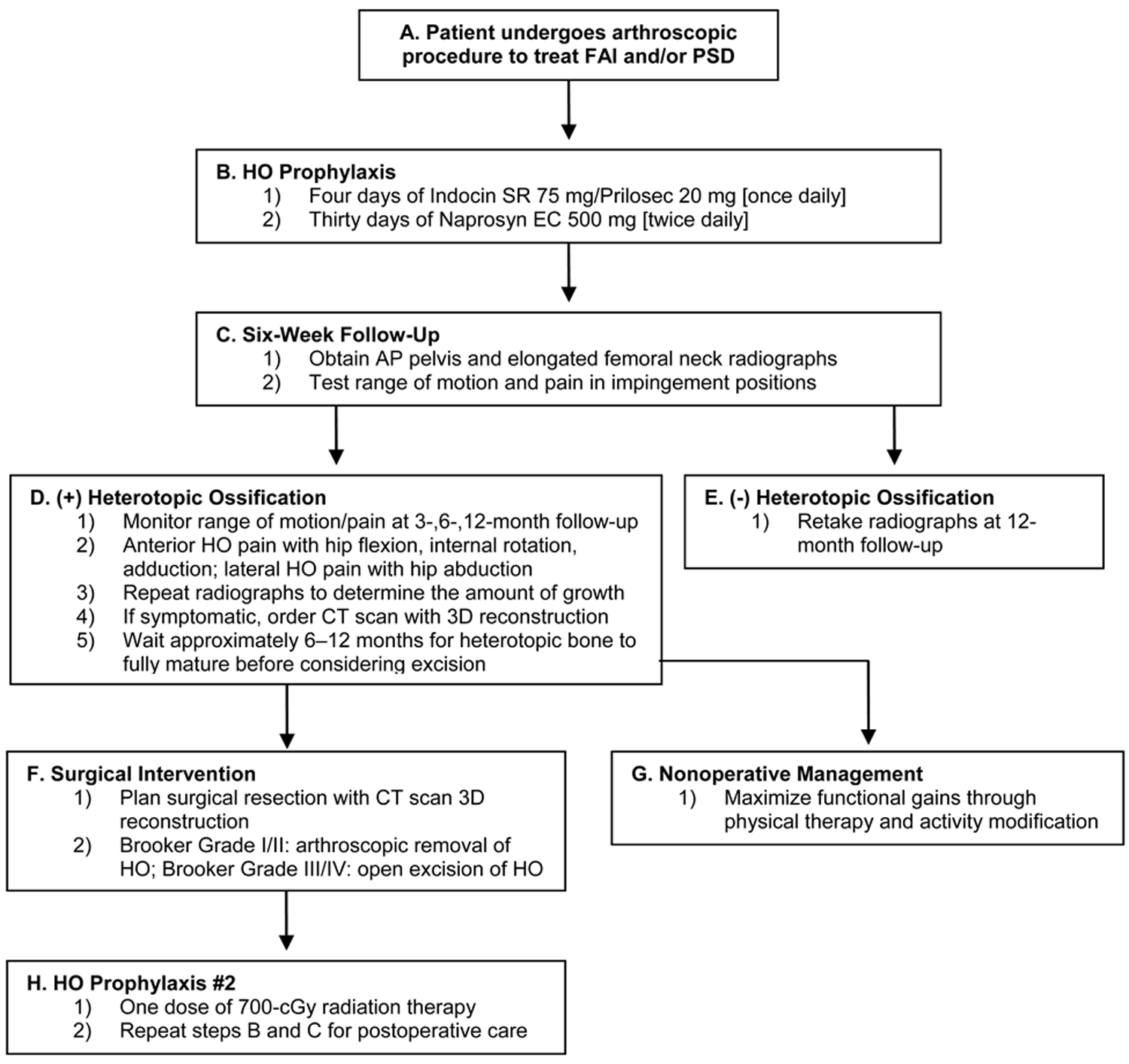
Flow chart of strategies.
The difference in the incidence of HO between open and arthroscopic procedures based on the current study must be interpreted with some caution. This study includes a substantial amount of surgical procedures addressing extracapsular soft tissue injury in the hip, including PSD and snapping hip disorder (19.6%). There is also an inherent difficulty in comparing the incidence of HO between closed and open approaches for treating FAI because there is a tendency among authors to focus only on significant complications rather than asymptomatic cases of HO that present as incidental radiographic findings. Moreover, differences in technique among surgeons performing either open, closed, or combined approaches most likely contribute to the variability in HO incidence and render an objective analysis difficult.
With regard to prophylaxis, the risk of postoperative HO was higher in patients who only received naproxen. Our initial NSAID protocol was 30 days of Naprosyn EC (Roche) 500 mg twice daily. During this time, there was an 8.3% (23/277) incidence of HO (Figure 6). The rate of HO decreased to 1.8% (6/339) when the protocol was switched to 4 days of Indocin SR (Merck) 75 mg followed by 30 days of Naprosyn EC (Roche) 500 mg twice daily (Figure 7).

Anteroposterior pelvis radiograph of a 27-year-old male hip with Brooker grade IV heterotopic ossification (HO) (arrow) taken approximately 9 months after an arthroscopic femoral osteochondroplasty and acetabular rim resection. The patient did not take any indomethacin for HO prophylaxis. Because of severe pain and stiffness, an open revision procedure was performed 9 months after the primary surgery. The patient received 1 dose of 700 cGy of radiation therapy immediately after surgery. At the time of writing, the patient has had no recurrence of HO, and his preoperative symptoms have been resolved.

Anteroposterior pelvis radiograph of a 48-year-old male hip with Brooker grade I heterotopic ossification (arrow) taken approximately 6 months after an arthroscopic femoral osteochondroplasty and acetabular rim resection. The patient took indomethacin for 4 days after surgery and had minimal to no symptoms.
Although multiple studies have compared the effectiveness of different NSAID protocols after hip replacement surgery and surgical treatment of acetabular fractures, there is no established regimen in the literature for HO prophylaxis after hip arthroscopy. The effectiveness of indomethacin as a prophylaxis for HO was first credited to Dahl 15 and has since been demonstrated in numerous studies.1,26,32,33,43 However, various reports exist that question its efficacy. Karunakar et al 23 reported no significant difference in the incidence of severe HO between patients receiving indomethacin and those receiving a placebo after surgical treatment of acetabular fractures. There is also no clear indication that indomethacin is more effective than naproxen in preventing HO. In a clinical trial, Vielpeau et al 45 demonstrated that naproxen was at least as effective as indomethacin as a prophylaxis for HO after hip arthroplasty.
Despite these conflicting reports, our data support the implementation of an indomethacin-based protocol for preventing HO after arthroscopic procedures of the hip. In patients with symptomatic HO after hip arthroscopy, we recommend a standardized treatment regimen (Figure 5). Range of motion exercises and physical therapy should be continued to maximize functional gains and obviate the need for revision surgery. If the patient continues to have pain or functional limitations, it is important to correlate the symptoms with the location of heterotopic bone. Heteropic ossification localized anterior to the joint in either the joint capsule, rectus femoris, or iliopsoas may result in continued impingement-type pain with flexion, adduction, and internal rotation of the hip. In contrast, HO located lateral to the joint may cause discomfort with hip abduction. Severe pain and stiffness caused by HO may ultimately warrant a revision procedure to excise ectopic bone growth in the hip. In this large clinical series, all symptomatic cases that required surgery had HO anterior to the hip joint. The data support the senior author’s clinical experience that anterior HO is associated with greater pain and symptomatic restriction in hip flexion. In addition, the 3 patients with Brooker grades I and II HO who underwent revision surgeries demonstrate that a relatively small amount of heterotopic bone can be highly symptomatic. For this reason, the decision to proceed with a revision surgery should involve a wide range of factors that include the patient’s clinical presentation and location of HO as well as the Brooker grade. Lastly, we have found that CT scans with 3-D reconstructions are helpful in localizing the lesion and planning surgical resection (Figure 8). Depending on the size and location of heterotopic bone, either an arthroscopic or open excision may be performed. After revision surgery, radiation therapy (1 × 700 cGy) is recommended to prevent recurrence of HO (Figure 5). Of these 7 revision cases with a minimum follow-up of 12 months, there were no cases of recurrent HO, and all patients had clinical improvement in their symptoms and range of motion. There were no differences in outcome comparing arthroscopic excision with open excision, and all patients were able to return to a preinjury level of function. Formal statistical analysis of these patients was not informative because of the small numbers and limited power; however, clear improvement in symptoms was seen in all symptomatic cases. The decision to proceed with open versus arthroscopic excision was based on the size and location of the HO.

Computed tomography (CT) scan of Brooker grade III heterotopic ossification. The CT scan can be a useful tool in preoperative planning for removal of heterotopic bone. In this instance, the bone is located anterior to the hip, most likely deposited in the iliopsoas muscle-tendon unit. Multidetector helical CT scans were obtained through the hip utilizing 0.625-mm slice thicknesses. Sagittal and coronal reformatted images were obtained. Three-dimensional volume–rendered images were obtained using GE Advantage Windows Workstation, Volume Viewer software.
In the current study, no increased morbidity associated with indomethacin was appreciated in terms of bleeding, gastric ulcers, decreased platelet function, and renal toxicity. Patients with gastrointestinal problems or NSAID allergies may have trouble tolerating indomethacin and naproxen and should consider radiation therapy as an alternative prophylactic strategy. Although there is no statistically significant or clinically important difference between the use of NSAIDs and radiation therapy for preventing HO, patients may be reluctant to voluntarily receive radiation therapy because of higher costs and risks that include malignancy, infertility, and genetic alteration.34,42,44
There are several important limitations associated with this study. Although the large consecutive series of hip arthroscopies provides a substantial amount of evidence to support our findings, the retrospective design of our study does not establish a clear causation. In addition to the modification of our HO prophylaxis protocol, the evolution of surgeon experience and possible subtle modifications of technique during this period may present confounding variables. Analysis of demographics did demonstrate equivalence of age, gender, and diagnosis between treatment groups. However, group 2 did have a greater number of labral repairs and small but significantly larger femoral deformity as defined by the alpha angle (P < .05). In this regard, patients receiving protocol 2 may have benefited from improving technical expertise of the senior author with atraumatic portal placement, minimization of muscular trauma, and cannula-based evacuation of osseous debris during osteoplasty. On the other hand, both increased femoral deformity and increased labral repair reflect greater technical challenge and would be expected to only increase the risk of HO formation in group 2. An ideal control group could also have included a group without chemoprophylaxis and one without the acute indomethacin prophylaxis, although this was and remains limited by the ethics of withholding prophylaxis in light of the known benefits and risk.
A future randomized controlled trial is advised to further build upon these preliminary findings. Future studies should continue to evaluate the effect of surgical technique and prophylaxis on rates of HO. These studies should also consider monitoring patient compliance. Because of gastritis and other side effects, patient compliance during long-term courses of strong NSAIDs such as indomethacin and naproxen may be as low as 50%. 20 This low level of compliance has the potential to distort findings on the effectiveness of any long-term prophylaxes.
Conclusion
In conclusion, symptomatic HO is an observed complication after hip arthroscopy. The incidence of HO was 1.8% in the group of patients who received indomethacin in conjunction with naproxen for prophylaxis, compared to 8.3% in the group that received naproxen alone. The majority of cases of HO (72.4%) occurred in male patients, and all cases occurred in the setting of osteoplasty performed for symptomatic FAI. Indomethacin-based NSAID protocols for HO prophylaxis should be considered after hip arthroscopy in this patient population.
