
Abstract
Background:
It is known from clinical and experimental studies that the healing potential of the anterior cruciate ligament (ACL) is extremely poor and that early phases of ligament healing require an augmented blood supply. MicroRNA (miRNA) is a type of small, noncoding RNA that negatively regulates gene expression, and miRNA (miR)-210 is reported to be crucial for cell response to hypoxia, vascular endothelial growth factor (VEGF)–driven endothelial cell migration, and formation of capillary-like structures.
Purpose:
The purpose of this study was to examine the effect of intra-articular injection of miRNA miR-210 on acceleration of ACL healing.
Study Design:
Controlled laboratory study.
Methods:
Two experiments were performed in this study. The ACLs of 12-week-old male LEW/CrlCrlj rats were partially transected. First, the temporal expression change of miR-210 after ACL injury was analyzed using real-time polymerase chain reaction (PCR) on day zero, and 1, 2, and 4 weeks after injury (n = 5 at each time point). Next, intra-articular injection of double-stranded (ds) miR-210 with atelocollagen was performed soon after injury. The control group was injected with control small interfering RNA (siRNA). Four weeks after injection, biomechanical and histological assessments of samples stained with H&E as well as Masson trichrome, and immunohistochemistry for VEGF, fibroblast growth factor 2 (FGF2), isolectin B4, and collagen type I, were performed. Real-time PCR analysis was also performed for quantitative evaluation of miR-210, VEGF-A, and collagen type I.
Results:
Real-time PCR analysis revealed that miR-210 expression was decreased soon after injury but gradually increased thereafter. Histological analysis confirmed that the transected area was covered with healing tissue in the miR-210 group but remained devoid of any tissue in the control group 4 weeks after injury. Biomechanical analysis confirmed the improvement of biomechanical properties in the miR-210 group; the ultimate failure loads 4 weeks after injection were 30.5 ± 3.1 N in the miR-210 group and 22.8 ± 3.1 N in the control group (P < .05). Real-time PCR analysis showed that endogenous miR-210, VEGF, and collagen type I were highly expressed compared with controls, and immunohistochemistry for VEGF, FGF2, isolectin B4, and collagen type I showed that VEGF and FGF2 were highly upregulated, and there were abundant blood vessels and fibrotic deposition in the miR-210 group.
Conclusion:
Injection of ds miR-210 was effective in promoting the healing of partially torn ACLs through enhancement of angiogenesis via upregulation of VEGF and FGF2.
Clinical Relevance:
It might represent a potential therapeutic approach for treatment of ACL injury.
The anterior cruciate ligament (ACL) is the major stabilizer of the knee and functions to limit rotation and anterior translation of the tibia. Injuries to the ACL have become increasingly prevalent and account for a large proportion of knee ligament injuries among young, active individuals. 4 A completely torn ACL rarely heals spontaneously, and most partial ACL tears eventually progress to complete rupture. 47 Hefti et al 22 reported no regeneration after complete ACL transection in rabbits, and very slow and incomplete regeneration after partial rupture. Similar results have been shown in other species. Failure of the ACL to heal causes instability of the knee, which usually leads to early osteoarthritis of the affected knee. 35 Thus, repair or reconstruction of the ACL after rupture is important for the stability of the knee and prevention of future osteoarthritis. At present, ACL reconstruction with intra-articular grafts is the most broadly accepted procedure for the young and active population.9,14 Anterior cruciate ligament reconstruction has been demonstrated to improve clinical instability of the knee joint, reduce knee laxity, and decrease risk of late meniscal tear and surgery.1,15,16,26,28,43 However, ACL reconstruction does not fully restore functional dynamic stability of the knee,20,46 and the return rates to preinjury activity level vary from 37% to 75%.14,19,51 It is hoped that stimulating healing of the native structure rather than replacing it will have a protective effect on the mechanics and subsequently the articular cartilage of the knee. 39
Several studies have been reported in an attempt to enhance the understanding of ligament development. It has been reported that synovial fluid of the knee with synovitis is a hypoxic condition,17,32 and that the blood supply of the ACL is restricted to small branches of the middle geniculate artery that diverge on the ligament surface.6,49 Furthermore, it is known that early phases of ligament healing require an augmented blood supply; however, angiogenesis and regeneration in the injured ACL occur at a very slow rate, and their magnitude increase does not reach a sufficient level for appropriate healing to occur. 5 Numerous studies have attempted to determine the effects of various growth factors and genes on the ligament engineering system with regard to cell proliferation, extracellular matrix (ECM) synthesis, neovascularization, and mechanical properties.31,36,37,44,50
MicroRNAs (miRNAs) are 20- to 22-nucleotide, small, noncoding RNA molecules that negatively regulate gene expression at the posttranslational level.2,3,11 Many miRNAs play roles in tissue specialization and tissue maintenance 34 ; they also take part in a variety of other cellular processes, including differentiation, proliferation, and apoptosis. The results of several therapeutic trials examining the regulation of endogenous miRNAs that are related to disease pathogenesis through in vivo administration of specific antisense oligoribonucleotides or double-stranded (ds) miRNAs have been reported.10,33,48 Recently, several groups have reported that miRNA (miR)-210 is a key player in cell response to hypoxia, and the relevant target is ephrin-A3 (EFNA3), a molecule that has essential functions in angiogenesis. 12 It has also been reported that miR-210 upregulation is a crucial element of endothelial cell response to hypoxia, which affects cell survival, migration, and differentiation. Even in normoxic conditions, its overexpression can stimulate the formation of capillary-like structures in vitro when cells are cultured in Matrigel, as well as vascular endothelial growth factor (VEGF)–driven cell migration.12,13,25 In consideration of these results, we hypothesized that miRNA-210 administration by intra-articular injection accelerates angiogenesis, thereby augmenting ACL healing.
The purpose of this study was to investigate the expression pattern of miR-210 in injured ACLs and to examine the effect of intra-articular injection of synthetic miR-210 on ACL healing in a rat model.
Materials and Methods
All procedures were performed according to the Guidelines for Animal Experimentation, Hiroshima University, and with approval of the Committee of Research Facilities for Laboratory Animal Sciences, Graduate School of Biomedical Sciences, Hiroshima University.
Animals
We used 12-week-old male LEW/CrlCrlj rats (Charles River Laboratories Japan, Tokyo, Japan) in these experiments. The rats were housed at the Laboratory Animal Center of Hiroshima University under standard diurnal conditions of light/dark. They were fed a standard commercial diet and given tap water ad libitum.
Anterior Cruciate Ligament Defect Model: Surgical Procedure
The rats were anesthetized with an intraperitoneal injection of pentobarbital sodium (40 mg/kg). The knee was exposed using a medial parapatellar approach with the patella laterally dislocated, and the ACL was identified at the knee joint in full flexion. The ACL from the right knee was transected with a scalpel at the midline to a depth of 1 mm from the anterior surface (Micro-Unitome; BD, Franklin Lakes, New Jersey), according to a previous report.27,39 To standardize the lesions, we marked the blade of the scalpel at a distance of 1 mm from the tip and operated on all animals under a microscope. The skin wound was then closed in layers. The rats were allowed unrestricted weightbearing and motion of their knees.
Preparation of ds miR-210 and siRNA-Atelocollagen Complex
We used ds miR-210 that was designed for intra-articular injection in the experimental group (sequences 66-CUG-UGC-GUG-UGA-CAG-CGG-CUG-A-87 and 87-AGC-CCC-UGC-CCA-CCG-CAC-ACU-G-66 labeled with FAM; Hokkaido System Science, Sapporo, Japan). We also prepared ds RNA molecules with no specific function for use as a control group (sequences 5-ATC-CGC-GCG-ATA-GTA-CGT-A-3 and 3-overhang dTdT/dTdT [sense/antisense] siRNA-negative control; B-Bridge International). Atelocollagen is a solution of highly purified type I collagen isolated from calf dermis by pepsin treatment (Koken, Tokyo, Japan). The ds miR-210 and atelocollagen complex was prepared as follows: an equal volume of atelocollagen (in phosphate-buffered saline, pH 7.4) and dsRNA solution (20 µg/15 µL) was combined and mixed by rotation at 4°C for 20 minutes. The control dsRNA and atelocollagen complex was identically prepared.
Experimental Design
Two experiments were performed in this study. In the first experiment, for analysis of temporal expression changes of the mature rat (rno)-miR-210 after partial resection of the anterior cruciate ligament, ACL-transected rats were evaluated immediately after surgery (day zero) and 1, 2, and 4 weeks after injury by real-time polymerase chain reaction (PCR) analysis (each time point, n = 5).
In the second experiment, the rats were randomized into 2 groups, miR-210 and control. In the miR-210 group, 30 µL of the miR-210 (20 µg/15 µL) and atelocollagen complex, prepared as described above, was injected into the right knee joint immediately after ACL transection, and an equal amount of control ds RNA and atelocollagen complex was injected into the control animals. For detection of distribution and induction of injected ds miR-210, ACL-transected rats that received an intra-articular injection of the complex containing atelocollagen and ds miR-210 labeled with FAM were evaluated by fluorescence microscopy 24 hours after injection. Five animals from each group were evaluated histologically (H&E and Masson trichrome stains), immunohistochemically (VEGF-A, fibroblast growth factor [FGF] 2, isolectin B4, and collagen type I), and biomechanically 4 weeks after surgery. For detection of gene expression of miR-210, VEGF-A, and collagen type I, they were also evaluated by PCR analysis 4 weeks after surgery in both groups (n = 5, both groups). For long-term follow-up, 5 animals from each group were evaluated histologically (H&E and Masson trichrome stains) 12 weeks after surgery. These results were evaluated without knowledge of treatment received (blinded). The rats were sacrificed by overdose of an intraperitoneal injection of pentobarbital sodium at each time point for evaluation.
Biomechanical Testing
The mechanical properties of the injured ACL in both groups were measured using a conventional tensile tester (1840NT/500; AIKOH Engineering, Osaka, Japan). All specimens were dissected free of all skin and subcutaneous muscle, ligament, and meniscal tissues, leaving the ACL intact, just before testing. The dissected specimens were set into a cylinder of polymethylmethacrylate cement at approximately 80° of knee flexion, which was designed so that the only relative motion that could occur between the tibia and femur was linear displacement nearly parallel to the axis of the ligament. The ultimate load to failure was measured at a cross-head speed of 200 mm/min until the bone–ligament–bone ACL complex failed. 28 In addition, the right knees of other rats without ACL injury (n = 5) were measured as normal ligaments.
Reverse-Transcription PCR and Quantitative Real-Time PCR
To examine the gene expression of miR-210, VEGF-A, FGF2, and collagen type I alpha 1 (Col1a1), real-time PCR was performed using SYBR Green (Invitrogen, Carlsbad, California) for VEGF-A, FGF2, and Col1a1, and using a TaqMan assay kit (Applied Biosystems, Foster City, California) for miRNA. Total RNA was isolated from the ACLs harvested from the experimental animals with Trizol reagent (Invitrogen), and total RNA yields were calculated and quality was determined using absorption spectrochemical analysis. One microgram of total RNA was reverse-transcribed using the QuantiTect Reverse Transcription Kit (Qiagen, Chatsworth, California) according to the manufacturer’s protocol. Reverse-transcription reactions contained a sample of total RNA, 50 nM stem-loop RT primer, 10× RT buffer, 100 mM of each dNTP, 50 U/µL MultiScribe reverse transcriptase, and 20 U/µL RNase inhibitor. The 15-µL reactions were incubated in a thermocycler (BioRad, Hercules, California) for 30 minutes at 16°C, 30 minutes at 42°C, and 5 minutes at 85°C, and they were held at 4°C. Real-time PCR was performed using a StepOne and Biosystems Real-time PCR System (Applied Biosystems, Foster City, California) in a 10-µL PCR mixture containing 1.33 µL reverse-transcription product, 2× TaqMan Universal PCR Master Mix, 0.2 µM TaqMan probe, 15 µM forward primer, and 0.7 µM reverse primer. Each SYBR Green reaction was performed with 1.0 µL template cDNA, 10 µL SYBR Green mix, 1.5 µM primers, and water to adjust the final volume to 20 µL. Primer sequences are as follows: VEGF-A forward CGT-CTA-CCA-GCG-CAG-CTA-TTG and reverse GTG-AGG-TTT-GAT-CCG-CAT-GAT; FGF2 forward CTG-TCA-CTC-TCA-GGC-AGT-C and reverse TGG-CTA-GGC-TAC-TAC-TAC-TAT-AC; Col1a1 forward CGA-CAG-AGG-CAT-AAA-GGG-TCA and reverse TAC-ACG-CAG-GTC-TCA-CCA-GTC-TC; and ACTB forward ACC-CTA-ACG-CCA-ACC-GTG-AAA and reverse TCA-TTG-CCG-ATA-GTG-ATG-ACC-TGA-C.
All reactions were incubated in a 96-well plate at 95°C for 10 minutes, followed by 40 cycles of 95°C for 15 seconds and 60°C for 1 minute, and performed in triplicate. The snoRNA135 or GAPDH gene was used as a control to normalize differences in total RNA levels in each sample. A threshold cycle (CT) was observed in the exponential phase of amplification, and quantification of relative expression levels was performed using standard curves for target genes and the endogenous control. Geometric means were used to calculate the ΔΔCT values and expressed as 2−ΔΔCT. The value of each control sample was set at 1 and used to calculate the fold-change of target genes.
Histologic and Immunofluorescence Analyses
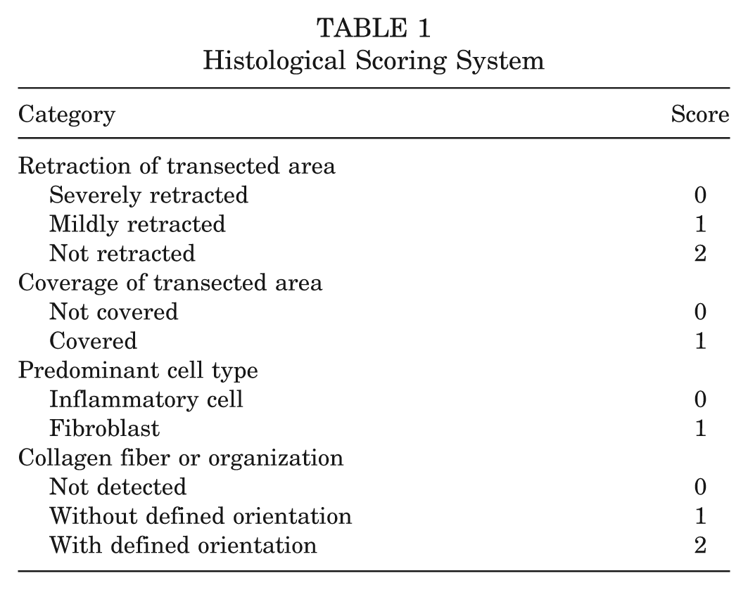
Frozen serial sections of 6-µm thickness were derived from the sagittal plane through the defect and prepared for histological analysis, and H&E and Masson trichrome stainings were performed. All specimens were given a histological score from 0 to 6 according to the grading scale, as described previously, 27 which was the sum of scores after considering the degree of retraction, tissue coverage, predominant cell type, and collagen fiber organization (Table 1). The best score (ie, least amount of damage) is 6, whereas the worst score is 0. All specimens were evaluated by 2 of our colleagues (T.N. and K.Y.) in blinded fashion without knowledge of the treatment received.
Histological Scoring System
For fluorescence microscopy of FAM-labeled miRNA-210 in ACL samples, 6-µm serial sections were mounted on saline-coated glass slides, air-dried, and fixed with 4.0% paraformaldehyde at 4°C for 5 minutes. Then, 4′,6-diamidino-2-phenylindole (DAPI) solution was applied for 5 minutes for nuclear staining.
Immunohistochemical Analysis
For immunofluorescence staining of VEGF-A, FGF2, isolectin B4, and collagen type I, 6-µm serial sections were mounted on saline-coated glass slides, air-dried, fixed with 4.0% paraformaldehyde at 4°C for 5 minutes, and immediately stained with rabbit polyclonal anti-VEGF or antibasic FGF antibody (Abcam, Cambridge, Massachusetts), goat polyclonal anti-collagen type I antibody (Santa Cruz Biotechnology, Santa Cruz, California), or fluorescein-labeled GSL I-isolectin B4 (Vector Laboratories, Burlingame, California). The secondary antibodies used were Alexa Fluor 488–conjugated or Alexa Fluor 568–conjugated goat anti-rabbit IgG for VEGF-A and FGF2, and Alexa Fluor 488–conjugated or Alexa Fluor 568–conjugated rabbit anti-goat IgG for collagen type I (all from Molecular Probes/Invitrogen, Carlsbad, California). DAPI solution was applied for 5 minutes to detect nuclear staining.
Statistical Analysis
Statistical significance was determined by 1-way analysis of variance. The Mann Whitney U test was used for the detection of differences between the 2 groups. A P value of <.05 was considered statistically significant. All data were calculated and are shown below as means ± standard deviations.
Results
There were no complications after surgery throughout the observation period, such as degenerative changes of the articular cartilage, wound or joint infection, progression to a complete ACL tear, formation of adhesive tissue, or limited range of motion of the joint.
Temporal Expression Changes of rno-miR-210 After Partial Resection of the ACLs
To confirm the level and temporal expression change of endogenous rno-miR-210 after partial resection of rat ACLs, we performed real-time PCR immediately after injury (day zero), and 1, 2, and 4 weeks after surgery. The expression of rno-miR-210 was significantly downregulated soon after injury but was gradually upregulated as time progressed, with statistically significant differences. There was an approximately 3-fold increase between day zero and 4 weeks (Figure 1).
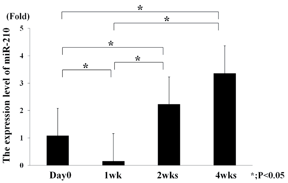
Temporal expression changes of mature rat (rno)-miR-210 after partial resection of rat anterior cruciate ligament by real-time polymerase chain reaction analysis (each time point; n = 5). SnoRNA-135 was used as an internal control. The expression of rno-miR-210 was significantly downregulated soon after injury but gradually upregulated as time progressed, with statistically significant differences. Data calculated as mean ± standard deviation. *P < .05.
Expression and Distribution of Injected ds miR-210 in Injured ACL
To detect the distribution of injected ds miR-210, transected ACLs that had received an intra-articular injection of the complex containing ds miR-210 labeled with or without FAM (20 µg of miR-210/15 µL volume) and atelocollagen were evaluated by fluorescence microscopy 24 hours after injection. In frozen sections, green fluorescence emission was observed in the synovial cells of the ACLs and the ligament itself 24 hours after injection. Specifically, strong expression of green fluorescence was observed in the synovial cells near the injured site and in high-power views, the expression of green fluorescence was observed in the cytoplasm of cells (Figure 2, A and B). By contrast, green fluorescence emission was not observed in the synovial cells of the ACLs after injection of nonlabeled ds miR-210 (Figure 2C).
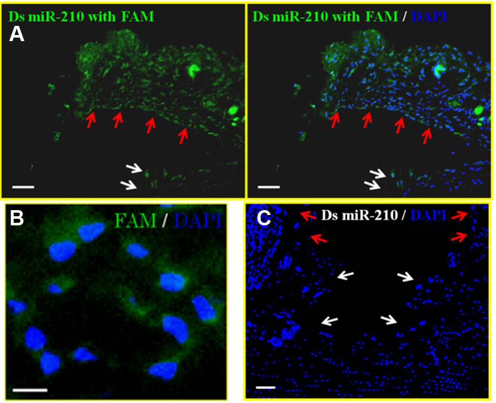
(A) The distribution of green fluorescence in the synovial cells of the ACL 24 hours after injection of the FAM-labeled ds miR-210/atelocollagen complex into the joint. Green fluorescence emission was observed, especially in the synovial cells and especially near the injured site in the ACL. Bar = 50 µm. (B) The expression of green fluorescence was observed in the cytoplasm of cells in high-power views. Bar = 10 µm. (C) The distribution of green fluorescence in the synovial cells of the ACL 24 hours after injection of the nonlabeled ds miR210/atelocollagen complex into the joint. Green fluorescence emission was not observed in the synovial cells of the ACLs after injection of nonlabeled ds miR-210. The red arrows indicate the synovium around the ACL; the white arrows indicate the site of resection of the ACL. Bar = 50 µm.
Gross and Histological Findings After Intra-articular Injection of ds miR-210
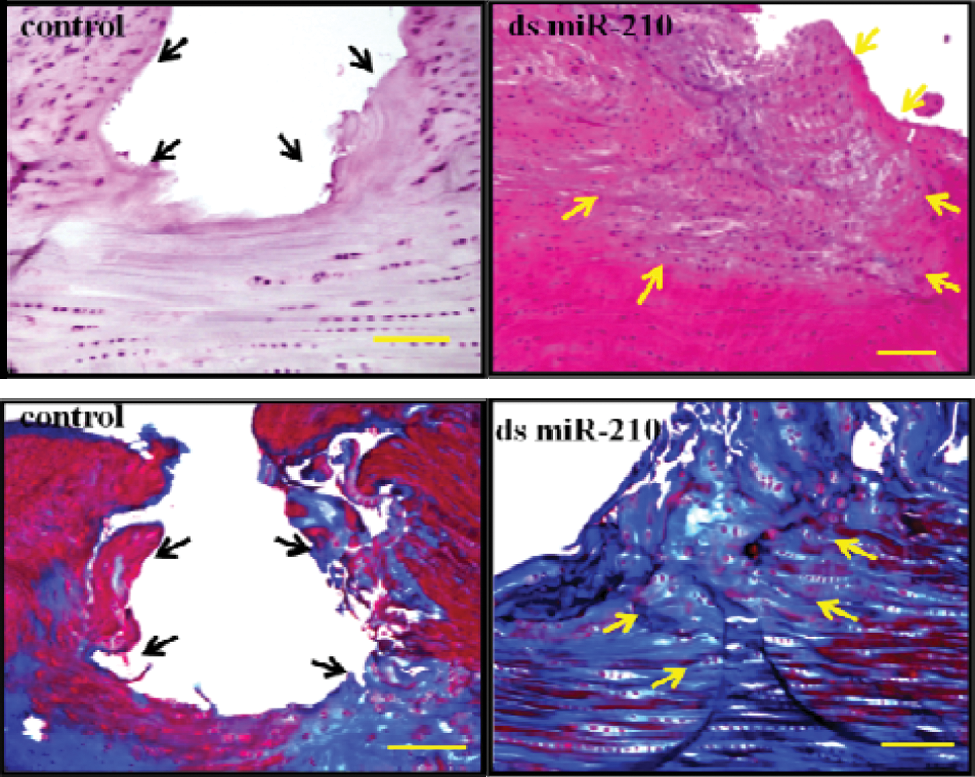
Macroscopic analyses of the injured ACLs 4 weeks after intra-articular injection of ds miR-210 showed that the transected area was severely retracted and was not filled in any of the control animals. By contrast, none of the specimens in the miR-210 group showed any retraction in the partially transected area (Figure 3A). Histological analyses of H&E staining at 4 weeks showed healing tissue with high cellularity around the injured ACL in the miR-210 group, whereas the transected ACLs in the control group demonstrated severe retraction and were not filled with any tissue. Masson trichrome staining demonstrated that the transected area was covered and filled with fibrous tissue in all specimens from the miR-210 group, whereas the transected area was uncovered in the control group (Figure 3B). The histological score in the miR-210 group (4.8 ± 0.37) was significantly higher than that in the control group (0.4 ± 0.24) 4 weeks after surgery (P < .05) (Table 2). To verify that the enhanced ACL healing was not the result of a nonspecific response such as unexpected vascular or immune response to the injected miRNA, 30 we used miR-140 (a cartilage-specific miRNA), and atelocollagen alone was injected into ACL-injured knee (each group, n = 5). However, ACL healing was not observed 4 weeks after the intra-articular injection of miR-140 or atelocollagen alone (Figure 3C).
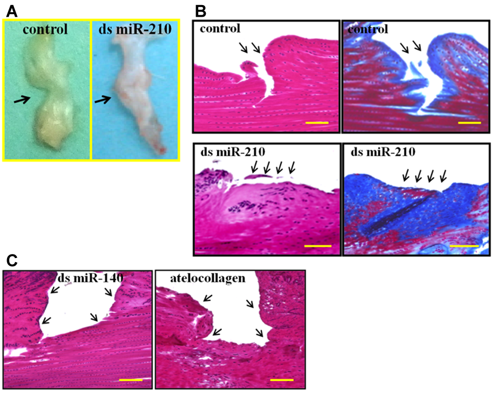
(A) Macroscopic findings of the ACL 4 weeks after intra-articular injection of ds RNA. (B) Histological findings of the ACL at 4 weeks. Left panels show H&E–stained samples and right panels show Masson trichrome–stained samples. Arrows indicate the site of resection of ACL. (C) Histological findings of ACL 4 weeks after injection of ds miR-140 and atelocollagen alone. Left panel indicates H&E staining of specimens in the miR-140 injection group, and right panel indicates H&E staining of specimens in the atelocollagen injection group. Arrows indicate the site of resection of the ACL. Bar = 200 µm.
Histological Scores for Each Group a
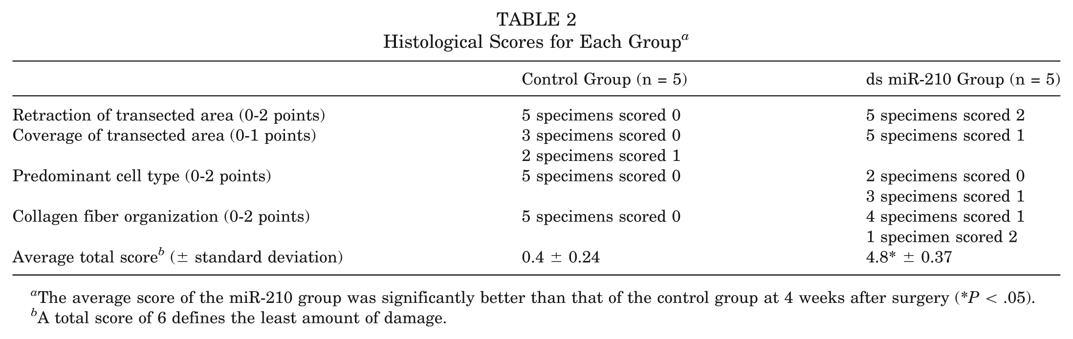
The average score of the miR-210 group was significantly better than that of the control group at 4 weeks after surgery (*P < .05).
A total score of 6 defines the least amount of damage.
Twelve weeks after injection, H&E staining showed that high cellularity in the healing tissue in the miR-210 group decreased, which indicated that healing tissue had become mature. In contrast, transected ACLs in the control group demonstrated severe retraction and remained not filled with any tissue. Masson trichrome staining demonstrated that the transected area was covered and filled with ligamentous tissue similar to normal ligament around the healing area in the miR-210 group, whereas the transected area remained uncovered in the control group (Figure 4).
Histological findings of ACL 12 weeks after injection. Upper panels show H&E–stained samples, and lower panels show Masson trichrome–stained samples. Arrows indicate the site of resection of the ACL. Bar = 200 µm.
Biomechanical Testing of ACL 4 Weeks After Intra-articular Injection
The ultimate failure load of the femur–ACL–tibia complex in the miR-210 group was significantly higher than that in the control group 4 weeks after intra-articular injection (P < .05). In both groups, the failure of the femur–ACL–tibia complex occurred at the transected area of the ACL in all specimens; the ultimate failure loads 4 weeks after injection were 30.5 ± 3.1 N in the miR-210 group and 22.8 ± 3.1 N in the control group. However, the ultimate failure load in both groups was significantly lower than that in the normal group (P < .05) (Figure 5).
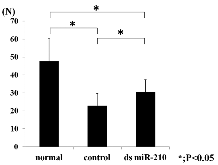
The ultimate failure load after partial resection of rat anterior cruciate ligament (n = 5). The ultimate failure load of the ds miR-210 group was significantly higher than that of the control group 4 weeks after surgery. Data were calculated as means ± standard deviations. * P < .05.
Gene Expression Analysis 4 Weeks After Intra-articular Injection
To examine gene expression–related ligament healing by upregulation of miR-210, real-time PCR and immunohistochemical analysis were performed. Real-time PCR analysis showed that the expression level of miR-210 and VEGF-A in the miR-210 group was significantly higher than in the control group (Figure 6A). Immunohistochemical analysis for VEGF-A and isolectin B4 indicated that VEGF-A was highly expressed in the miR-210 group, which corresponded to an increased number of blood vessels in the miR-210 group as compared with controls (Figure 6B). In the miR-210 group, expression of FGF2 was increased around the resected site compared with the control group (Figure 7A). Furthermore, in the control group, collagen type I expression was sparse around the resected site, whereas abundant collagen type I deposition was observed around the resected site in the miR-210 group (Figure 7B). Real-time PCR analysis revealed that the expression levels of Col1a1 in the miR-210 group were significantly higher than in the control group (Figure 7C).
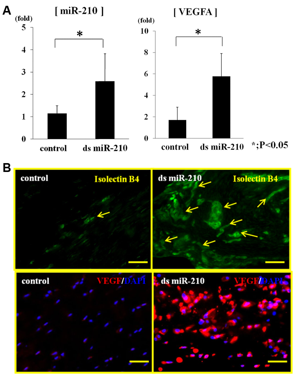
Gene expression analysis 4 weeks following intra-articular injection. (A) Expression of mature rno-miR-210 and vascular endothelial growth factor (VEGF)-A 4 weeks after injection by real-time polymerase chain reaction analysis (n = 5). The expression levels of miR-210 and VEGF were significantly higher in the miR-210 group compared with those in the control group (P < .05). (B) Immunohistochemical analysis of isolectin B4 and VEGF in the ACL 4 weeks after injection of miR-210 or control ds RNA into the knee joint. Immunohistochemical analysis indicates that VEGF was highly expressed in the miR-210 group, which correlated with the increased number of blood vessels in the miR-210 group as compared to controls. Arrows indicate the blood vessels in the synovium. Upper bar = 50 µm; lower bar = 25 µm.
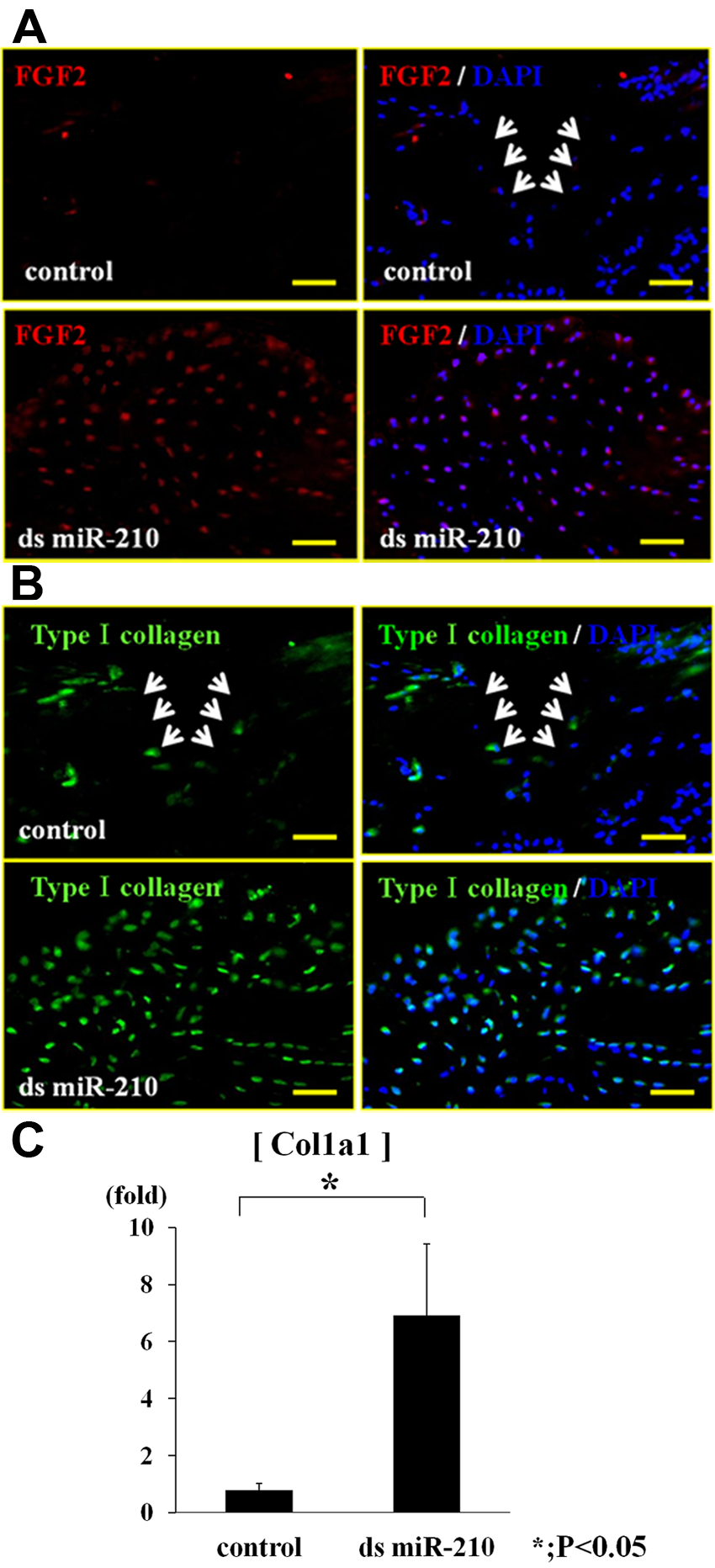
(A) Immunohistochemical analysis of fibroblast growth factor 2 expression in the ACL 4 weeks after injection of miR-210 or control ds RNA into the knee joint. Arrows indicate the site of resection of the ACL. In the miR-210 group, there was high expression of fibroblast growth factor 2 around the resected site compared with that in the control group. Bar = 50 µm. (B) Immunohistochemical analysis of collagen type I expression in the ACL 4 weeks after injection of miR-210 or control dsRNA into the knee joint. Arrows indicate the site of resection of ACL. In the control group, expression of collagen type I was sparse around the resected site, whereas abundant collagen type I deposition around the resected site was observed in the miR-210 group. Bar = 50 µm. (C) Expression of collagen type I alpha 1 four weeks after injection by real-time polymerase chain reaction analysis (n = 5). The expression levels of collagen type I alpha 1 were significantly higher in the miR-210 group compared with those in the control group (P < .05).
Discussion
We hypothesized that healing of the injured ACL could be accelerated by changing the external environment at an early stage in the healing process through angiogenesis by upregulation of miR-210. Therefore, the decrease in the expression of miR-210 after ACL injury was first proven, and then the miR-210 levels were augmented by intra-articular injection of exogenous miR-210, which subsequently resulted in accelerated ACL healing. The intra-articular structures of the ACL have limited healing capacity. When injured ACLs are examined histologically, there is a gap at the rupture site that remains open. 22 It has been reported that factors that affect ligament healing and the healing response include the surrounding tissues, blood supply, nutrient delivery, biomechanical forces, and synovial fluid, as well as the supply of growth factors. It is also known that angiogenesis, the formation of new blood vessels from a preexisting vessel, is an essential step in the process of tendon healing; however, angiogenesis and regeneration in the injured ACL occur at a very slow rate, leading to insufficient healing. 5
Recently, miRNAs have attracted immense attention because of their crucial role in human disease, and they have been proposed as potential new therapeutic targets. Several therapeutic trials including our studies to regulate miRNA in vivo have been undertaken.40-42 It has been reported that miR-210 is a crucial element of the endothelial cell response to hypoxia and that the targets of miR-210 are ephrin-A3, E2F3, NPTX1, RAD52, ACVR1B, MNT, CASP8AP2, FGFRL1, and HOXA-1, which have essential functions in cell survival, migration, and differentiation.8,18,24,38,45,52 Hu et al 23 demonstrated that local injection of ds miR-210 generated in a minicircle vector into the myocardium can improve angiogenesis, inhibit apoptosis, and improve cardiac function in mice.
In this study, it was revealed that the expression of miR-210 was significantly downregulated soon after injury to normal ACLs, but it gradually upregulated as time progressed, with statistically significant differences. The injected ds miR-210 was taken up by the synovial cells of the ACL, specifically those near the injured site, which enhanced the healing process after ACL injury, as confirmed by histological and immunohistochemical analyses. Four weeks after injection of ds miR-210 into ACL-transected rats, the injured area was filled with fibrous collagenous tissues, and the histological score and mechanical strength were improved postoperatively with statistical significance as compared with controls. Moreover, 12 weeks after injection, healing tissue in the miR-210 group revealed more maturity compared to that at 4 weeks.
Our results demonstrated that angiogenesis was upregulated in the healing site by VEGF-A. Furthermore, it was revealed that overexpression of miR-210 in the torn ACL could enhance FGF2 and collagen type I in the healing process. Previous studies have shown that miR-210 can improve tubulogenesis, 12 and some growth factors including FGF have the capacity to stimulate both proliferation and expression of extracellular matrix proteins for tissue engineering of tendons and ligaments, 21 findings that support our data. A recent study also showed that miR-210 can prevent mesenchymal stem cell apoptosis, 29 as well as modulate mitochondrial respiration, iron metabolism, and reactive oxygen species generation. 7 These reports indicate that a variety of complex responses at both the cellular and organism levels are activated, including endothelial cell proliferation, migration, and angiogenesis. Therefore, miR-210 delivery into the knee joint may have several pleiotropic effects on several organs including the ligament itself, the synovium, and surrounding tissues, in addition to the proangiogenic roles that were investigated here. From our results and these previous reports, we postulate that the miR-210 and atelocollagen complex introduced into the injured ACL by intra-articular injection could upregulate the expression of VEGF-A, FGF2, and other angiogenetic factors, leading to acceleration of the healing potential of the injured ACL.
This is the first study to evaluate the therapeutic administration of ds miRNA for injured ACLs. There are some limitations to this study. First, the defect was created by a knife cut, with little or no damage to the fascicles that were proximal and distal to the injury. This method is clearly different from a clinical rupture, in which many of the fascicles are thought to sustain tensile injury along their length prior to rupture. Second, the underlying mechanisms including the identities of target genes in the ACL healing pathway are still largely unknown. Administration of miRNA would be expected to bring about more effects via regulation of many gene networks, which would be an advantage of miRNA therapy. Although further comprehensive studies are needed to determine its suitability as a therapeutic agent, the current study showed that intra-articular injection of ds miRNA in vivo into an injured joint is a potential new treatment strategy.
In conclusion, the present study demonstrated that intra-articular injection of ds miR-210 could promote the healing of partially torn ACLs through enhancement of angiogenesis. Although the mechanism underlying the promotion of healing of the torn ACL has not been elucidated, the results of this study suggest that administration of synthetic miRNA would be a feasible treatment option for an injured ACL.
Footnotes
One or more of the authors has declared the following potential conflict of interest or source of funding: This work was supported by a grant-in-aid to M. Ochi for scientific research from the Ministry of Education, Culture, Sports, Science and Technology, Japan (No. 21249079).