






Abstract
Background:
There are only 2 reported cases of arthroscopic retrograde osteochondral autograft transplantations regarding tibial plateau cartilage defects.
Purpose:
To present a detailed description of arthroscopic retrograde osteochondral autograft transplantation of the tibial plateau as well as to provide its advantages and disadvantages in comparison with other techniques.
Study Design:
Case series; Level of evidence, 4.
Methods:
Four patients (3 men and 1 woman; mean age, 31.2 years) suffering from tibial plateau cartilage lesions underwent surgery. In each case, the lesions were caused by sports injuries. There were 3 lateral and 1 medial tibial plateau defects. The International Knee Documentation Committee (IKDC) score and Knee injury and Osteoarthritis Outcome Score (KOOS) were recorded preoperatively and postoperatively. Radiological assessment was made by plain radiographs, CT arthroscans, and MRI.
Results:
The mean follow-up was 55 months (range, 52-60). The mean preoperative IKDC score was 53.5 (range, 37-66), while the mean postoperative IKDC score at final follow-up was 95.4 (range, 93.1-97.7). Regarding the KOOS calculation, there was significant improvement concerning each parameter after surgery. All patients were satisfied with the surgical procedure and returned to their previous activity level. Postoperative imaging showed very good adaptation and incorporation of the osteochondral autografts.
Conclusion:
Treatment of tibial plateau cartilage defects with arthroscopic retrograde osteochondral autograft transplantation could be performed on a routine basis in clinical practice. The results were encouraging and showed good incorporation of the graft, a minimal failure rate, and satisfactory functional outcomes of patients.
Keywords
Cartilage lesions that follow an initial joint injury are often underestimated. Their natural history could dramatically affect the activity level and quality of life of patients, especially in the case of an active young person. Many techniques have been developed trying to restore osteochondral defects as well as to promote the regeneration of hyaline cartilage.2,7,15 The use of osteochondral autograft transplantation (OATS) is becoming increasingly popular.10,12 Treatment of tibial plateau cartilage defects is challenging, and the literature on the subject is relatively poor.
We report the results of 4 patients with isolated osteochondral lesions of the tibial plateau that occurred during sports activities. All patients underwent arthroscopic surgery using the retrograde autograft transplantation technique. The aim of this study was to evaluate the clinical and radiological treatment outcomes of tibial plateau cartilage lesions with the use of this arthroscopic-assisted procedure.
Materials and Methods
In 2007, 4 patients suffering from cartilage lesions of the tibial plateau underwent surgery in our department. A mechanism of high-energy trauma was the cause in all cases: 2 patients had a ski accident, 1 had a football injury, and 1 patient had a dancing injury. The first patient with the ski accident was classified as level 6 according to the Tegner activity scale (recreational sports), and the second one was classified as level 8 (competitive sports, downhill skiing). The patient playing football was classified as activity level 7 (recreational sports), and the patient with the dance injury was classified as activity level 7 as well (recreational sports). Inclusion criteria were any isolated osteochondral defect of the tibial plateau at stage 4 according to the International Cartilage Repair Society (ICRS) classification system. Moreover, only injuries related to sports activities were included in the study. Exclusion criteria were tibial plateau cartilage defects with concomitant knee lesions at the time of surgery such as ligamentous injuries or osteoarthritic changes and injuries that did not take place during sports activities.
There were 3 lateral and 1 medial tibial plateau defects with a diameter of 10, 10, 10, and 8 mm, respectively. Regarding their medical histories, 1 patient underwent an arthroscopic cartilage debridement in the past, 1 patient had an old lateral tibial condyle fracture that was treated nonoperatively, and 1 patient had medial and lateral partial meniscectomies of the involved knee. The fourth patient had no former intervention or injury in his knee. There were 3 men and 1 woman with a mean age of 31.2 years (range, 17-41). Three left and 1 right knees were operated on, while the average time interval, between the day of the accident and the day of surgery, was 22.5 months (range, 11-31) (Table 1).
Characteristics of Patients and Cartilage Defects a
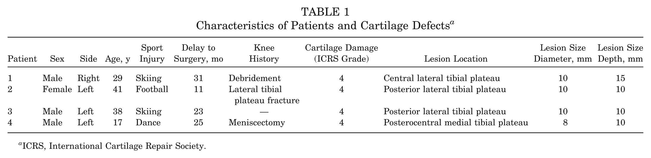
ICRS, International Cartilage Repair Society.
All patients were assessed, preoperatively and postoperatively, through a detailed clinical examination. The International Knee Documentation Committee (IKDC) score and Knee injury and Osteoarthritis Outcome Score (KOOS) were both recorded before the surgery and at the final follow-up. All operations were performed by the same orthopaedic surgeon (P.D.) using the OATS (Arthrex, Naples, Florida). Radiological evaluation was assessed by plain radiographs, computed tomography (CT) arthroscans, and magnetic resonance imaging (MRI).
Surgical Technique
All surgical procedures were performed under general anesthesia. The anterolateral and anteromedial portals were installed, and routine diagnostic arthroscopic surgery was performed. The absence of any concomitant lesion was verified. An initial assessment of the defect size was done with an arthroscopic probe. Debridement of the damaged osteochondral site was made up to the border of normal vital cartilage. With the aid of the specific drill guide (OATS), a 2.4-mm guide wire passed the center of the lesion. Then, a tunnel was drilled through the tibia, using a core reamer. The appropriate angular calibrator (OATS) was placed to estimate the angle and the orientation of the graft (Figure 1). The size of the osteochondral plugs was 10 mm in diameter in 3 cases. In 1 case, the diameter defect was 8 mm. The length of each autograft was 20 mm long. They were harvested from the most peripheral, proximal, and medial parts of the femoral trochlea under anteromedial mini-arthrotomy. For 3 cases, the chondral surface of the autografts had an obliquity of 20°. In the fourth case, the inclination was 30°. The osteochondral plug was inserted by a retrograde approach, through the tibial tunnel, under arthroscopic control (Figure 2). If the initial stability of the graft was insufficient with the press-fit technique, a bioabsorbable screw was used to fill the space below the plug. This was done only in 1 case, where a 10 × 17–mm screw (Arthrex) was used. The tibial cortical window was closed with periosteum. Congruity of the osteochondral plug with the surrounding articular surface was verified under direct arthroscopic vision (Figure 3).
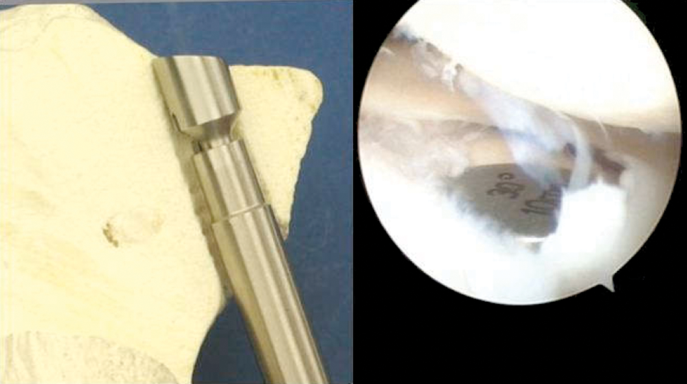
Placement of the angular calibrator, estimating the angle and the orientation of the graft. Image copyright Arthrex GmbH; reprinted with permission.
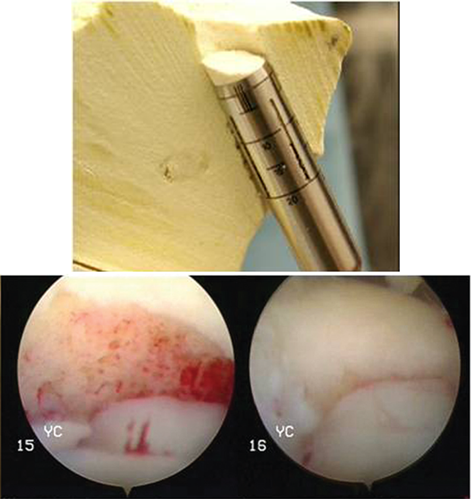
Insertion of the osteochondral plug by a retrograde approach. Image copyright Arthrex GmbH; reprinted with permission.
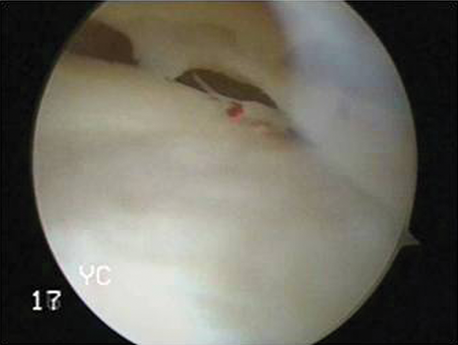
Final position of the graft, showing congruency with the surrounding articular cartilage.
The patients began passive movements of the knee and isometric quadriceps exercises from the first postoperative day. Weightbearing was not allowed for the first 6 weeks after surgery. Total active range of motion began after 6 weeks. The patients were permitted to bear full weight at 3 months after surgery.
Results
The mean follow-up was 55 months (range, 52-60). Data were obtained from all cases, preoperatively and postoperatively, using the IKDC score and KOOS. According to the IKDC examination form, patients were classified as grade A before and after surgery. The mean preoperative IKDC score was 53.5 (range, 37-66), while the mean postoperative IKDC score, at the final follow-up, was 95.4 (range, 93.1-97.7). Regarding the calculation of KOOS, there was significant improvement concerning each parameter after surgery. All patients were satisfied with the surgical procedure and returned to their previous activity level as demonstrated by the Tegner activity level scale (Table 2).
Preoperative and Postoperative Values of Functional Scores a
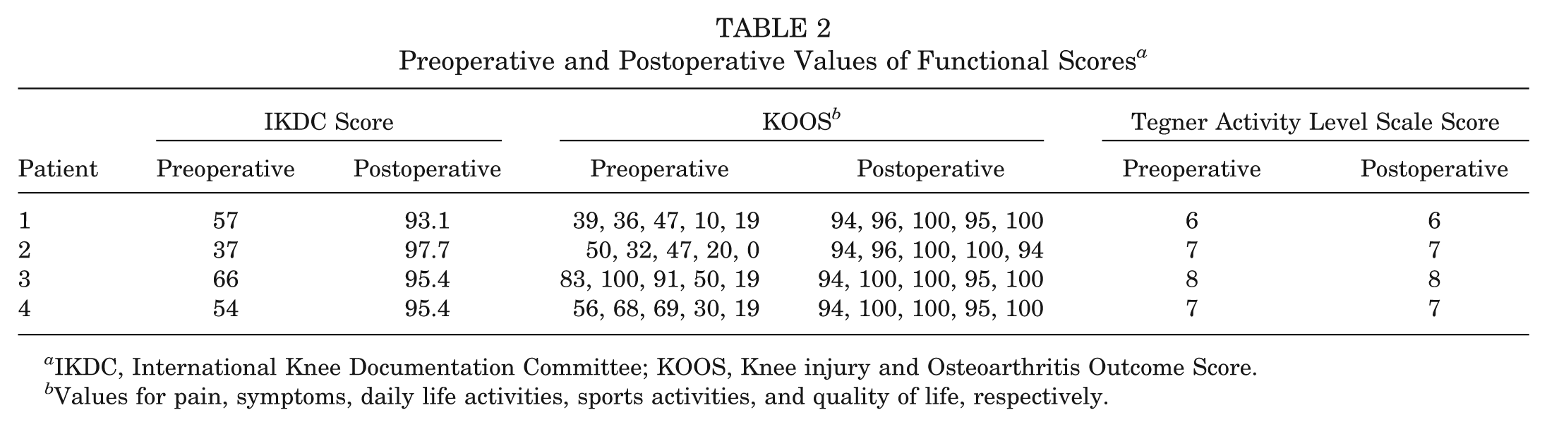
IKDC, International Knee Documentation Committee; KOOS, Knee injury and Osteoarthritis Outcome Score.
Values for pain, symptoms, daily life activities, sports activities, and quality of life, respectively.
In 1 case, a secondary intervention was performed, 8 months after surgery, to remove the bioabsorbable screw. It was induced because of pain and tenderness due to irritation from the hardware. During this procedure, the arthroscopic control revealed a perfectly integrated graft and no irregularities with the adjacent native cartilage. Moreover, the donor site healed with fibrocartilage satisfactorily. Postoperative imaging, at the final follow-up, showed very good adaptation and incorporation of the osteochondral autografts (Figures 4 and 5). No further complications were recorded in all cases.
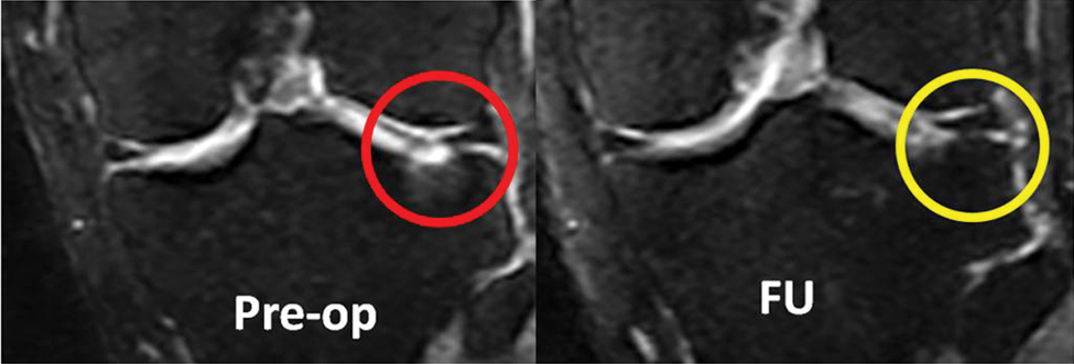
Knee magnetic resonance imaging scans of the third patient. Preoperative image on the left; circle shows the cartilage defect. Postoperative image on the right; circle shows the healed defect.
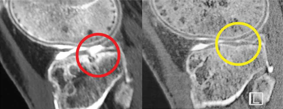
Knee computed tomography arthroscans of the second patient. Preoperative image on the left; circle shows the cartilage defect. Postoperative image on the right; circle shows the healed defect.
Discussion
Numerous techniques have been developed trying to restore the congruity of an injured joint such as the Pridie perforation, microfracture, and abrasion arthroplasty, for example, but there are no evidence-based results and no consensus to suggest the ideal treatment. 1 Nevertheless, their results were insufficient because they led to the formation of fibrocartilage tissue, which had inferior biomechanical properties compared with the normal original hyaline cartilage. 4 Some experts have used osteochondral allografts to treat full-thickness cartilage defects and have reported satisfactory results.11,14,18 However, their indications are restricted to treatment of large bone defects, and there is always a risk of disease transmission and allergic reactions. The new era of cartilage repair involves autologous chondrocyte implantation either alone or matrix induced. Several authors have reported open or arthroscopic therapeutic modalities using cultured chondrocytes with good results.3,16,20 However, according to other surgeons, they do not appear to provide superior results, and their routine use is limited because of high cost, longer operating time, and higher technical difficulties.6,8
There are a significant number of studies using osteochondral autografts in the treatment of cartilage lesions of the femoral condyles and talar dome and, more recently, in the treatment of osteochondritis dissecans of the elbow.5,9,17 The management of tibial plateau cartilage defects is more demanding because of difficulties in their approach. A wide arthrotomy may be needed, and extensive soft tissue detachments or tibial tubercle osteotomy can be inevitable to manage these lesions. Moreover, good visualization of the chondral defects, especially when they are located posteriorly on the tibial plateau, could be complicated using an antegrade arthroscopic technique.
The surgical technique used in our series provides all the benefits of an arthroscopic approach along with those of autograft transplantation, preserving the hyaline cartilage of the graft. Using the retrograde position of the osteochondral graft made it possible to access the tibial plateau cartilage lesions located more posteriorly. The position of the grafts and their congruity with the adjacent cartilage were confirmed accurately with the arthroscopic optical magnification. Choosing the cartilage surface of the proximal medial part of the femoral condyle was based on the anatomic characteristics. Its rounded shape was judged to match better with the convexity of the lateral tibial plateau. The lesion in the medial tibial plateau was 8 mm, so we believed it would be no significant mismatch between the autograft and the surrounding tibial cartilage.
The angles of inclination were estimated by using standard angle guides for the best fit and correct position of the bone plug. Matsusue et al 13 were the first to give a very detailed presentation of this innovative technique. They showed how important the inclination angle of the osteochondral plug was. They offered a specific method to calculate this angle, using a lateral radiograph of the knee during preoperative planning. We did not use this method on a routine basis, judging that image magnification could add difficulties of finding the accurate orientation of the defect, which might lead to miscalculations.
In accordance with the previous authors, 13 a large autograft was used instead of small multiple grafts to avoid further difficulties of positioning and to achieve more stable fixation of the graft. Indeed, good quality characteristics of the osteochondral graft were verified at the radiological and arthroscopic (1 case) postoperative evaluations, while there was no fracture or collapse of the osteochondral plug. The incorporation of the graft as determined by radiographs and CT and MRI scans resulted in a satisfactory functional outcome, and patients returned to their previous sports activities.
Regarding the graft fixation, a press-fit technique remains the optimal method, but sometimes, it is difficult to achieve, and there is always the risk of graft collapse. Matsusue et al 13 used bioactive ceramic fillers, while Ueblacker et al 19 used a diagonal bioabsorbable screw. We used an interference bioabsorbable screw only in 1 case. However, this screw caused soft tissue irritation, tenderness, and pain, leading to its removal.
Ronga et al 16 have presented a case report using arthroscopic autologous chondrocyte implantation for the treatment of a cartilage defect in the lateral tibial plateau. The possibility of restoring a homogeneous hyaline-like joint surface, without irregularities, and no donor site morbidity were mentioned as the main advantages of this technique. However, important disadvantages remain the high cost and the fact that a 2-step surgical procedure is required to harvest, culture, and perform the implantation of the grafting material.
In general, the technique used in the present study offers the advantages of a minimally invasive, 1-staged, and cost-effective procedure. Although technically demanding, this method can be reproducible in daily clinical practice without the use of complex radiological calculations. Using the native hyaline cartilage of the patient, it is feasible to treat full-thickness articular cartilage injuries of the tibial plateau and restore the congruity of the knee joint. Moreover, drilling and reaming of the defect during preparation of the bone tunnel, where the retrograde graft will be elevated, could offer the effect of bone marrow stimulation techniques. Careful consideration is needed regarding the inclination and congruity of the osteochondral graft, but the use of specific guides minimizes the risk of failure and complications. Applying properly the specific instrumentation, the graft can be fixed with a press-fit technique without using additional hardware that could complicate the procedure.
One limitation of this study is the small number of patients; however, the rarity of these lesions has to be considered. Furthermore, this is the first study in the literature presenting a series of tibial plateau osteochondral defects treated with this technique and evaluated after a relatively long follow-up. Another limitation is the use of this technique to restore small osteochondral defects only (maximum, 10 mm in diameter). Its application in multiple and larger lesions could have a high risk of failure because of bone weakening and difficulties to achieve sufficient cartilage congruity, but further biomechanical and clinical studies are necessary to prove it. A final limitation is the lack of control groups that permit the comparison of the present technique with different well-established methods used for the treatment of cartilage defects. However, re-examination after 5 years postoperatively revealed no complications, and all the patients were able to resume their previous level of sports activity.
In conclusion, the treatment of tibial plateau cartilage defects with arthroscopic retrograde OATS could be performed on a routine basis in clinical practice. The results were encouraging and showed good incorporation of the graft, a minimal failure rate, and satisfactory functional outcomes in patients. A young active person with a lesion of ICRS grade 4, with good quality hyaline cartilage and a stable knee, seems to be the ideal candidate on whom to apply this technique. Randomized controlled studies with a larger sample of patients can be valuable to verify these promising results and show the efficacy of this technique in competitive high-level athletes.
Footnotes
The authors declared that they have no conflicts of interest in the authorship and publication of this contribution.