
Abstract
For as long as athletes have been competing, injuries from competition have resulted. Sports medicine has a rich and storied history with significant contributors from many different countries and civilizations. Over time, we have honored the contributions of important figures in sports medicine with the use of eponyms. However, the continued use of eponyms in medicine has been called into question by a number of authors. They cite inaccuracies in definition and context, lack of descriptive value, and the possible celebration of unsavory characters. However, eponyms are pervasive in the medical literature. They bring color and character and allow us to honor those who came before us. Furthermore, eponyms can hide some distressing aspects of a disease. This review of eponyms in sports medicine provides an opportunity to celebrate our predecessors, recognize the international flavor of sports medicine, and promote accurate use of eponyms for the future.
So mind your Q’s and mind your P’s; Say “Thank you, sir,” and “If you please.” Then some day, in the future dim, You too may be an eponym.
19
From the Olympic Stadium to the Roman Coliseum, from the ball courts of South America to the polo fields in Persia, to the modern day football stadiums, basketball arenas, and ice rinks, humans have fostered and possessed a rich and timeless fascination with sports. Wall paintings of wrestling matches have been found in Egyptian tombs dating back to nearly 2000
Some authors believe that the first recorded sports injury was documented by Homer in the Iliad. 44 In this book, Homer mentions a grueling boxing match between Epeus and Euryalus: “The two men now being girt went into the middle of the ring and immediately fell to; heavily indeed did they punish one another and lay about them with their brawny fists.” Other scholars believe that the Bible, Genesis 32:24-25, references the first documented sporting injury: “And Jacob was left alone; and there wrestled a man with him until the breaking of the day. And when (the man) saw that he prevailed not against (Jacob), he touched the hollow of his thigh; and the hollow of Jacob’s thigh was out of joint and he wrestled with him.” 53 Regardless of which injury was documented first, one thing is clear—for as long as humans have participated in sport, injuries have followed.
In a previous article in The American Journal of Sports Medicine, Appelboom et al 1 identified several factors responsible for the merger of sports and medicine in ancient Greece. The first was the teachings of Hippocrates and Herodicus, which helped transfer the care of athletes from the hands of priests to physicians. Second was the tremendous knowledge gained during periods of war from the treatment of battlefield injuries. Some of the early Olympic games were unbelievably violent, with broken bones, profuse bleeding, and loss of consciousness being the standard. Previous military exposure provided early sports medicine physicians with valuable experience in treating early, gruesome athletic injuries. Finally, in ancient Greece during periods of peace, sports and athletic performance attracted great interest and energy and achieved high status. The desire to compete and win created an opportunity for pioneering sports medicine physicians to treat injury and improve performance.
From ancient Greece to modern times, sports medicine has been fostered by a range of figures on different continents. Throughout time, we have honored the wisdom, knowledge, and sometimes the mistakes of those who came before us to shape practice today. Influential sports medicine physicians are routinely remembered with eponyms to honor their memory and contributions to our field. Unfortunately, several authors have recently challenged the continued use of eponyms. Thompson and Grant, 57 in their work to clarify the use of the term Barton fracture, called to “discontinue eponymic terminology and refer to these fractures [Barton’s] specifically according to their anatomical considerations.” Other authors have suggested that eponyms are “meaningless. Serving to mystify rather than clarify.” 60 Academic purists take the argument even further, and their desire for absolute accuracy has resulted in their call to abolish the use of eponyms in the medical literature. 65 Organ and Sojka, 48 in their paper titled “The Eponym Problem” published 50 years ago in the American Journal of Surgery, highlighted numerous disadvantages of the use of eponyms, including multiple eponyms for one structure or entity, lack of descriptive value, and multiple inaccuracies in definition and historical context.
The word eponym derives from the Greek word eponymos, which means “to name after.” It is often assumed that the eponym is named for the person who first described or devised the pathological condition, procedure, or physical examination maneuver. However, a careful review of the medical literature will reveal that this is not always the case. Dr Mark Ravitch, a pediatric surgeon and medical historian, reminds us when studying eponyms, “Given an eponym, one may be sure (1) that the man so honored was not the first to describe the disease, the operation or the instrument, or (2) that he misunderstood the situation, or (3) that he is generally misquoted, or (4) that (1), (2) and (3) are all simultaneously true.”30,50
While the use of eponyms does have some disadvantages, there are many reasons why they not only should be continued, but should be embraced. First, eponyms are everywhere. It is not a realistic endeavor to abolish their use in medical or mainstream culture. There are countless eponyms in daily use. Judith Whitworth, the director of medical research in Canberra, Australia adds, “Eponyms bring color to medicine, they provide a convenient shorthand for the profession and the community alike, and they embed medical traditions and culture in our history.” 63 Medicine is built on the premise that we remember and honor those who came before us. We stand on the accomplishments of our predecessors. What better way to remember the contributions of the clinicians who came before us?
The use of eponyms is not a static or permanent event. With new information and improved understanding, inaccurate eponyms can be identified and discarded. For example, Hurler syndrome was once known as gargoylism. Prior to that, it was called a lipochondrodystrophy due to the erroneous belief that the disorder resulted from an abnormal accumulation of lipid material in cells. Later this material was discovered to be a mucopolysaccharide, rendering the name misleading and inaccurate. Names can and do change with time.
Eponyms can also be useful substitutes in the right situation. Isn’t it easier to say one suffers from Ehlers-Danlos than the previous term India rubber man? 67 Should we choose to call people lepers or is it more humane to say they suffer from Hansen’s disease? The use of eponyms can conceal some of the more distressing aspects of a disease or illness.
Some suggest that all eponyms should be discontinued because some eponyms use the names of those who on further inspection are found to be vile. We disagree. History is filled with unsavory and immoral characters. We remember the names of evil dictators, kings, rulers, and tyrants. To remember them is not the same as to celebrate them. Careful review and study of history allow us to choose who to despise and who to celebrate.
A few articles over the years have attempted to preserve and study the history of eponyms in orthopaedics; however, none have specifically addressed the field of sports medicine.30,58 In this article, commonly used eponyms in orthopaedic sports medicine are reviewed. We note the very international nature of sports medicine, as surgeons from the United States, Canada, France, Great Britain, Ireland, Argentina, and Sweden are included. This review is intended to preserve a portion of the history of sports medicine, remind us of the international nature of our field, and promote the accurate use of these musculoskeletal eponyms in the future.
Eponyms in Anatomy and Pathology
Bankart Lesion/Bony Bankart Lesion
Arthur Sydney Blundell Bankart (1879-1951) was a prominent British surgeon. The son of a physician, he became an influential figure early in his career. Known for efficient operating, Bankart practiced 3 surgical specialties at the same time (orthopaedic surgery, neurosurgery, and pediatric surgery). He was a founding member of the British Orthopaedic Association and served his country as a surgeon in both World Wars.
Dr Bankart is mostly remembered for his work on anterior shoulder instability, 3 although he was not the first to describe its pathological characteristics. This was done by Francis Mitchell Caird in 1887. 12 In 1906, the prominent German surgeon Georg Clemens von Perthes described the first surgical procedure to address shoulder instability. 49
Bony injury to the glenoid cavity has been named after Bankart as well, even though he wrote in the British Journal of Surgery, “I have never seen recurrent dislocation associated with fracture of the glenoid cavity or with any other bony abnormality, and I would suggest that, if such an association occurs at all, it must be very rare indeed” 2 (Figure 1). Currently, a tremendous amount of emphasis has been placed on recognizing bone loss in the glenoid after an anterior shoulder dislocation to guide management and surgical decision making. Failure to recognize a significant bony Bankart lesion has been associated with a high failure rate after arthroscopic stabilization. 11
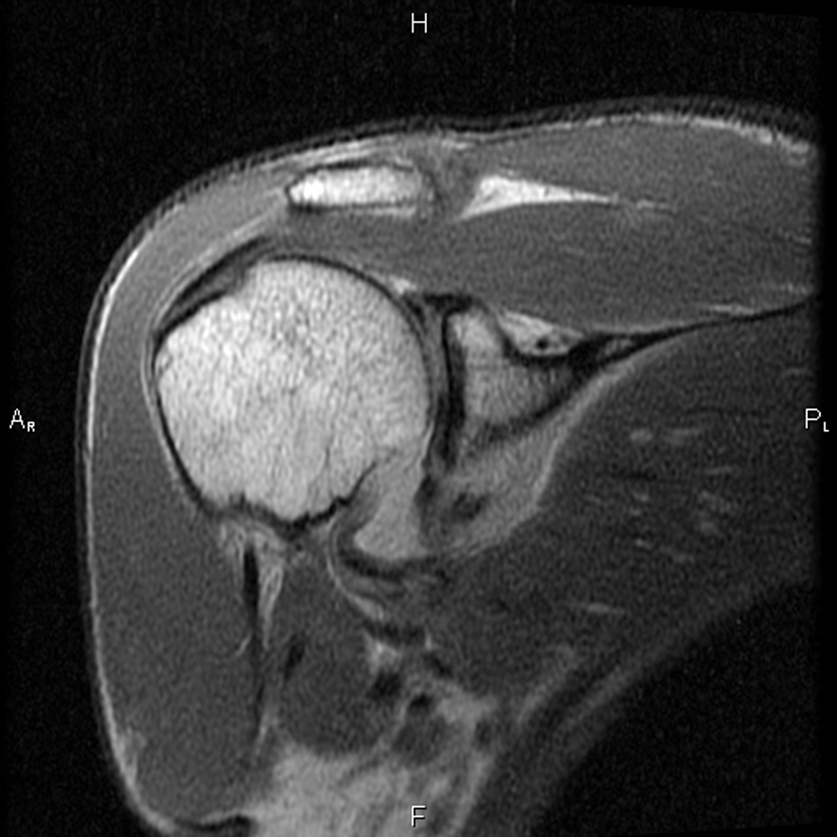
T1 coronal magnetic resonance image of a bony Bankart lesion.
Bennett Lesion
George Eli Bennett (1885-1962) was president of both the American Academy of Orthopaedic Surgeons (1939-1940) and the American Orthopaedic Association (1941-42). A superb athlete, he played semi-pro baseball at the age of 16. 7 After graduation from the University of Maryland (College Park, Maryland) in 1908, he continued his training at the Hospital for the Ruptured and Crippled (New York, New York). Bennett joined the staff at Johns Hopkins Hospital (Baltimore, Maryland) in 1910 and remained on staff until 1947. 23 He made many contributions to the field of orthopaedics but was best known for his work in sports medicine. In a paper in 1941 that appeared in JAMA, he described a mineralization of the posteroinferior glenoid seen in throwing athletes. 6 This lesion has been termed a Bennett lesion, although the pathological significance of this lesion is controversial. There remains considerable debate as to the cause and treatment of this lesion.43,66
Buford Complex
Don Buford (1966- ) is currently in private practice in the Dallas, Texas, area. As a medical student at the University of California, Los Angeles, Buford watched multiple arthroscopy videos as part of a summer research project at SCOI (Southern California Orthopedic Institute, Van Nuys, California). Noting the thickened middle glenohumeral ligament and the large sublabral foramen, he asked Stephen Snyder whether this was normal or a variant. Buford and Snyder then first reported their findings of the Buford complex in an abstract for the AOSSM in 1992 and then as a paper in Arthroscopy in 1994. 64 Dr Buford’s father was Don Buford Sr, a switch hitting Major League Baseball player inducted into the Baltimore Orioles’ Hall of Fame.
Gerdy Tubercle
Pierre Nicolas Gerdy (1797-1856) was a French physician, anatomist, and physiologist. Born to peasants in the vineyards of northern France, Gerdy received his medical training in Paris. Gerdy never did well on exams and with his initial clinical avenues closed, he focused on anatomy and physiology. As a teacher of anatomy, Gerdy excelled and quickly gained notoriety and a position within the Department of Medicine. His opportunity came with the publication of work at the Sociètè de l’Ecole de Mèdecine, in which he demonstrated that arteries and veins had intrinsic regulatory function and were not simple inert conduits. 4 This publication allowed him to pursue a surgical career.
Gerdy suffered from tuberculosis his entire career. Despite this limitation, he made numerous contributions to the medical literature on a wide array of topics. He wrote about the physiological characteristics of nerve sensation, gait analysis, abscesses, hernias, wound care, arsenic poisoning, and of course anatomy. 4 Several structures in the human body bear his name; however, the most commonly referenced is Gerdy tubercle, a prominence on the lateral side of the tibia where the iliotibial band attaches, as do fibers of the tibialis anterior muscle. Gerdy tubercle is commonly used as a landmark on lateral approaches to the knee. In complicated posterolateral corner knee injuries, an osteotomy through Gerdy tubercle has been described to help with the exposure. 22
Insall-Salvati Ratio
John N. Insall (1930-2000) was considered by many to be the father of modern day total knee replacements and associated techniques. 33 A skilled surgeon, he made many important contributions to surgery of the knee. He was instrumental in the design of the total condylar prosthesis in the 1970s, and later he helped develop mobile bearing inserts and fixed bearings to allow for increased knee flexion. He championed a new surgical approach to the knee, the quadriceps snip. Insall also made important contributions to the treatment of patellofemoral instability and chondromalacia. His articles appeared in orthopaedic journals spanning more than 4 decades. A founding member of the Knee Society and its president in 1987, Insall also served as editor of the publication Surgery of the Knee (now in its fifth edition).
Eduardo A. Salvati (1939- ) is a hip and knee surgeon practicing at the Hospital for Special Surgery in New York. He trained in Argentina and Italy before proceeding to New York for a fellowship in hip and knee arthroplasty. A past president of the American Hip Society and Secretary of the International Hip Society, Dr Salvati has enjoyed a long and prolific academic career in hip and knee surgery.
The Insall-Salvati ratio is determined from a lateral radiograph of the knee. The greatest diagonal length of the patella (LP) is compared to the length of the patellar tendon (LT). Variation of greater than 20% represents an abnormal patellar position (alta or baja). 31
Jones Fracture
Sir Robert Jones (1857-1933) was the nephew of Hugh Owen Thomas, considered by most to be the father of British orthopaedics. Known for his strong work ethic, Jones, after training with his uncle, was appointed head surgeon for the medical effort to help care for more than 20,000 workers of the Manchester Ship Canal Project. 26 This fostered his interest in fracture care and honed his surgical skills. Jones published the first clinical radiograph in 1896 when he used x-ray to help localize a bullet in the third metacarpal of a young boy. He founded the British Orthopaedic Society in 1894 and the British Orthopaedic Association in 1918. Jones was also heavily involved in providing medical care for the British Army in World War I. His later career was dedicated to helping correct deformities in children. 54
Fractures at the base of the fifth metatarsal distal to the metatarsal tuberosity in the metaphyseal-diaphyseal junction are identified as true Jones fractures (Figure 2). It is often recommended to fix these fractures in athletes with a solid screw of maximum diameter. Jones was the first to report on this fracture with a case series of 6 patients in the Annals of Surgery in 1902 after he sustained the fracture himself while dancing (his radiograph is case 1 in the publication). 34 A recent trend in the treatment of Jones fractures in the athletic population is to fix these with a large screw to help minimize the chance of nonunion and refracture in an active, highly demanding population. 42
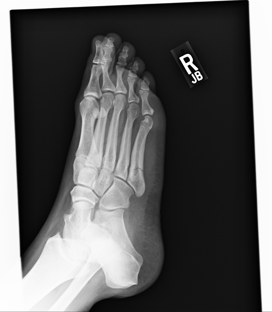
Oblique radiograph of a true Jones fracture.
Lisfranc Joint and Lisfranc Fracture-Dislocation
Jacques Lisfranc de Saint-Martin (1787-1847) was a French surgeon and gynecologist who served as Napoleon’s most trusted physician. He was the chief of surgery at Hôpital de la Pitie (Paris, France) and trained under Guillaume Duputryen. 30 Incredibly dedicated to his profession and to operating, Lisfranc had inscribed on his gravestone “Surgery is bright when operating, but it is still brighter when there is no blood and mutilation and yet leads to the patient’s recovery.” 21 The Lisfranc injury or fracture is a ligamentous or fracture dislocation involving the tarsometatarsal joints (TMT) of the midfoot. During Lisfranc’s lifetime, this injury typically occurred when a soldier or horseman would fall while riding, having his foot trapped in the stirrup. In the present day, Lisfranc injuries continue to be seen in equestrian events but also in football, windsurfing, motor vehicle accidents, and falls. 61
Lisfranc detailed a surgical procedure in 1815 for amputation of the foot at the TMT level, commonly performed on the battlefield. 36 Because of his work nearly 200 years ago, the TMT and injuries to this joint of the foot bear his name.
Segond Fracture
Paul Ferdinand Segond (1851-1912) was a professor of surgery at the University of Paris (Paris, France). The son of the anatomist Louis-Augste Segond, he is known for work in the knee as well as in the field of gynecology. He published a cadaveric study in 1879 which demonstrated that internal rotation and varus stress to a flexed knee resulted in an avulsion of the lateral tibial plateau at the insertion of the mid-lateral capsule. 52 This injury has become synonymous with an injury to the anterior cruciate ligament (ACL) in 75% to 100% of cases. 14 Recently, case reports of “reverse” Segond fractures have been described on the medial side of the knee associated with injury to the posterior cruciate ligament and medial meniscus.18,27
Stener Lesion
Bertil Stener (1920-1999) was a Swedish orthopaedic surgeon who made several significant contributions to orthopaedics during his long and distinguished career. Upon completion of medical school, he became a master illustrator. Winning awards for his drawings, he joined the Anatomy Department at Uppsala as an instructor. His surgical career began in 1963, when he left Uppsala to join the Department of Orthopedics in Gothenburg. Stener’s work in orthopaedics focused on 3 areas in which he made substantial contributions: ligamentous injuries to the thumbs and fingers (especially in skiers); the neurophysiological features of the ligamento-muscular reflexes; and tumor surgery. 25 Known as a gifted and skilled surgeon, he was one of the first to describe total spondylectomies in tumor surgery involving the spine.
In sports medicine, Stener is remembered for his illustrations and descriptions of traumatic injuries to the abducted thumb commonly seen in skiers. Stener, however, was not the first to describe injury to the ulnar collateral ligament (UCL) of the thumb. Campbell, 13 in his classic article, discussed chronic UCL laxity seen in Scottish gamekeepers. Stener discussed a specific and correctable form of UCL injury seen when the aponeurosis from the adductor pollicis is interposed between the torn ends of the ulnar collateral ligament (Figure 3). Because of the interposed tissue, the UCL cannot heal and the metacarpophalangeal joint remains unstable. 56 This has been called a Stener lesion.

Magnetic resonance image of the interposed adductor aponeurosis between the torn ends of the ulnar collateral ligament.
Eponymous Physical Examination Maneuvers
Codman Exercises
Ernest Amory Codman (1869-1940) is considered the father of evidence-based medicine and was an important contributor to the areas of shoulder surgery and the treatment of bone sarcomas. He challenged his peers to follow their results to ensure that patients were having successful outcomes. Codman referred to this as the “end result idea.” 37 Together with Edward Martin, a gynecologist from Philadelphia, Codman created the American College of Surgeons in 1910 to champion surgical outcomes. He also reported the first known rotator cuff repair in 1910. Codman devoted a substantial part of his career to the study of the shoulder, much of which was reported in his 1934 publication The Shoulder: Rupture of the Supraspinatus Tendon and Other Lesions in or About the Subacromial Bursae. In this book, he outlined his exercises for rehabilitation of the shoulder, known as Codman exercises.
Codman’s contributions to medicine extended far beyond the shoulder. He was responsible for the first cancer registry created in the United States. In a collaboration with his friend Harvey Cushing, Codman developed the “ether record,” a standard anesthesiologist record for every surgical case. In addition, he was responsible for the creation of the Hospital Standardization Committee, which over the years transformed to JCAHO (the Joint Commission for Accreditation of Hospital Organizations). 37
Hawkins Test
Richard J. Hawkins (1942- ) is a shoulder specialist practicing in South Carolina. He is a founding member of the American Shoulder and Elbow Surgeons, former president of the Canadian Academy of Sports Medicine, and director of the Sports Medicine Council of Canada. He currently directs the Steadman Hawkins Sports Medicine Fellowship in Spartanburg, South Carolina. He has authored more than 200 articles and published 7 books on orthopaedics. A native Canadian, he wrote a paper on impingement syndromes in athletes that appeared in the American Journal of Sports Medicine in 1980. In this article, he described a physical examination maneuver to confirm coracoacromial impingement. When the clinician has the patient forward flex to 90° and forcibly internally rotates the shoulder, the greater tuberosity is driven under the coracoacromial ligament, reproducing the painful shoulder. 28 This test has become a routine part of the shoulder examination used by many sports medicine physicians.
Jobe Relocation Test
Frank W. Jobe (1925- ) is an American orthopaedic surgeon residing in Los Angeles, California. He has served as an orthopaedic consultant to the Los Angeles Dodgers since 1964. He is most famous for performing the first UCL reconstruction on Dodgers’ pitcher Tommy John on September 25, 1974. This procedure allowed John to return to the Dodgers in 1976 and pitch for 14 more seasons in the major leagues. The success of this reconstruction revolutionized the sport. Previously a tear of the UCL was considered career ending. In addition to devising the UCL reconstruction, Jobe was a founding member of the American Shoulder and Elbow Surgeons and served as its president in 1985-1986. He is cofounder of the Kerlan-Jobe Clinic and Fellowship (Los Angeles, California); he has authored more than 140 publications.
Jobe’s primary academic interest has been care for the throwing athlete. He has championed the idea that anterior microinstability is the cause for the dead arm syndrome in pitchers. The Jobe Relocation Test (JRT) has been advocated as a sensitive way to detect this subtle anterior instability. 32 The examiner performs an anterior apprehension test, with the patient supine on the table. With a positive apprehension test (arm in abduction and external rotation), the humerus is stabilized and a posteriorly directed force is applied to the humeral head to prevent anterior subluxation. 20 If this posterior force relieves the sense of apprehension, the JRT is considered positive.
McMurray Test
Thomas Porter McMurray (1888-1949) was an Irish orthopaedic surgeon born in Belfast. He trained with Sir Robert Jones (see above) at Royal Southern Hospital in Liverpool. 58 At the age of 26, he held consulting staff positions at the Royal Liverpool Children’s Hospital (Liverpool, UK) and the David Lewis Northern Hospital (Liverpool, UK). 23 He gained considerable experience and clinical wisdom from his assistance with British efforts during the First World War. After the war he published many articles and book chapters. He was known as a master surgeon and teacher, and it was reported that he was able to make even the most complex operation appear simple. 23 McMurray served as the president of the British Orthopaedic Association in 1940.
McMurray published a paper in 1942 in the British Journal of Surgery in which he described an examination maneuver to detect a tear of the meniscus. The McMurray test was originally described with the patient supine with the knee flexed to 90°, so that the heel was nearly touching the buttocks. 40 The examiner grabs hold of the ankle and to test for medial meniscus tear extends the knee while placing a valgus stress and externally rotating the tibia. To examine the lateral meniscus, the flexed knee is extended while internally rotating the tibia. A pop, click, or pain with these maneuvers is suggestive of a tear. Today, many sports medicine physicians continue to use the McMurray test, especially when combined with other physical examination maneuvers such as palpating for joint line tenderness and the Thessaly test, to help diagnose a tear of the mensicus. 35
Neer Impingement Test
Charles Summer Neer II (1917-2011) was an orthopaedic shoulder surgeon born in Oklahoma. A third generation physician, he graduated from University of Pennsylvania Medical School (Philadelphia, Pennsylvania). His residency started at the Presbyterian Hospital in New York City, but this was interrupted by service in World War II. Dr Neer later concluded his training at the New York Orthopedic Hospital (New York, New York) in 1949. He joined the faculty at Columbia Presbyterian and the New York Orthopaedic Hospital, remaining until his retirement in 1990.8,10 He quickly became a national and international figure in shoulder surgery, and surgeons routinely traveled to New York to learn from him. His scholarly contributions to the knowledge and care of the shoulder were second to none. He was a pioneer in shoulder arthroplasty, developed a classification system for proximal humerus fractures, recognized that surgical treatment for distal clavicle fractures with coracoclavicular ligament disruption achieved better results than nonoperative treatment, developed a humeral based capsular shift for multidirectional instability, and championed anterior acromioplasty as a successful alternative to acromionectomy for impingement. 8 In addition, he founded the American Society for Shoulder and Elbow Surgeons (ASES) and the Journal for Shoulder and Elbow Surgery.
The Neer impingement test is used to help identify patients with coracoacromial impingement syndrome. Pain is elicited in these patients by forward flexion and internal rotation of the humerus. This brings the greater tuberosity in contact with the undersurface of the anterior acromion.
O’Brien Test
Stephen J. O’Brien (1955- ) is an orthopaedic surgeon practicing at the Hospital for Special Surgery in New York. He is a previous Neer award winner for his research on the shoulder (1989). O’Brien has authored more than 20 book chapters and 80 peer-reviewed articles. His primary area of interest is the treatment of the biceps and labral complex. In 1998, in the American Journal of Sports Medicine, he published findings regarding an examination maneuver to reproduce symptoms in the superior labrum. 47 To perform this examination, the patient is instructed to flex the shoulder 90° with the elbow extended. The arm is then adducted 10° to 15° and maximally internally rotated. The patient then resists a downward force applied by the examiner. The examination is repeated a second time with the patient supinating the arm and the downward force is again applied (Figure 4). Relief of pain with supination of the arm is considered a positive test and concerning for a superior labral abnormality (SLAP lesion).

O’Brien test to detect a SLAP tear.
Ober Test
Frank Roberts Ober (1881-1960) was an orthopaedic surgeon born in Maine. After graduating from Tufts Medical School (Boston, Massachusetts), he completed his residency at Boston Children’s Hospital (Boston, Massachusetts). With the outbreak of the First World War, Ober became the head of the orthopedic section of a French hospital for 2 years. 41 When he returned stateside, Ober was professor at Harvard Medical School (Cambridge, Massachusetts) and returned to work at Boston Children’s. He directed the state of Vermont’s polio rehabilitation effort and was surgeon in chief of the New England Peabody Home for Crippled Children. Ober served as the president of the Massachusetts Medical Society in 1941-1942 and was president of the American Orthopaedic Association in 1943.
In 1936, Ober published an article in the Journal of Bone and Joint Surgery describing an “abduction sign” to check for contraction of the fascia lata/iliotibial band (ITB). 45 To perform this test, the patient must lie on his or her side to minimize lumbar lordosis. The leg is flexed at 90° at the knee; the examiner grasps the ankle with one hand and steadies the hip with the other. If a tight ITB is present, the hip will remain more or less abducted and will not drop down. 45 The modified Ober test is performed in the same fashion, except the knee is in full extension. 59 Both the modified and original Ober test can also be used to help the patient stretch a tight ITB.
Stinchfield Test
Frank E. Stinchfield (1910-1992) was an orthopaedic surgeon born in Warren, Minnesota. He received his undergraduate degree from the University of North Dakota (1932) after transferring from Carleton College (Northfield, Minnesota). 16 He finished his medical school at Northwestern University in Chicago (1935), where he remained for the start of his residency training. He also spent time as a resident at Columbia-Presbyterian (New York, New York) on the fracture service, the Mayo Clinic (Rochester, Minnesota), and the Campbell Clinic (Memphis, Tennessee). 16 Stinchfield enlisted and served in the US Army with distinction during World War II. He was promoted to colonel of 2 hospitals in Europe and was named orthopaedic consultant to the Surgeon-General headquarters to the Allied Forces.16,17 Upon completion of his military duties, Stinchfield returned to become an associate professor of orthopaedics at the New York Orthopedic Hospital (NYOH) at Columbia-Presbyterian. He remained at NYOH for the remainder of his surgical and educational career. After less than a decade, he became chairman of Orthopedics at Columbia-Presbyterian, a job he held for 20 years. In 1968, he helped introduce the Charnley hip to the NYOH and also founded the Hip Society.
A master educator, clinician, and scientist, Stinchfield enjoyed a prolific academic career. He held many important leadership positions during his tenure including president of the American Academy of Orthopaedic Surgery, the American Board of Orthopedic Surgery, and the International Hip Society. 16 He published more than 100 articles, mostly on arthroplasty, and was responsible for educating 8 former residents who became chairmen of orthopaedic departments in North America. 17
The Stinchfield test is used to help the clinician detect intra-articular hip injury. It is performed by having the patient lie supine and elevate his or her leg against gentle manual resistance. Pain elicited with this maneuver will radiate from the sensory innervations of the hip capsule involving the femoral, obturator, and sciatic nerves. 38
Eponymous Surgical Procedures
Bristow Procedure for Shoulder Instability
Walter Rowley Bristow (1883-1947) trained under Robert Jones and was president of the British Orthopedic Association. He served in the British Army in both World Wars and was brigadier in charge of all orthopaedic services during World War II. 46 Bristow also served as the president of the Orthopedic Section of the Royal College of Medicine in Britain. The paper credited with describing Bristow’s surgical procedure for anterior instability was written in 1958 by one of Bristow’s pupils, A. J. Helfet. 29 Many changes to the original description have been made over the years, but the hallmarks of this nonanatomic surgical procedure to minimize anterior instability involve transfer of the tip of the coracoid to the anterior inferior glenoid and augmentation of the dynamic sling of the subscapularis. 51 This procedure remains an option in surgical decision making for anterior instability in the shoulder with significant bone loss. Concerns about this reconstruction include that it is nonanatomic, there is potential for loss of external rotation, and if additional surgery is required, scarring and altered anatomy make for an extremely challenging revision.
Tommy John Reconstruction (Ulnar Collateral Ligament Reconstruction)
Thomas Edward John (1943- ) is a former left-handed Major League Baseball pitcher prominent as the first athlete to have a UCL reconstruction. The surgery was performed by Dr Frank Jobe (see above) in 1974 when John had a 13-3 record and was guiding the Dodgers to the National League pennant. The surgery was performed on September 25, 1974. John missed the entire 1975 season rehabilitating his left arm. In 1976 he returned to the Dodgers and posted a 10-10 record and was named the NL Comeback Player of the Year. He continued to pitch for 14 more seasons, winning 288 games (164 after the surgery), the seventh highest total for a left-handed pitcher in Major League Baseball history.
Weaver-Dunn Procedure for Acromioclavicular Injury
James K. Weaver (1929- ) is an orthopaedic surgeon currently practicing in Colorado. After graduation from the University of Colorado Medical School (Denver, Colorado), he did an internship at the University of California, San Francisco before returning to Colorado for residency. He joined the faculty at the University of New Mexico upon completion of his military service at Elgin Air Force base. He served as chairman at the University of New Mexico (Albuquerque, New Mexico) for 5 years in the late 1960s.
Harold K. Dunn (1937- ) is an orthopaedic surgeon residing in Utah. He grew up on his family’s ranch in New Mexico. A cowboy at heart, Dunn attended college after receiving a calf roping scholarship. In college, he developed a passion for science leading to a career in medicine. He completed his medical training at the University of New Mexico and Baylor (Waco, Texas) before arriving at the University of Utah (Salt Lake City, Utah) in 1969. His primary academic interests are adult reconstruction and spine. Dunn became the second chairman of orthopaedics at Utah in 1981, a post he held for nearly 25 years.
Weaver and Dunn combined to write an article published in the Journal of Bone and Joint Surgery in 1972 on the treatment of acromioclavicular injuries. Their technique involved resecting the distal 2 cm of the clavicle, removing the coracoacromial ligament from the acromial side, passing the ligament into the medullary canal through drill holes in the superior clavicle, and securing the reduction. 62 Biomechanical studies have demonstrated this procedure to have inferior results to more modern anatomic reconstructions, and despite modification, this technique has fallen out of favor. 5
Conclusion
Since the beginning of early civilizations, humans have remained fascinated and enthralled with sports. With the invention and expansion of sport, the development of sports injuries followed. Sports medicine physicians have dedicated their careers to the care of athletes, enabling these participants to enjoy sports and play at as high a level as possible. Over time, we have honored those medical pioneers, and their contributions to the practice of sports medicine, with the continued use of eponyms. Rather than abolish the use of eponyms in the literature, we argue to embrace them. Moving forward, we should be more careful, diligent, and thorough about describing new sports medicine injuries, physical examination maneuvers, and procedures so that the name can be remembered accurately. We agree with Dr McKusick, who in an editorial in Medicine in 1988 wrote, “An entity has not arrived until a name has been devised for it.” 39
Footnotes
Acknowledgements
The authors thank Joseph C. McCormick III, MD, Michael K. Popkin, MD, and James P. Sostak II, MD, for their input.
One or more of the authors has declared the following potential conflict of interest or source of funding: Dr Larson is a consultant for Smith & Nephew and AZ Surgical; he is also a stockholder in AZ Surgical.
An online CME course associated with this article is available for 1 AMA PRA Category 1 Credit™ at ![]() . In accordance with the standards of the Accreditation Council for Continuing Medical Education (ACCME), it is the policy of The American Orthopaedic Society for Sports Medicine that authors, editors, and planners disclose to the learners all financial relationships during the past 12 months with any commercial interest (A ‘commercial interest’ is any entity producing, marketing, re-selling, or distributing health care goods or services consumed by, or used on, patients). Any and all disclosures are provided in the online journal CME area which is provided to all participants before they actually take the CME activity. In accordance with AOSSM policy, authors, editors, and planners’ participation in this educational activity will be predicated upon timely submission and review of AOSSM disclosure. Noncompliance will result in an author/editor or planner to be stricken from participating in this CME activity.
. In accordance with the standards of the Accreditation Council for Continuing Medical Education (ACCME), it is the policy of The American Orthopaedic Society for Sports Medicine that authors, editors, and planners disclose to the learners all financial relationships during the past 12 months with any commercial interest (A ‘commercial interest’ is any entity producing, marketing, re-selling, or distributing health care goods or services consumed by, or used on, patients). Any and all disclosures are provided in the online journal CME area which is provided to all participants before they actually take the CME activity. In accordance with AOSSM policy, authors, editors, and planners’ participation in this educational activity will be predicated upon timely submission and review of AOSSM disclosure. Noncompliance will result in an author/editor or planner to be stricken from participating in this CME activity.