
Abstract

The physis can be the site of acute trauma and chronic overuse injuries in young athletes. Stress injuries resulting from overuse have been described at the physes of the proximal humerus, the distal radius, the proximal ulna, the distal femur, and the proximal tibia.2-4,8,22 Although traumatic fractures affecting the distal tibial physis are common in adolescent athletes,6,10,12,14,18,19 to our knowledge, there is no previously published report of an overuse stress injury affecting this physis secondary to athletic activity. We describe a young female recreational dancer and gymnast who had a 2-month history of progressive ankle pain presenting with stress-related changes of the distal tibial physis that appeared similar to those that have been described at the distal radial physis in the profile of gymnast wrist.
Case Report
A 9-year-old white female recreational gymnast and dancer had a 2-month history of progressive right ankle pain. The patient was very active, taking gymnastics classes once per week, dance classes twice per week, dance or gymnastics class at school daily, and occasional rock wall climbing. She did not recall any traumatic events occurring before her pain began in the previous 2 months, but she did report that 3 weeks before presentation she fell off of the balance beam onto both legs from a height of 6 inches, landing on her right ankle. This fall only mildly increased the patient’s pain, but she continued to bear weight as normal, and the pain returned to baseline after a few hours. The patient continued to participate in dance or gymnastics daily until the pain gradually became severe with weightbearing and bothered her. She was in good health with no history of endocrine or metabolic disorders. She had not begun menses and was normal for her age group for height and weight.
The patient’s examination was notable for mild swelling at the medial malleolus of the right ankle, tenderness to palpation at the distal tibial physis, and an antalgic gait. There was no tenderness at the joint line or along the fibula. The ankle range of motion was normal with no restriction of dorsiflexion or plantar flexion. There was normal subtalar motion, and the anterior drawer sign and inversion stress test finding were both negative. The patient was neurologically intact for sensation and motor strength throughout her lower extremities. Conventional radiographs showed widening of the distal tibial physis medially and, just proximal to the physis, cystic changes in the metaphysis along with a band of sclerosis (Figure 1). They also showed soft tissue swelling, which was more prominent on the medial aspect of the ankle. Additionally, there was some irregularity at the fibular physis, with a well-corticated osseous fragment adjacent to the lateral malleolus, possibly representing a previous avulsion fracture.
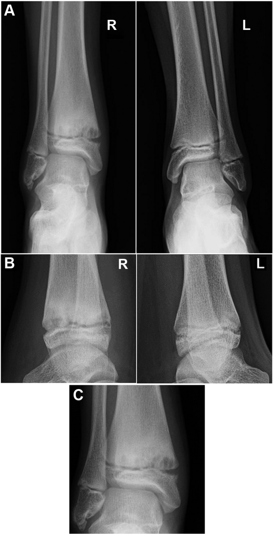
Radiographs at initial presentation. (A) Bilateral anteroposterior views. (B) Bilateral lateral views. (C) Magnification of the anteroposterior radiograph of the right ankle, showing metaphyseal cystic changes and physeal widening.
The patient’s ankle was put in a cast, and the patient was advised not to bear weight on this ankle for 6 weeks. After the cast was removed, the patient’s ankle was placed in a walking boot, and she was allowed progressive weightbearing. She had some recurrence of pain with progressive weightbearing and was not able to return to activities without restrictions until 6 months after presentation. The patient was evaluated 30 months after initial presentation and was found to have equal leg lengths, no ankle or leg deformity, and normal motion at the ankle joint. Radiographs taken 6 weeks after her initial presentation showed continued cystic and sclerotic changes of the metaphysis without improvement, although the physeal widening was decreased. Minimal improvement of the patient’s metaphyseal changes was noted on anteroposterior and lateral radiographs obtained 3 months after initial presentation (Figure 2), but marked improvement was seen at 6 months, although metaphyseal changes were still present. In radiographs obtained 30 months after presentation, the physeal widening and metaphyseal cystic changes seen on previous radiographs had resolved (Figure 3).
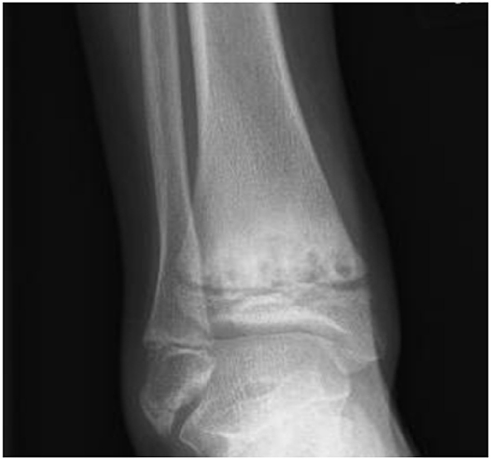
Anteroposterior radiograph of the right ankle 6 weeks after initial presentation.
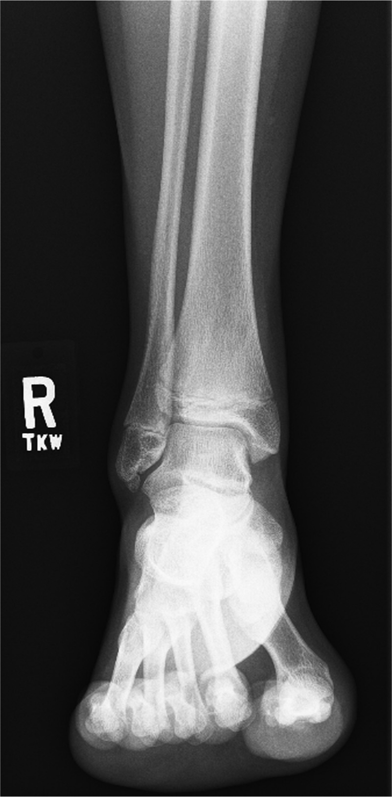
Anteroposterior radiograph of the right ankle 30 months after initial presentation.
Discussion
Although the tibia is often reported to be the most common location of physeal stress fractures in athletes, 20 to our knowledge, no previous report has documented a stress lesion or epiphysiolysis of the distal tibial physis secondary to athletic activity. Although this patient was involved in dance and gymnastics, both activities caused her pain when weightbearing. We know of only 1 other report of an epiphyseal stress lesion at the distal tibial physis within the English literature. 11 In that case, a 15-year-old boy sustained a stress fracture to his distal tibial physis after resuming weightbearing following a subtalar arthrodesis that was performed on the same ankle. 11
Injuries affecting the physes of long bones are reported to comprise 15% to 18% of all pediatric acute fractures2,21,24 and 6% of all pediatric athletic injuries. 16 These reported incidences may be lower than the actual incidence of physeal injuries because physeal injuries can sometimes be difficult to diagnose. 21 The physis is important functionally, serving as the site of growth of long bones in skeletally immature persons; however, it is biomechanically weaker than the surrounding bone and ligaments, making it prone to injury.2,8,16,19 Furthermore, the susceptibility of the physis to injury is increased during times of rapid growth such as the prepubertal growth period.1-3,8,22
The susceptibility of the physis for stress lesions in active athletic adolescents has been well documented.2-5,8,12,22,23 Physeal injuries are usually caused by shearing or avulsion forces that separate the epiphyseal plate at the zone of hypertrophy. 24 In addition to these forces, injury to the epiphyseal or metaphyseal blood supply may also contribute to physeal injury, marked by widening at the physis. 13 Of note, the distal tibial physis specifically begins closure at ages 12 to 14 years in girls and 15 to 18 years in boys, with closure initially occurring centrally, followed by medially and finally laterally. 27
Acute, traumatic injuries affecting the distal tibial physis account for approximately 10% of all physeal injuries, 21 but our patient had no history of trauma before the onset of pain. Most acute fractures affecting the distal tibial physis are Salter-Harris types II and III, which represent approximately 45% and 25%, respectively, of fractures at this physis.21,26 Salter-Harris type I fractures are observed less frequently, with a reported incidence ranging from 5.6% to 15% of all distal tibial physis fractures.14,21,26 In addition to the type of force applied at the ankle, skeletal maturity may also play a role in determining which type of Salter-Harris fracture occurs in a patient. Spiegel et al 26 found that patients with type I Salter-Harris fractures were typically younger than those with type II fractures, with average ages of 10.5 and 12.6 years, respectively. Although our patient was of the right age for a Salter-Harris type I fracture, the history and radiograph results were not consistent with that type of acute injury.
Similarly, the radiographic appearance of the physis in our patient was more consistent with a repeated overuse physeal injury than that seen in acute Salter-Harris type I fractures. Radiographically, physeal stress injuries are characterized by widening of the physis, sclerosis of 1 or both sides of the physis, cystic changes at the metaphysis, a beaked effect of the distal aspect of the epiphysis, or haziness within the usually radiolucent area of the growth plate.2,4,5,8,23 The radiographic findings in our patient were more consistent with these changes, which are similar to those seen in Little League shoulder, in the elbow of young throwing athletes, and in gymnast wrist. Laor et al 15 showed similar radiographic findings in a series of pediatric athletes with proximal tibial stress injuries. Other diagnoses in the differential diagnosis of this radiographic appearance include chronic infection; healing, acute, traumatic Salter-Harris fracture; tumor; underlying metabolic disease; and congenital metaphyseal chondrodysplasia.5,23
The recovery from this injury was consistent with that from a chronic physeal stress lesion. Our patient’s symptoms had nearly resolved by 2 months, at which point she began resuming some activity but had recurrence of pain and was not entirely symptom free until 7 months after initial presentation. The recovery period for Little League shoulder has been reported to be approximately 3 months, whereas that for gymnast wrist has been reported to be 4 weeks to 6 months.4,8
Similarly, the recovery of the physis radiographically in our patient was consistent with a chronic physeal stress lesion. The radiographic irregularities seen with Little League shoulder may take 8 weeks to 1 year to reossify, whereas healing of acute type I fractures typically occurs at 2 weeks. 4 With gymnast wrist, it has been noted that recovery may not be visible until the affected arm is rested and that the time to healing depends on the extent of damage.5,23
The distal tibia is an uncommon location for a stress fracture in patients of any age. In 1 study of 3657 stress fractures affecting the lower extremity, only 4 were localized to the distal tibia, and none of those fractures affected the physis. 28 There have been several documented cases of distal tibial stress fractures occurring after ankle arthrodesis and after a more proximal fracture within the tibia, mostly in adults.9,11,17,25,29 In all of those cases, the distal tibial stress fracture occurred after some other structural change affecting the same leg. Stress fractures occur when stress is repeatedly applied to a bone that has not yet remodeled sufficiently to accommodate to this stress, 7 so perhaps the structural changes that preceded the stress fractures in the aforementioned cases altered the balance of musculoskeletal forces acting on the bone.
In summary, the history, examination results, radiographic appearance, and recovery of this patient are all consistent with a stress lesion of the distal tibial physis secondary to athletic activity. Although obviously rare, in light of the insidious onset of ankle pain in dancers and gymnasts, this diagnosis should be entertained.
Footnotes
The authors declared that they have no conflicts of interest in the authorship and publication of this contribution.