
Abstract
Over the past 2 decades there has been a profound shift in our perception of the role of the meniscus in the knee joint. Orthopaedic opinion now favors salvaging and restoring the damaged meniscus where possible. Basic science is characterizing its form (anatomy) and functionality (biological and biomechanical) in an attempt to understand the effect of meniscal injury and repair on the knee joint as a whole. The meniscus is a complex tissue and has warranted extensive basic science, translational, and clinical research to identify techniques to augment healing and even replace the meniscus. The application of quantitative magnetic resonance image sequencing to the meniscus and articular cartilage of the affected compartment promises to add a quantifiable outcome measure to the body of clinical evidence that supports restoration of the meniscus. This article discusses the recent advances and outcomes in the pursuit of meniscal restoration with particular focus on the use of augmentation strategies in meniscal repair, meniscal imaging, and translational strategies.
Keywords
Distributing load, delivering congruency, enhancing stability, and contributing to lubrication and nutrition are major functions of the normal meniscus. 58 Once these functions are impaired, chondroprotection is disrupted, increasing the risk of degenerative change and increased morbidity. Over 1,000,000 meniscal procedures are performed in the United States annually and this number is growing. 99 Surgical intervention for the injured meniscus is currently driven by mechanical symptoms and/or pain, with symptomatic relief being the key concern of the patient. However, an emerging clinical and scientific body of evidence suggests that meniscus deficiency may predispose the knee to premature osteoarthritis and that restoring the meniscus may also be in the interest of the long-term health of the knee. 59
The clinical association between osteoarthritis and meniscectomy was first established by Fairbank in 1948. 25 Roos et al 80 have demonstrated a relative risk of 14 (increasing prevalence of 1400%) for osteoarthritis of the knee at 21 years after meniscectomy. Total removal of the meniscus increases peak contact stresses in the affected compartment, and this may account for an estimated 4% cartilage loss per annum (greater on the lateral side).52,102 Furthermore, the increase in contact stress is directly proportional to the amount of removed meniscal tissue, and restoration of partial lesions has been shown to approximate contact mechanics on the affected plateau.11,30 Therefore, the goal of treatment for meniscal deficiency is to provide symptom relief in the short term while restoring form and function in the medium to long term. Partial meniscectomy may be the only surgical option for select patients, but it is important to consider nonoperative measures where possible. In a multicenter randomized controlled study of symptomatic patients with medial meniscal tears and coexisting mild to moderate osteoarthritis, there was no subjective difference in pain or functional outcome between 2 groups: one assigned to arthroscopic partial meniscectomy, the other to physical therapy alone. 47 This was reported in the context of considerable crossover from the nonoperative group to the meniscectomy group during the study, suggesting that nonoperative measures may not be ideal for every candidate and that continued clinical follow-up is warranted.
In this article, the benefit of restoring a functioning meniscus is discussed in the context of existing strategies and evolving basic science. The role of magnetic resonance imaging (MRI) of the meniscus has expanded to assessment of tissue quality in the acute setting and the evolving biology of repair and scaffold/allograft integration. Magnetic resonance imaging allows for noninvasive assessment on the macro- and microarchitectural level and is discussed as a quantifiable facet of clinical outcome in meniscal surgery.
Meniscal Repair and Imaging
Specific tear and patient characteristics are critical for clinical decision making with regard to meniscal repair. 67 A comprehensive overview of indications for meniscal repair has recently been published by Maak et al. 57 Patient-related indications include positive symptoms of meniscal injury (instability, locking, effusion, grinding, and pain), physical findings on knee examination (joint line tenderness, effusion, limitation of motion), and provocative signs (eg, pain with squatting). Contraindications to repair include poor tissue vascularity and degenerative changes, advanced patient age, poor patient compliance with rehabilitation, and the presence of either knee instability or osteoarthritis. Magnetic resonance imaging is important for characterizing both the tear pattern and tissue quality. Tear-specific indications include positive symptoms directly attributable to the tear, reducibility of the tear, and favorable tear characteristics (eg, single vertical tear in one plane in the red-red region; red-white tear in the middle third region). Tear characteristics currently deemed unsuitable for repair include chronicity of the tear, smaller longitudinal tears (<10 mm), radial tears limited to the inner two-thirds of the meniscus (avascular region), and degenerative tears. Degenerative tears are identified on MRI as increased signal intensity due to the increased water mobility associated with myxoid degeneration (in contrast to the typical uniformly low signal intensity of the normal meniscus afforded by the restriction of water motion and very short relaxation times associated with the type I collagen network). Regardless of tear pattern or patient characteristics, such tears are notoriously difficult to repair owing to both poor tissue quality and limited intrinsic healing ability.
Meniscal repair is undertaken to ultimately restore the native biomechanical properties of the macroarchitecture and in doing so, recreate the circumferential hoop stresses. As with other soft tissue reconstruction, consider the initial repair as temporizing the biomechanical demand while recreating a repair site that facilitates healing for lasting functionality. Thus, the type of meniscal tear influences our thought process on the role and mechanism of repair. While radial tears may limit the function of the meniscus to distribute load, some circumferential vertical or horizontal tears may have residual functional potential. Regarding healing at the repair site, vascularity of the repair strongly influences outcome. Peripheral, well-vascularized “red-red” zone tears have a good propensity for healing, as do many of the tears at the “red-white” junction. Tears in the inner “white-white” zone are relatively unlikely to heal (Figure 1). 4
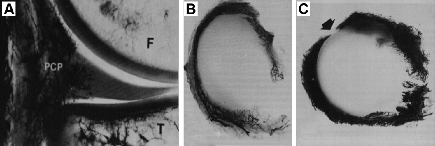
(A) A 5-mm thick frontal section of the medial compartment of the knee (Spalteholtz ×3). Branching radial vessels from the perimeniscal capillary complex (PCP) can be seen penetrating the peripheral border of the medial meniscus. F, femur; T, tibia. This image demonstrates a sagittal view of meniscal vascularization. 4 Superior aspect of the (B) medial and (C) lateral menisci after vascular perfusion with India ink and tissue clearing using a modified Spalteholtz technique. Note the vascularity at the periphery of the menisci as well as at the anterior and posterior horn attachments. The absence of peripheral vasculature of the posterior lateral corner of the lateral meniscus (arrow) represents the area of passage of the popliteal tendon. 4
The techniques available for meniscal repair include both open and arthroscopic techniques; specifically, outside-in, inside-out, and all-inside approaches. While tear characteristics, location, and operator experience may influence the approach used, there has been an increased shift to all-inside techniques and the use of internal fixation devices where possible, given their relative ease of application and minimal exposure. Contemporary all-inside systems have decreased the operative time and eased repair, but both biological and biomechanical factors are likely to be more important to outcome than the precise technique. Some studies have raised concerns regarding failure, stiffness, and displacement properties of all-inside repair implants. 55 However, overall they have demonstrated comparable outcomes to inside-out repair for peripheral unstable longitudinal “bucket-handle” tears in a recent systematic review. 33 Recent inclusion of MRI assessment with clinical outcome in studies comparing all-inside techniques to inside-out or outside-in approaches provides objective evidence of efficacy and represents a valuable outcome measure for further study in this area. 18
Literature charting the outcome of all-inside techniques has been consistently confounded by varying rates of concurrent anterior cruciate ligament (ACL) reconstruction. 55 Meniscal tears found in association with ACL injury and repaired at the time of ACL reconstruction have a particularly good prognosis for healing. There is also an association with increased healing after tibial plateau fractures. 81 These observations are likely multifactorial in mechanism but have been suggested to relate to biological augmentation of the repair from factors in the bone marrow released within the joint, as well as the slower rehabilitation protocol. Overall, the best results for repair tend to be seen in acute tears. 95 Older patients (>30 years) have been shown to have a higher incidence of retears, although in a separate study these were found to occur significantly later, possibly relating to an interplay of biology and demand on the repair.7,23
Evaluating the efficacy of repair, second-look arthroscopy has been the most dependable way to determine meniscal healing. Utilizing noninvasive advanced imaging, it has been shown that on MRI examination, T2-weighted sequences had higher specificity and accuracy, while proton density and T1-weighted sequences had higher sensitivity. 61 In a study of 36 subjects comparing fluid-sensitive MRI to contrast-enhanced conventional arthrography as the standard, MRI had a sensitivity of 0.83, specificity of 1.0, positive predictive value of 1.0, and negative predictive value of 0.92. In the 8 of 36 patients who underwent second-look arthroscopy due to clinical symptoms of meniscal dysfunction, MRI proved more accurate than arthrography in discriminating partial from complete healing. 93 These techniques have subsequently been used to assess differential healing rates of various repair techniques. 94 While noncontrast MR techniques have shown to be efficacious in assessing meniscal repair, the appeal of quantitative MR techniques, providing a direct measurement of tissue relaxation time and a standard of deviation, are more appealing for statistical analysis. The tissue relaxation times of the intact meniscus reflect its ultrastructure, with very short relaxation times that are not measurable using standard MR pulse sequences. Ultrashort echo time (UTE) MRI allows for direct measurement of T2* of fibrocartilage, a reproducible parameter that correlates with the orientation of the collagen in the fibrocartilage (Figure 2). 31 In a recent study of an ovine model of meniscal repair, quantitative T2* values correlated with the results of quantitative multiphoton microscopy measurements of collagen structural integrity (Potter et al, unpublished data). In the future, the diagnostic value of MRI could be improved by a combined application of different, and ideally, quantitative sequences.
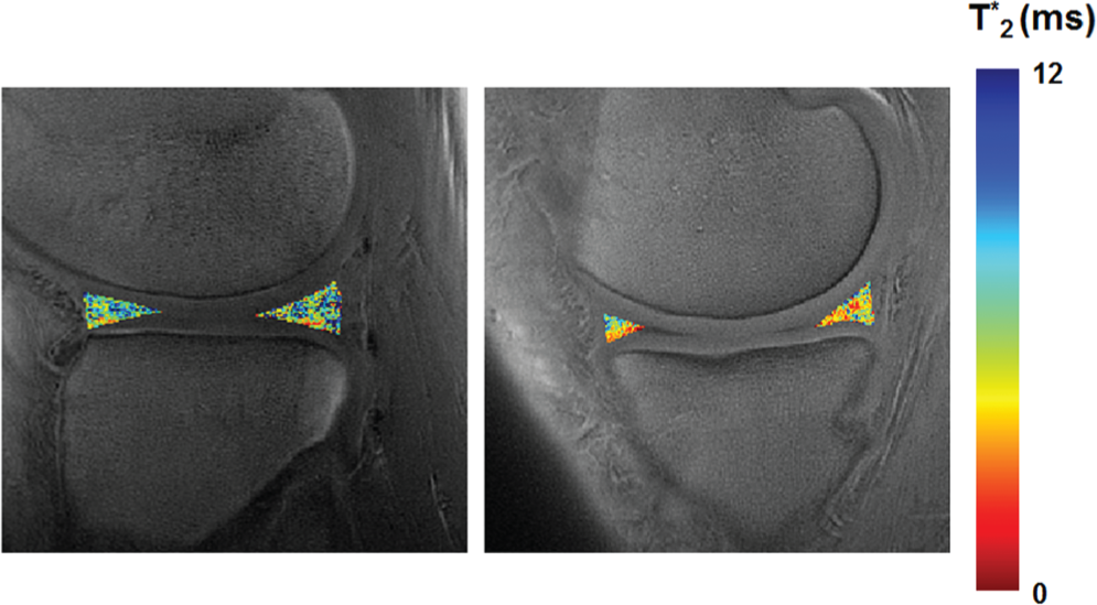
Ultrashort echo time (UTE) images of a patient 6 months after lateral meniscal repair (left) shows more heterogeneity and prolonged T2* values compared with the intact medial meniscus (right).
Biological Augmentation of Repair
There is growing interest in extending the success of the aforementioned repair techniques to tears with unfavorable characteristics. 69 Biological augmentation strategies attempt to overcome the inherent limitations in healing related to poor vascularity and heterogeneous cellularity by promoting chemotaxis, cellular proliferation, and matrix production at the repair site. Mechanical techniques that stimulate healing by creating vascular access and stimulating cells (peripheral meniscal stem cells and fibrochondrocytes), cytokines, and bone marrow cells in the vicinity are representative of earlier augmentation strategies. 5 These include rasping (synovium and meniscus) and radial trephination of the meniscus. 110 These techniques were developed using animal models and applied clinically with varying clinical outcomes overall. 67 The use of exogenous fibrin clot was developed in animal models and has shown some success in humans, despite the technical difficulties of maintaining the clot in situ at the repair site.6,46 Meniscal and synovial rasping and abrasion are minimally invasive in the context of the index procedure and have demonstrated some advantage. 91 Growth factors for meniscal repair have been validated in vitro for cell migration and proliferation with matrix production. 58 Specifically, fibroblastic growth factor–2 (FGF-2) and connective tissue growth factor (CTGF) have demonstrated enhancement of meniscal repair in vivo in a rabbit model. However, in an effort to enhance vascularization of the repair site, vascular endothelial growth factor (VEGF) did not show an advantage in a sheep model.41,51,66
In terms of cell-based therapy, the use of differentiated cells (chondrocytes and fibrochondrocytes) and undifferentiated mesenchymal stem cells (MSCs) in meniscal repair has been promising. Specifically, improved healing of meniscal defects has been documented in the presence of chondrocytes seeded on scaffolds, allogenic meniscal tissue, and fibrin glue.83,104 The menisci form as a condensation of MSCs in utero, and tissue engineering (TE) strategies attempt to resurrect that potential. 3 Mesenchymal stem cells are believed to have additional trophic effects and may function to produce cytokines at the repair site. 16 Animal models using MSCs have demonstrated increased healing and integration of meniscal lesions.44,109 Interestingly, in a rabbit model, Zellner et al 107 showed superior meniscal repair in hyaluronan scaffolds seeded with undifferentiated MSCs compared with a precultured prochondrocytic MSC group, thus supporting the concept that they may, in their undifferentiated form, also act in the facilitation of repair rather than simply as a pluripotent cell source. Moriguchi et al 63 demonstrated reproducible healing of avascular tears in the miniature swine model using scaffold free tissue–engineered constructs derived from synovial mesenchymal stem cells.
Indeed, the application of scaffolds and hydrogels to meniscal repair provide a 3-dimensional (3D) structure across the repair site and act as a vehicle for cells, growth factors, and minimally manipulated biological products. Narita et al 65 describe gelatin hydrogels with impregnated FGF in the rabbit model implanted at the tear site while Piontek et al 70 describe a novel all-inside tear-wrapping technique outside the repair with a collagen membrane.
While translating cellular and growth factor advances to clinical care may be exciting, it is also fraught with practical and regulatory difficulty. As a result, there is increasing interest in the addition of minimally manipulated biological products such as platelet-rich plasma (PRP) or bone marrow aspirate concentrate (BMAC). PRP contains various growth factors, including transforming growth factor beta–1 (TGF-ß1), FGF-2, platelet-derived growth factor (PDGF), and VEGF, all of which are involved in varying levels in cell differentiation, chemotaxis, angiogenesis, and extracellular matrix production. Ishida et al 45 demonstrated in vitro that meniscal cells cultured in PRP had increased expression of mRNA of extracellular matrix proteins relative to the controls. Full-thickness meniscal tears created in the avascular region of rabbits showed superior histological repair when treated with PRP using a gelatin hydrogel delivery system. Bone marrow aspirate concentrate provides another point of care option for enhancing repair by increasing the concentration of cells and growth factors in marrow aspirate in a low-cost, low-risk manner that may provide some of the benefits of stem cell therapy while meeting regulatory oversight. Ultimately, randomized controlled trials are required to provide the evidence to support the use of biologics in meniscal surgery. Quantitative biological characterization of these products is key in data interpretation as these products can vary significantly depending on processing.17,103
Meniscal Allograft Transplantation
Meniscal allograft transplantation (MAT) is the only current therapeutic option for total meniscal replacement. Careful consideration of perioperative factors relating to the graft, patient assessment, and ongoing clinical study continue to refine the indications for this procedure that is no longer considered experimental (Table 1). 56 Meniscal allografts are available as cryopreserved, deep frozen (fresh frozen), and fresh for implantation. Lyophilized grafts are no longer used clinically, owing to inferior material properties and clinical results. 106
Clinical Overview of Meniscal Allograft Transplantation a
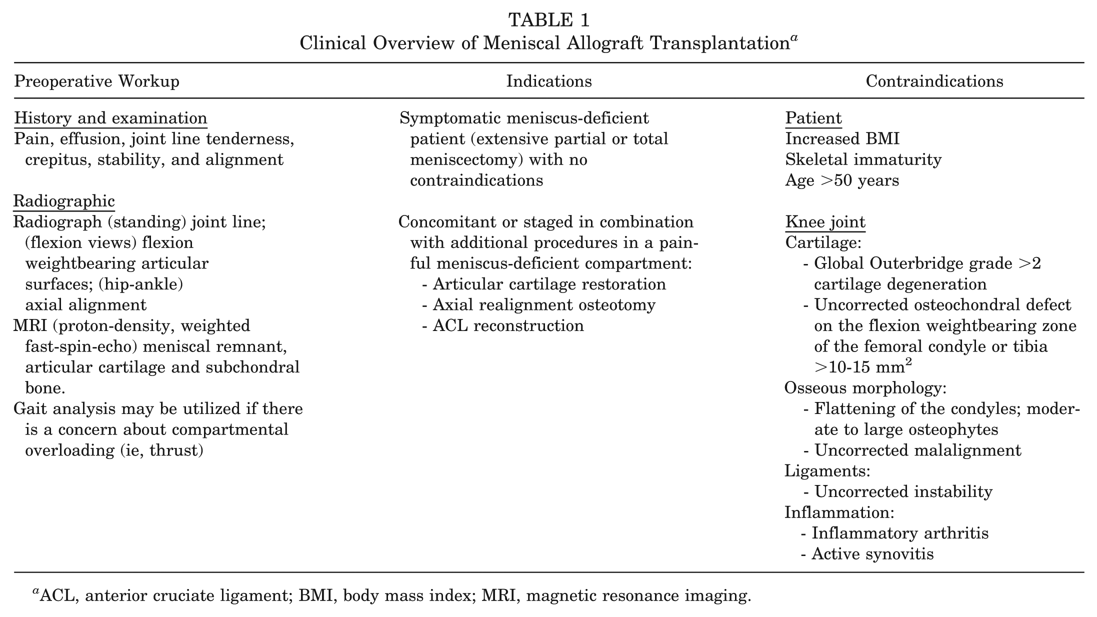
ACL, anterior cruciate ligament; BMI, body mass index; MRI, magnetic resonance imaging.
In considering the available allografts, focus has been directed toward the role of viable graft cells, immunogenicity, and sterilization, each with implications on graft processing. Transplantation of viable donor meniscal cells within the allograft has not demonstrated a clear advantage; the allografts are repopulated by host cells (of various phenotypes) and very few donor cells persist. 76 Regarding immunogenicity of the allograft, it is theorized that deep freezing may denature histocompatibility antigens. However, B lymphocyte and cytotoxic T cells have been described in deep frozen allografts in human studies. 76 There has only been 1 case of frank rejection in a cryopreserved, nontissue antigen-matched allograft, but concern exists about a subclinical immunoreactivity in the graft that may impair integration. 56 Disease transmission from meniscal allografts and adjoining bone remains an important aspect of this surgery to the surgeon and patient. In 1990, the estimated risk of HIV from a frozen connective tissue allograft was 1 to 8 million. 56 Secondary sterilization with gamma irradiation has improved the bacterial sterilization. However, doses required to inactivate HIV are substantially higher and cause deterioration of the material properties of the allograft; therefore, they are not applied routinely. 56
From a technical standpoint, meniscal allografts may be inserted with or without attached bone. The meniscus may be implanted with separate bone plugs attached to the horns, using a common bone bridge attached to both horns, or on a hemi-plateau. 76 There are 2 main fixation techniques for arthroscopic-assisted meniscal transplantation: fixation with bone plugs and suture fixation through transosseous tunnels. A technique describing reinsertion to the meniscal root has also been described. 53 While clinical data support both fixation techniques, bone plug fixation has been shown to better recreate biomechanical and contact pressures in cadaveric experimental models and in silica finite element model analysis (Maher et al, unpublished data).2,40,60,96 Sizing of the graft is critical: Smaller grafts may fail to restore the joint congruence and larger grafts are prone to extrusion. The graft is secured to the capsule using standard meniscal repair techniques, and the meniscotibial attachments are not routinely restored. Ligamentous stability and axial alignment are critical factors in recreating load sharing across the compartments in treatment of the entire knee (Figure 3).
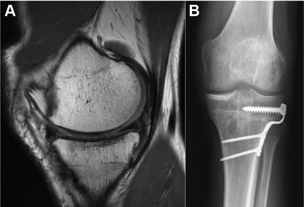
(A) An MRI of a medial meniscal allograft using bone plug fixation with concomitant high tibial osteotomy realignment procedure and revision ACL reconstruction using Achilles tendon graft (12 months out, asymptomatic, good functional outcome). Note the slight extrusion of the degenerated anterior horn but good position and signal within the posterior horn. (B) Standing radiograph at 12 months after high tibial osteotomy and meniscal replacement.
To date, histological examination after MAT has demonstrated biological integration that is variable in terms of cellular infiltration and associated matrix production in the short term.76,77 Critically, there has not been a qualitative or quantitative histologic response that predicts success of the implant, rather an absence of cellular proliferation has been seen in failed implants. 20 From a clinical perspective, most of the evidence on MAT outcome is level 4 evidence and has been quantified in a recent meta-analysis in terms of Coleman score (45.9 ± 8.4), putting it in the range of other cartilage studies. 24 Published data must be interpreted in the context of the case heterogeneity that exists, such as indications, graft type selection, fixation, sizing, and procedure. Overall, the majority of studies report improvement in knee symptoms in the short term and medium term (Table 2). This remains true despite findings of varying degrees of graft shrinkage, degeneration, and extrusion seen with MRI, as this often does not correlate with symptoms. In that regard, a consensus regarding clinical failure of MAT is needed in the literature as this remains variable, ranging from degrees of extrusion to conversion to knee arthroplasty.
Medium- to Long-term Outcome of Meniscal Allograft Transplant a
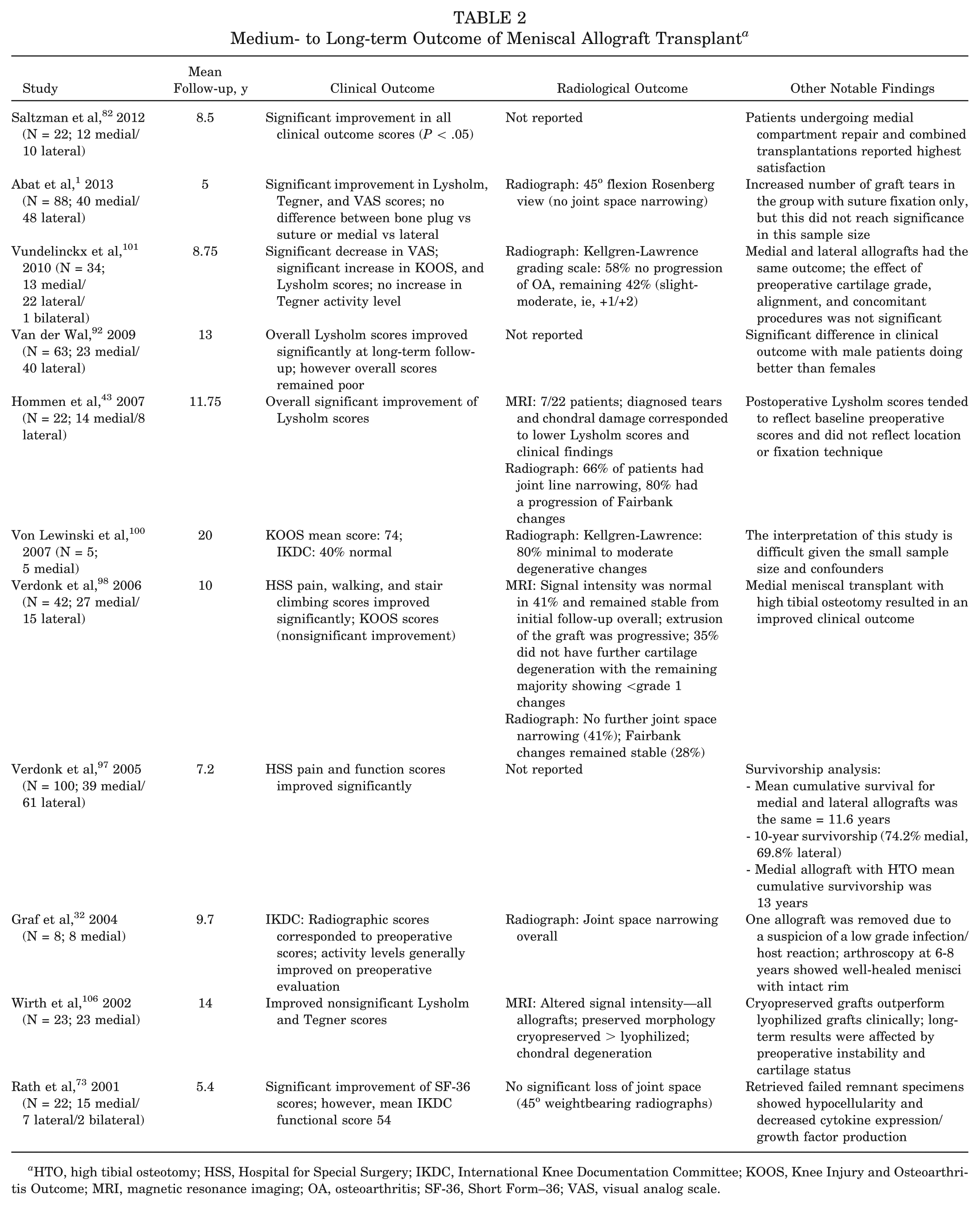
HTO, high tibial osteotomy; HSS, Hospital for Special Surgery; IKDC, International Knee Documentation Committee; KOOS, Knee Injury and Osteoarthritis Outcome; MRI, magnetic resonance imaging; OA, osteoarthritis; SF-36, Short Form–36; VAS, visual analog scale.
Currently, study is ongoing to investigate the intraoperative in vivo contact mechanics of patients undergoing MAT (Rodeo et al, unpublished data). Articular cartilage contact stress measurements (obtained in the operating room) will be correlated with measurements of cartilage contact and deformation derived from MRI done with axial load across the joint in an effort to more fully evaluate the transplanted compartment and the subsequent integration of the allograft and health of the articular cartilage. The use of quantitative MRI allows evaluation of hyaline cartilage composition and microstructure before the development of more advanced morphologic changes. Such evaluation should include both assessment of collagen orientation (T2 mapping) and proteoglycan (T1rho, sodium imaging, or delayed gadolinium MRI of cartilage [DGEMRIC]). 70 It is hoped that these data will help to develop accurate predictors of outcome and the use of MRI as a noninvasive surrogate marker of biomechanical function.
Meniscal Substitutes
Although they are not available for use in the United States (US), there are 2 meniscal substitutes available for clinical use elsewhere. The collagen meniscus implant (CMI) is commercially available and widely used in the European Union (EU). It was approved by the US Food and Drug Administration (FDA) in December 2008, but this approval was withdrawn in October 2010. Actifit is a synthetic biodegradable polyurethane meniscal scaffold indicated for both medial and lateral partial meniscal replacement (Actifit Orteq Limited, London, UK). It is currently available in the EU, and FDA evaluation is ongoing. The indication for use of either of these substitutes is the treatment of painful, irreparable partial meniscal defects as both implants require an intact peripheral vasularized rim and horns for fixation. Both constructs are biodegradable and facilitate tissue regeneration by providing a 3D scaffold for infiltrating cells to lay down extracellular matrix (ECM) and to support new meniscal tissue formation.
Collagen meniscus implant is a scaffold that is composed of the ECM components of type I collagen and glycosaminoglycans (GAGs): chondroitin and hyaluronic acid. It is chemically cross-linked with formaldehyde and sterilized using gamma radiation. From clinical studies, histological findings at the early timepoints demonstrate that CMI functions as a meniscal scaffold with remodeling within the construct. Biopsies have shown new blood vessels and collagen fibrils and progressive replacement of CMI with immature collagen.74,79,90 On electron microscopy, Reguzzoni et al 74 further defined the regeneration by demonstrating parallel lacunae walls with collagen fibrils, blood vessels, and fibroblast-like cells. At the 5-year timepoint, Steadman and Rodkey 87 observed fibrocartilage and organized extracellular matrix in all of the patients that they biopsied without evidence of infection, inflammation, or immune reaction. An estimation of meniscal defect fill in their study of 8 patients was reported as 69%. Regarding clinical outcome, recently published data in the medium to long term has shown encouraging results (Table 3). Monllau et al 62 demonstrated significant improvement in pain and function without development or progression of degenerative knee disease in most patients, despite implant shrinkage at a minimum follow-up of 10 years. Zaffagnini et al 105 have recently reported good results with meniscal scaffold in the lateral compartment at 2 years follow-up. However, a well-defined collective picture of the clinical outcomes of CMI is difficult given the methodological research quality of some studies and the variation in postoperative evaluation techniques. 39
Clinical Outcome Studies of Meniscal Substitutes: Medium- to Long-term for CMI and All Outcomes for Actifit (Short Term) a
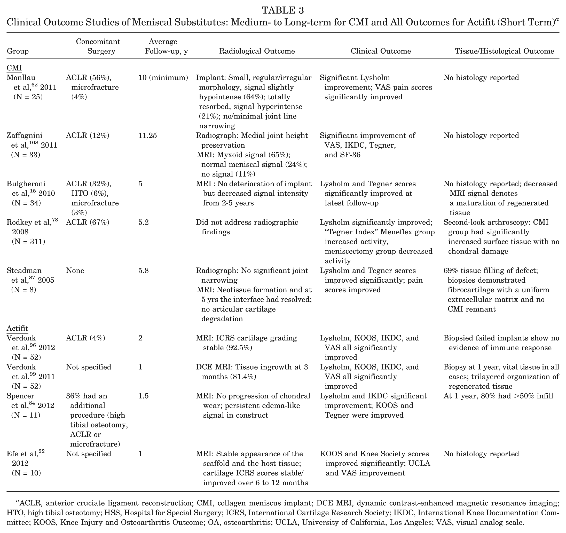
ACLR, anterior cruciate ligament reconstruction; CMI, collagen meniscus implant; DCE MRI, dynamic contrast-enhanced magnetic resonance imaging; HTO, high tibial osteotomy; HSS, Hospital for Special Surgery; ICRS, International Cartilage Research Society; IKDC, International Knee Documentation Committee; KOOS, Knee Injury and Osteoarthritis Outcome; OA, osteoarthritis; UCLA, University of California, Los Angeles; VAS, visual analog scale.
Actifit is a slowly degrading (5 years) polymer-polycaprolactone and urethane acellular scaffold. 84 Tissue growth was first investigated clinically at 3 months using double contrast-enhanced CT confirming tissue ingrowth. 102 Biopsy samples taken at 1 year demonstrated a trilayered tissue organization pattern based on the presence or absence of vessels, ECM characteristics, and cell structure in 81% of patients. The tissue was viable with no evidence of an immune reaction and it resembled native meniscus. Tissue ingrowth began superficially and the maturation process progressed into the deeper layers, supporting previously reported data from animal studies that demonstrated active fibrovascular infiltration from the synovium in dogs.49,105 Verdonk et al96,99 reported on 52 patients with irreparable meniscal defects over 2 years, building on previously published data. Their results confirmed clinical and statistically significant (P < .01) improvement in all clinical scoring systems. Stable or improved International Cartilage Research Society (ICRS) grades were observed in 92.5% using dynamic, contrast-enhanced MRI imaging. Ultimately, continued follow-up is required to ascertain whether meniscal substitute technology can improve the longer-term health of the postmeniscectomy knee, but at the present time it does seem to be associated with promising results.
In addition, a prospective, multicenter, open label, nonrandomized study evaluating the treating of medial meniscus deficiency with the NUsurface meniscus implant is currently ongoing in Europe and Israel. This is a free-floating ultra-high molecular weight polyethelene (UHMWPE)–reinforced polycarbonate urethane meniscal device (NUsurface, AIC, Memphis, Tennessee) that approximates the shape of a discoid meniscus and requires an intact peripheral rim for insertion. It was investigated in the preclinical setting using a sheep model and found to exert some chondroprotective effects while reproducing native loading patterns of the meniscus in a finite element (FE) analysis. 111
Rehabilitation After Meniscal Surgery and Managing the In-Season Athlete
Rehabilitation of the knee after meniscal intervention requires both patient- and procedure-related considerations. The restored meniscus must function within the knee joint as an organ 54 and as part of a kinetic chain that has regained muscular control, proprioception, and confidence without compromising the biomechanics or the healing response of the intervention. 12 In this regard, close communication between the surgeon, patient, and physical therapist are essential to achieve realistic milestones without compromising meniscal healing or function.
Unfortunately, the option of partial meniscectomy and staged meniscal restoration for the “in-season” athlete is not available to surgeons in the US. It is important to note in the setting of partial meniscectomy, that while symptom-free mobility and an expedient return are the goals of the patient, articular cartilage may need time to adjust to the altered mechanical loading in the postmeniscectomy compartment. 13 Careful and graduated return to full weightbearing should only occur under the close guidance of the physician and associated physical therapy team as altered loading has been associated with cartilage degeneration. 50 In this regard, it is worth counseling the athlete that a 4- to 6-week timepoint is a realistic target and that premature return to sport may be potentially damaging in the medium to long term. For this recovery we should accept that return to sport may take longer in some individuals, and thus it is critical to evaluate each patient individually.
After meniscal restoration (repair/replacement), the obvious concern is for the integrity of the repair and allowing sufficient time for the meniscus to heal or integrate. Excessive weightbearing is prevented in the early period to prevent high compressive and shear forces that can disrupt repair sites in transplantation and radial tear repair. 68 In the senior author’s institution, rehabilitation after repair consists of a double upright hinged brace to allow 0° to 90o of flexion for the first 4 weeks, early weightbearing in full extension, weightbearing without the brace beginning at 4 to 6 weeks, and no high impact athletics for 4 to 6 months. There has been a trend to individualize rehabilitation protocols, and certainly the movement toward accelerated, integrated protocols may facilitate return to sport “in-season” without compromising the career of the athlete. Identifying “at-risk” individuals using physical assessment tools and an understanding of the effect of preexisting meniscal injury will further improve this process without compromising the outcome.14,19
From a translational perspective, we should incorporate our improved understanding of the mechanobiology of soft tissue to optimize healing in the postoperative period. It has been long known in orthopaedic surgery that mechanical forces could drive tissue formation such as bone formation in response to load. Recently, with the advent of enabling biophysical and molecular technologies, we are beginning to understand how individual cells transduce mechanical force into biochemical signals. In turn, this knowledge of mechanotransduction at the cellular level is beginning to clarify the role of mechanics in soft tissue healing with current literature suggesting that tensile strain on fibrochondrocytes may reduce inflammatory factors and increase GAG and collagen production.10,27 However, eliciting these anabolic effects in the acute phase may be hampered by pro-inflammatory cytokines (eg, Tumor Necrosis Factor–alpha) that can modulate the response of meniscal cells to mechanical signals. 26
Tissue Engineering and Translational Replacement Strategies
The TE strategies for the meniscus have followed significant basic science advances in articular cartilage research. Scaffolds (synthetic, extracellular matrix-constituent, and tissue-derived) and hydrogels for meniscal tissue regeneration each have their own inherent advantages. Regarding their incorporation into meniscal substitutes, finite element (FE) models have demonstrated that circumferential and axial/radial moduli are important determinants of the contact pressure distribution by the native meniscus and thus should be matched in any replacement technology. 40
A significant advantage of synthetic scaffolds is the relative ease of manipulation of their biomechanical properties. Electrospun poly(ϵ-caprolactone) (PCL) scaffolds have also shown promise for a TE solution for the meniscus and may provide an appropriate microarchitectural environment for meniscal cells. These nanofibers may provide increased biomechanical advantage of the temporizing structure and of the regenerated meniscus by helping to organize and optimize the 3D orientation of ECM deposition in the circumferential direction while facilitating cellular infiltration through the scaffold’s fibers.8,21,28 At the same time, 3D woven PCL scaffolds have conferred increased biomechanical properties in a composite preparation with cartilage-derived matrix, obviating the need for growth factor augmentation and may certainly be applied to meniscal restoration. 64 Gunja et al36-38 have conducted work using nonwoven poly-L-lactic acid (PLLA) scaffolds exploring the positive, synergistic effects of bFGF and hypoxia, hydrostatic pressure, and transforming growth factor–ß (TGF-ß) and coculture (chondrocytes and meniscal cells) using in vitro models.
Ultimately, it is hoped that cellular infiltration and matrix deposition may restore some of the viscoelastic properties of meniscal tissue where the fluid phase carries a significant amount of the load. 85 The biocompatibility and biomimetic properties of ECM constituent scaffolds have led to their use, despite their inferior biomechanical profiles. Balint et al 9 have reported the use of a collagen scaffold reinforced by a network of degradable tyrosine-derived polymer fibers that has demonstrated circumferential tensile strength stiffness and hoop stress behavior under compressive loading that mimics the normal meniscus.
Hydrogels (natural or synthetic) are biochemically versatile and can be used as cell and growth factor delivery systems. 58 Although their biomechanical properties can be tailored somewhat by substrate concentration and composition, they often fall short of native meniscal properties. To overcome this problem, Holloway et al 42 proposed a fiber-reinforced composite containing UHMWPE fibers in a poly vinyl alcohol (PVA) hydrogel and varied the composition and orientation of the fibers to mimic native meniscal properties for total meniscal replacement. Upon implantation in a dynamic cadaveric model, this construct was seen to restore contact mechanics to a comparable level as MAT. 34 Anatomically shaped alginate hydrogels have been fabricated using extracted geometries from MRI and microcomputed tomography (microCT) and demonstrate improved biochemical and mechanical properties after seeding with fibrochondrocytes and supplementation with IGF-1. 72
Tissue-derived scaffolds under current investigation include small intestinal submucosa (porcine) and silk scaffolds. The use of xenogenic meniscal tissue is currently being investigated in animal models and may provide a solution in partial meniscal replacement. Indeed, processing of ovine meniscal tissue to improve porosity and interconnectivity as a xenogenic meniscal scaffold has potential. 84
The design of a successful meniscal scaffold, while temporizing a degree of biomechanical demand should also incorporate features amenable to cellular infiltration at the meniscal tissue interface. These include pore-size and interconnectivity while maintaining a near native meniscal compression modulus to exert a profibrochondrogenic effect.48,49 Mesenchymal stem cells, fibrochondrocytes, and chondrocytes have been successfully incorporated into cell-scaffold complexes and demonstrate promising results in both in vitro and in vivo studies. The overwhelming issue with the latter 2 cell types is availability. The use of autologous fibrochondrocytes/chondrocytes requires a 2-step operative process. Given the relative scarcity of these cells in the tissue, it then requires monolayer expansion, which can result in down-regulation of genes involved in matrix production. 35 Allogenic and xenogenic cell sources have shown promise in animal models and while they may overcome the discrepancy in supply and demand, they are fraught with translational issues.75,104 Mesenchymal stem cells, on the other hand, have been successfully isolated from adipose tissue, muscle, and bone marrow and subsequently used in soft tissue engineering. 16 The next generation of genetically manipulated MSCs may further enhance the regenerative process by promoting growth factor production and directed chondrogenic differentiation.88,89 As the final cell type for discussion, synoviocytes have often been overlooked in meniscal regeneration and repair augmentation. 29 This is despite the fact that meniscal repair has been shown to be facilitated by fibroblasts from the surrounding synovium and joint capsule that are believed to undergo fibrocartilaginous metaplasia at the repair site. 3
Cell chemotaxis, maintenance of cellular phenotype, and inducing matrix production are critical for the biological integration of a meniscal scaffold. The roles of biophysical and biochemical signaling are currently under review in this regard. Both the microarchitectural environment and biomechanical properties of a scaffold material act as biophysical cues for seeded and infiltrated cells (Makris). In addition, the effect of mechanical loading on cellularized constructs, meniscal explants, and scaffoldless-assembly meniscal tissue is being investigated in an attempt to understand and utilize the resultant cellular responses; these may have a role in the maintenance of cell phenotype, matrix production, and overall improved vital role of cell phenotype maintenance and differentiation. Fibroblastic growth factor–2 has been shown to recover the monolayer expansion of meniscal cells, whereas TGF-ß1 has caused meniscal cells to take on a more chondrogenic phenotype. 58 Growth factor supplementation has demonstrated improved cellular migration, proliferation, and matrix production, as discussed, but important translational considerations include mode of delivery, localization of effect, and timing of growth factor release. The use of innovative imaging markers in future studies may prove efficacious in tracking cell migration and matrix production.
Conclusion
Orthopaedic opinion has swayed rightfully toward restoration of the meniscus. As a tissue, the meniscus is exquisitely complex in both its form and function. The expansion of meniscal repair to previously irreparable avascular and complex tears is a result of our increased knowledge of the biomechanics and biology and a move to biological augmentation, where possible. Meniscal allograft transplantation is no longer experimental and it is hoped that continued bench-top and clinical research will improve outcome. Exciting discoveries in tissue engineering are approximating native meniscal biomechanical properties and it is hoped that they will transition to clinical practice soon. Novel quantitative MRI sequencing allows for objective, noninvasive assessment of tissue repair and regeneration at a biochemical and cellular level. Ultimately, as this area moves forward, the creation of individual, institutional, and multicenter registries with a focus on patient safety and standardized quantifiable outcome measures will allow accurate evaluation of the restoration of the meniscus.
Footnotes
Acknowledgements
The authors acknowledge Suzanne Maher, PhD, Tony Chen, PhD, Hongsheng Wang, PhD, Hongqiang Guo, PhD, and Clifford Voigt, MD, from the Department of Biomechanics, Hospital for Special Surgery, New York, New York.
One or more of the authors has declared the following potential conflict of interest or source of funding: for H.G.P., (1) funding from the National Institutes of Health NIH RC1AR058255-01 (no-cost extension, 2012), Evaluation of an MRI Biomarker for Meniscal Repair (CO-P1) and (2) institutional research support from General Electric Healthcare.
An online CME course associated with this article is available for 1 AMA PRA Category 1 Credit™ at ![]() . In accordance with the standards of the Accreditation Council for Continuing Medical Education (ACCME), it is the policy of The American Orthopaedic Society for Sports Medicine that authors, editors, and planners disclose to the learners all financial relationships during the past 12 months with any commercial interest (A ‘commercial interest’ is any entity producing, marketing, re-selling, or distributing health care goods or services consumed by, or used on, patients). Any and all disclosures are provided in the online journal CME area which is provided to all participants before they actually take the CME activity. In accordance with AOSSM policy, authors, editors, and planners’ participation in this educational activity will be predicated upon timely submission and review of AOSSM disclosure. Noncompliance will result in an author/editor or planner to be stricken from participating in this CME activity.
. In accordance with the standards of the Accreditation Council for Continuing Medical Education (ACCME), it is the policy of The American Orthopaedic Society for Sports Medicine that authors, editors, and planners disclose to the learners all financial relationships during the past 12 months with any commercial interest (A ‘commercial interest’ is any entity producing, marketing, re-selling, or distributing health care goods or services consumed by, or used on, patients). Any and all disclosures are provided in the online journal CME area which is provided to all participants before they actually take the CME activity. In accordance with AOSSM policy, authors, editors, and planners’ participation in this educational activity will be predicated upon timely submission and review of AOSSM disclosure. Noncompliance will result in an author/editor or planner to be stricken from participating in this CME activity.