






Abstract
Background:
Although platelet-rich plasma (PRP) has been used in rotator cuff repair, most authors have been unable to report the advantages of this method in clinical trials.
Hypothesis:
The use of PRP promotes better functional and structural results in arthroscopic rotator cuff repair.
Study Design:
Randomized controlled trial; Level of evidence, 1.
Methods:
This was a prospective, randomized, double-blind study with 2 groups of 27 patients each (PRP group and control group). Complete supraspinatus tears with retraction of less than 3 cm were subjected to arthroscopic single-row repair; at the end of the surgical procedure, liquid PRP prepared by apheresis was given to the patients in the PRP group with autologous thrombin. The outcomes were assessed by the University of California at Los Angeles (UCLA) and Constant scales, visual analog scale (VAS) for pain, and magnetic resonance imaging (MRI) before and 3, 6, 12, and 24 months after surgery. The significance level was 5%.
Results:
The 2 groups of patients exhibited significant clinical improvement (P < .001). Between the preoperative assessment and 24-month follow-up, the mean UCLA score increased from 13.63 ± 3.639 to 32.70 ± 3.635 and from 13.93 ± 4.649 to 32.44 ± 4.318 in the control and PRP groups, respectively (P = .916). The mean Constant score increased from 47.37 ± 11.088 to 85.15 ± 9.879 in the control group and from 46.96 ± 11.937 to 84.78 ± 14.048 in the PRP group (P = .498). The mean VAS score varied from 7.00 ± 1.939 and 6.67 ± 1.617 before surgery to 1.15 ± 1.916 and 0.96 ± 2.244 at the 24-month assessment in the control and PRP groups, respectively (P = .418). The only difference was in the mean UCLA score at 12 months, with 30.04 ± 4.528 in the control group and 32.30 ± 3.506 in the PRP group (P = .046). The control group exhibited 1 case of a complete retear and 4 partial retears, and the PRP group exhibited 2 cases of partial retears (P = .42).
Conclusion:
Platelet-rich plasma prepared by apheresis and applied in the liquid state with thrombin did not promote better clinical results at 24-month follow-up. Given the numbers available for analysis, the retear rate also did not change.
The prevalence of rotator cuff tears is 20.7% in the overall population and rises to 50% among patients older than 80 years. 45 Although arthroscopic rotator cuff repair is associated with significant functional improvement,7,11,17,24,37 the retear rate varies from 8% to 94%.5,7,11,17,20,24,37
Deficient tendon-to-bone healing is considered a relevant cause of retears.6,18 After repair, the rotator cuff does not recover its original histological traits, but its fixation involves the formation of scar tissue, 32 which exhibits less biomechanical strength. 19
Platelet-rich plasma (PRP) has been used in the treatment of rotator cuff tears to improve clinical results and the retear rate. It is a volume of autologous plasma that exhibits a platelet concentration above baseline levels 27 and is rich in growth factors.14,25,28,42,43 There are 2 methods to prepare PRP: apheresis 44 and centrifugation. 25 Although the latter produces PRP in a more practical and less expensive manner, 25 the former yields significantly higher platelet concentrations.15,43
To date, 12 comparative studies have assessed the effect of PRP on rotator cuff repair, ‖ of which 8 were randomized studies.2,8,20,23,30,33,34,40 Platelet-rich plasma has been reported to induce both better 30 and poorer21,40 functional outcomes. Favorable3,20,23 and unfavorable 4 results have also been reported relative to tendon structural integrity, and there is not yet a consensus on the efficacy of PRP use. Two systematic reviews with meta-analyses9,46 showed no clinical differences between PRP and control groups but demonstrated fewer retears in small and medium lesions treated with PRP. Three studies21,23,26 have assessed PRP prepared by apheresis, and only 1 was randomized. 23
The aim of the present study was to assess the effect of thrombin-activated PRP prepared by apheresis on the arthroscopic repair of isolated small- and medium-sized supraspinatus tears.
Materials and Methods
Trial Design
We conducted a single-center, prospective, 1:1 randomized, parallel, therapeutic clinical trial in which both participants and examiners were blinded. The study was approved by the local research ethics committee. The study was registered and approved by the National Institutes of Health (ClinicalTrials.gov identifier: NCT01029574).
Participants
The inclusion criteria were as follows: complete supraspinatus tears confirmed by magnetic resonance imaging (MRI) without a history of trauma and retraction of less than 3 cm. Cases with tears in other tendons, fatty degeneration (grade 3 on the Fuchs scale 16 ), glenohumeral arthrosis, previous shoulder surgery, psychiatric or rheumatological diseases, fibromyalgia, spine affections, or platelet count lower than 150,000/mm3 were excluded. Participants were also excluded when the following were observed during surgery: subscapular tears requiring repair, infraspinatus tears, not fully reparable tears, or the need to convert to open surgery.
PRP Preparation
Platelet-rich plasma was prepared after anesthesia to ensure appropriate blindness. The Haemonetics MCS+ 9000 blood cell separator and 994-CFE apheresis set (Haemonetics Corp) were used to prepare PRP. Approximately 400 mL of blood was directed through peripheral access to the continuous blood cell separation device at 5800 rpm for 15 minutes. Sodium citrate (10%) was used as an anticoagulant agent. The device used in the present study can separate approximately 40 mL of PRP. Platelet-poor plasma (approximately 360 mL) was returned to the participants using the same peripheral access. Autologous thrombin was prepared using 10 mL of PRP and 0.4 mL of 10% calcium chloride. After gel formation, the thrombin-rich supernatant was collected. The material needed for the later application of activated PRP was placed in syringes: two 10-mL aliquots of PRP as well as 2 fractions of thrombin and 10% calcium chloride (1.5 and 0.8 mL, respectively).
Surgical Procedure
The same surgeon performed all 54 surgical procedures. Surgery was performed with the participants in the beach-chair position under general anesthesia and interscalene block. Acromioplasty was performed in all cases, as was debridement without decortication of the greater tuberosity. Single-row repair was performed with absorbable double-loaded suture anchors (Twinfix, Smith & Nephew Endoscopy) and simple stitches. The number of anchors and the need for additional side-to-side sutures were decided at surgery. Procedures on the biceps tendon were performed in cases with partial tears, subluxation, and complete dislocation. Tenotomy was performed in patients older than 60 years and tenodesis in younger patients. Whenever needed, biceps tenodesis was performed with 1 of the anterior suture anchors above the rotator cuff.
PRP Application
In the PRP group, the arthroscopic portals were sutured after the end of the surgical procedure with the exception of the posterior portal, which was left open to provide visibility. A Jelco 14-gauge intravenous catheter was introduced through an area covered by intact skin, that is, not through any portal. The needle entry site was located between the anterior and lateral portals (Figure 1). The needle was placed at the tendon-to-bone interface (Figure 2). Next, the infusion of normal saline solution was interrupted, and the fluid remaining in the subacromial space was aspirated. Subsequently, the platelet concentrate was applied. Ten milliliters of the collected PRP was mixed with 1.5 mL of the previously prepared autologous thrombin and 0.8 mL of 10% calcium chloride and was injected in liquid form through the needle positioned as described above. A total volume of 24.6 mL was applied, comprising 20 mL of PRP, 3 mL of thrombin, and 1.6 mL of calcium chloride. An assistant blocked the sutured portals with his fingers to minimize leakage of the injected material. The technique used in the present study to apply PRP was based on Randelli et al. 31 The assistant stopped blocking the portals 5 minutes after the application of PRP. Finally, the posterior portal was closed using the same technique as used previously.
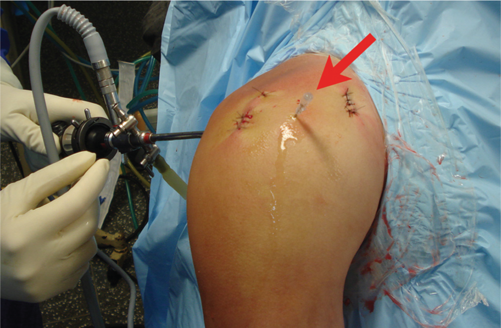
Needle inserted through intact skin after portal closure (arrow).
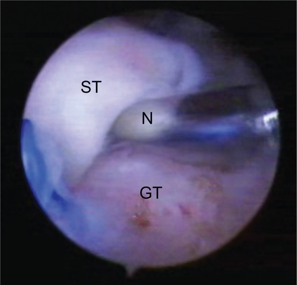
Needle (N) placed between the repaired tendon and bone bed. GT, greater tuberosity; ST, supraspinatus tendon.
Rehabilitation
The analgesic regimen was standardized for all patients. During the hospital stay, the participants were given intravenous medication comprising 1 analgesic agent (2 g dipyrone every 6 hours), 1 nonsteroidal anti-inflammatory drug (NSAID) (100 mg ketoprofen every 12 hours), and 1 opioid analgesic agent (100 mg tramadol every 8 hours). After discharge, medication was prescribed per the oral route, comprising 1 NSAID (200 mg celecoxib every 12 hours) and 1 analgesic agent (500 mg acetaminophen every 6 hours) on a continuous basis and 1 opioid analgesic agent (30 mg codeine every 6 hours) to be taken in case of very severe pain. The standardized analgesic regimen was indicated for 7 days and subsequently modified on an individual basis.
The shoulder was immobilized for 6 weeks. No motion was allowed during the first 3 weeks; passive exercise was permitted thereafter. Active assisted and active free exercises were started after week 6, when sling use was discontinued. Muscle strengthening was started at week 12.
Outcomes
Functional assessment as measured by the University of California at Los Angeles (UCLA) shoulder rating scale was established as the primary outcome. The secondary outcomes included functional assessment according to the Constant shoulder scale, pain as measured by a visual analog scale (VAS), and the retear rate assessed by MRI. All of the clinical analyses were performed before surgery and 3, 6, 12, and 24 months afterward. In addition, the VAS score was obtained on days 1 and 7. The number of complications was also assessed.
The MRI scans were obtained using a 1.5-T magnet (Signa HDxT, GE Healthcare) with a dedicated shoulder coil. The shoulder imaging protocol included oblique coronal proton density images (repetition time/echo time [TR/TE], 2800/38 ms; field of view [FOV], 14 cm; slice thickness, 3.5 mm; intersection gap, 0.4 mm; matrix, 320 × 256); axial, oblique coronal, and oblique sagittal fat-suppressed T2-weighted images (TR/TE, 3400/50; FOV, 14 cm; slice thickness, 3.5 mm; intersection gap, 0.4 mm; matrix, 256 × 256); and oblique sagittal T1-weighted images (TR/TE, 780/15; FOV, 14 cm; slice thickness, 3.5 mm; intersection gap, 0.4 mm; matrix, 320 × 256). Neither intra-articular nor intravenous gadolinium was used for any of the examinations. The images were analyzed by a musculoskeletal radiologist with 12 years of experience.
The MRI scans were assessed for the tendon state according to the Sugaya classification. 36 Furthermore, the incidence of complete retears and the overall retear rate (addition of partial and complete retears) were compared. Magnetic resonance imaging was performed before surgery and 3, 6, and 12 months afterward.
Sample Size
The sample size was calculated based on the UCLA score. Based on the concept of the minimal clinically important difference, the results for the PRP group ought to be superior by 2 points compared with the control group. Following Sugaya et al, 36 the SD was established as 3.7 in that calculation. With 80% of statistical power, and a (2-tailed) significance level of 5%, 54 participants proved to be required (27 in each group).
Randomization
The participants’ allocation was formulated by a simple randomization strategy using a tool available at http://www.random.org/coins/. Study participants were randomized by a physician who was not involved in the study. The results of the randomized allocation were kept in sealed brown envelopes, which were attached to the clinical records of the patients. The envelopes were only opened at the time of surgery, after the participant was anesthetized.
Blinding
The physical therapist who performed the functional assessment, the physical therapists involved during the stage of rehabilitation, the radiologist who performed the MRI analysis, the participants, and the statistician were all blinded as to which of the groups the participants were allocated.
Statistical Analysis
Clinical parameters at 24 months were analyzed by the Mann-Whitney test. A repeated-measures Student t test was used to compare the groups according to the UCLA, Constant, and VAS pain scores at other time points. The Friedman test was used to perform the post hoc analysis. Postoperative MRI results were analyzed by the χ2 or Fisher exact test. The significance level was established as P < .05. No imputation method was used for missing data, and all the participants completed follow-up.
An intention-to-treat analysis was performed. Statistical analysis was performed using SPSS version 20.0 software (SPSS Inc).
Results
Participant Flow
A total of 211 patients were assessed at our service between September 2008 and April 2012 regarding their eligibility for the present study. A total of 135 participants did not meet the inclusion criteria, and 1 refused participation. Therefore, 75 participants were subjected to randomization. Twenty participants were excluded: subscapular tears were observed in 17 participants during surgery, the tendon was not fully reparable in 2 participants, and conversion to open surgery was performed in 1 participant. One further participant was excluded in the follow-up period because of the inability to perform the postoperative protocol (intracranial surgery on postoperative day 15). Therefore, the data of 54 participants were analyzed, with 27 participants in each group. A flowchart representing the participants included in the study is depicted in Figure 3.
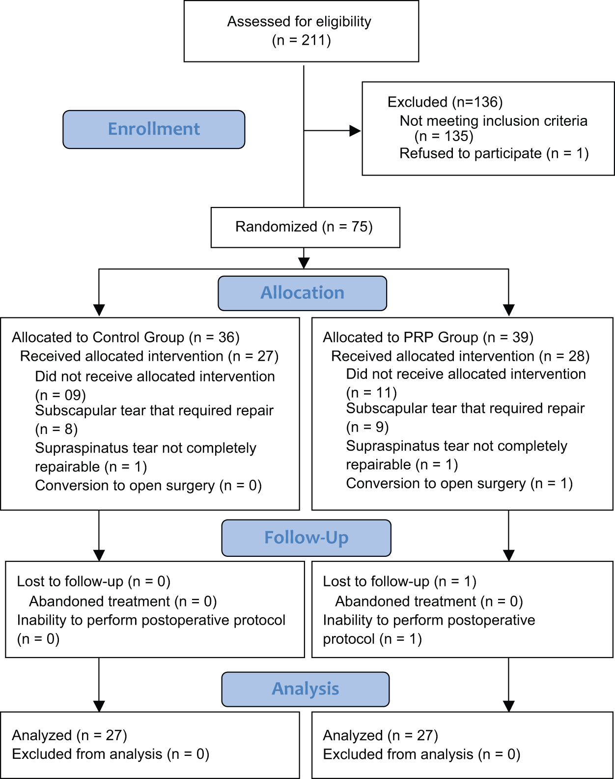
Flowchart of participants.
Baseline Data
The baseline characteristics of the participants in the groups were compared, and the results are described in Tables 1 and 2. No investigated variable exhibited significant differences.
Baseline Categorical Data a
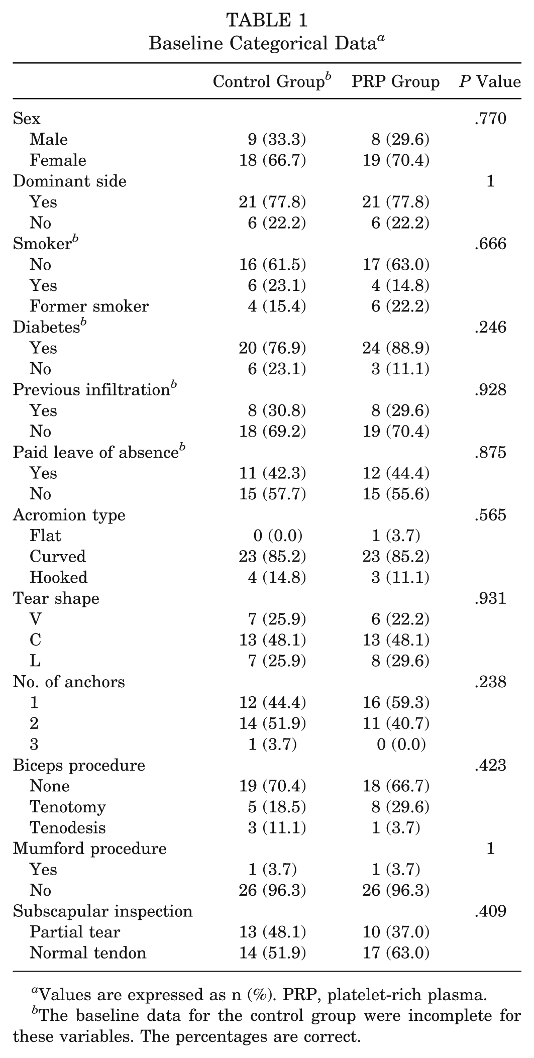
Values are expressed as n (%). PRP, platelet-rich plasma.
The baseline data for the control group were incomplete for these variables. The percentages are correct.
Baseline Continuous Data a

PRP, platelet-rich plasma.
The baseline data for the control group were incomplete for these variables.
Clinical Assessment
UCLA Score
The preoperative mean UCLA scores were 13.63 ± 3.639 in the control group and 13.93 ± 4.649 in the PRP group, without statistically significant differences (P = .795). Neither of the assessments at 3 and 6 months produced statistically significant differences (P = .356 and .288, respectively).
At 12 months, the mean scores of the control and PRP groups were 30.04 ± 4.528 and 32.30 ± 3.506, respectively; this difference was statistically significant (P = .046). At 24 months, the mean scores were 32.70 ± 3.635 in the control group and 32.44 ± 4.318 in the PRP group, with no significant difference (P = .916). The corresponding data are described in Table 3. The Friedman test revealed that both groups exhibited significant improvements throughout the follow-up time (P < .001).
Functional Assessment: UCLA Score a

PRP, platelet-rich plasma; UCLA, University of California at Los Angeles.
Constant Score
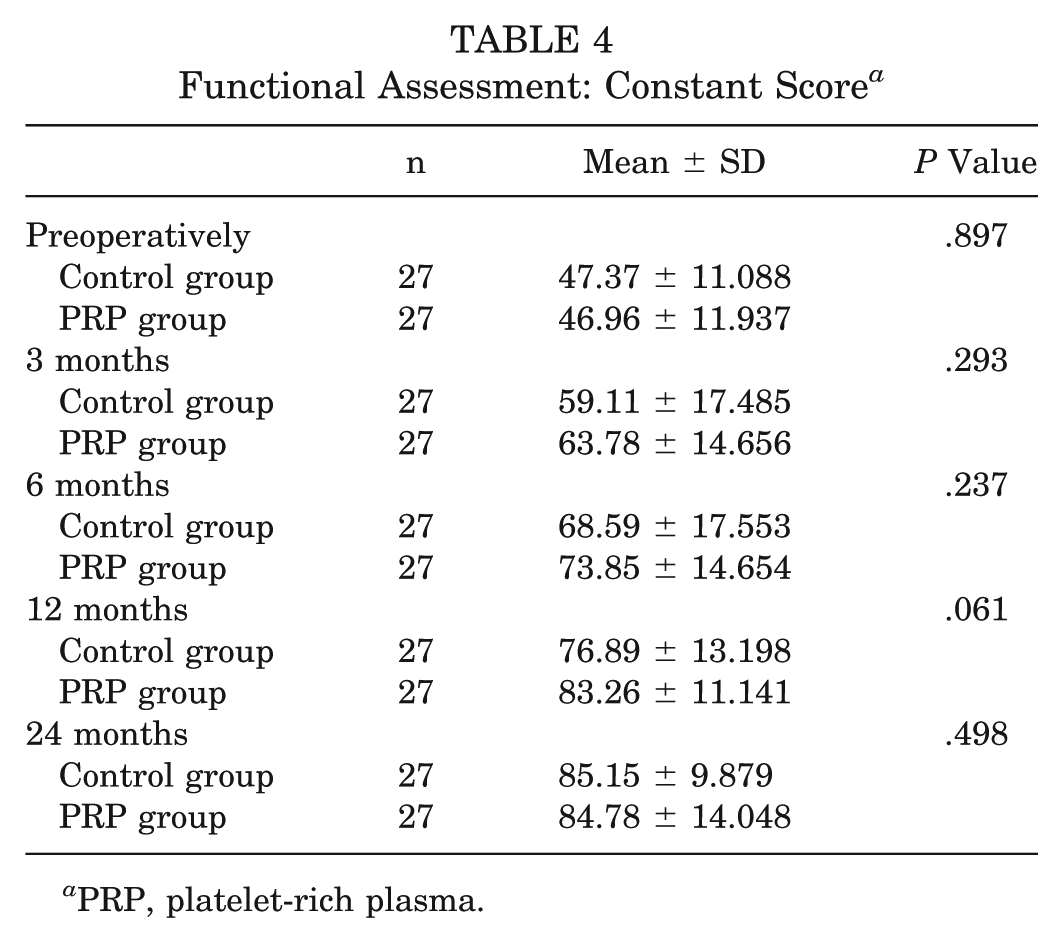
The preoperative mean Constant scores were 47.37 ± 11.088 in the control group and 46.96 ± 11.937 in the PRP group, without statistically significant differences (P = .897). The assessments at 3, 6, and 12 months did not reveal statistically significant differences (P = .293, .237, and .061, respectively).
At 24 months, the mean scores were 85.15 ± 9.879 in the control group and 84.78 ± 14.048 in the PRP group, with no significant difference (P = .498). The corresponding data are described in Table 4. The Friedman test revealed that both groups exhibited significant improvements over the follow-up period (P < .001).
Functional Assessment: Constant Score a
PRP, platelet-rich plasma.
VAS Score
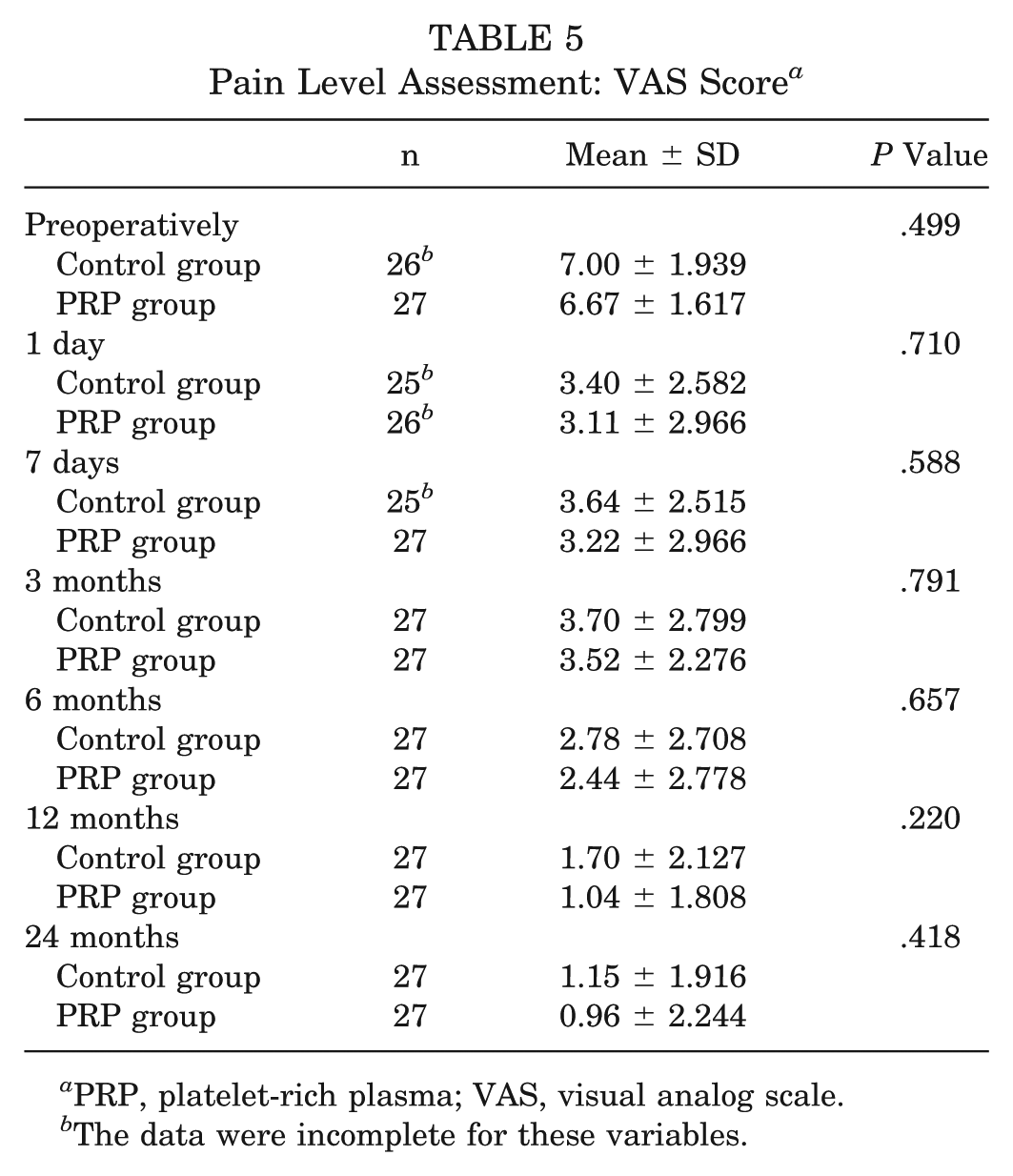
The preoperative mean VAS scores were 7.00 ± 1.939 in the control group and 6.67 ± 1.617 in the PRP group, without statistically significant differences (P = .499). The assessment during the early postoperative period (days 1 and 7) did not reveal statistically significant differences between the groups (P = .710 and .588, respectively). The assessments at 3, 6, and 12 months revealed no statistically significant differences (P = .791, .657, and .220, respectively).
At 24 months, the mean scores of the control and PRP groups were 1.15 ± 1.916 and 0.96 ± 2.244, respectively; this difference was not statistically significant (P = .418). The corresponding data are described in Table 5. The Friedman test indicated that both groups exhibited significant improvements during the follow-up time (P < .001).
Pain Level Assessment: VAS Score a
PRP, platelet-rich plasma; VAS, visual analog scale.
The data were incomplete for these variables.
MRI Assessment
Tendon Characteristics
At the 12-month assessment, 11.1% of the participants in the control group and 29.6% of the participants in the PRP group exhibited normal signals and sufficient thickness (Sugaya type I). The incidence of partial retears (Sugaya type III) was 4-fold higher in the control group at the 3-month assessment and 2-fold higher at the 6- and 12-month assessments compared to the PRP group. This variable did not exhibit statistically significant differences between the groups at any investigated time point (Table 6).
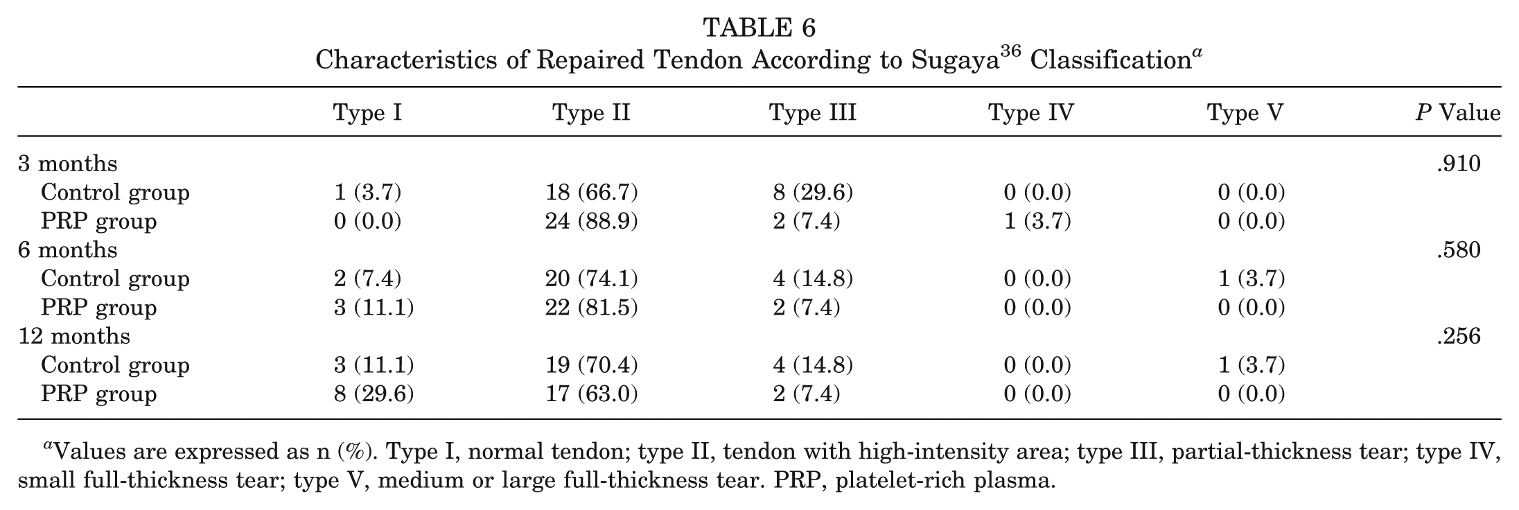
Values are expressed as n (%). Type I, normal tendon; type II, tendon with high-intensity area; type III, partial-thickness tear; type IV, small full-thickness tear; type V, medium or large full-thickness tear. PRP, platelet-rich plasma.
Retears
Only 1 case of a complete retear occurred in the control group; this difference was not statistically significant (P > .999). We performed further analyses and added the cases with complete and partial tears together to determine the overall incidence of retears. Thus, 8 and 3 cases were identified in the control and PRP groups at the 3-month assessment, respectively (P = .175), and 5 and 2 cases were identified in the control and PRP groups at the 6- and 12-month assessments, respectively (P = .420) (Table 7).
Anatomic Integrity of Repaired Tendon a

Values are expressed as n (%). PRP, platelet-rich plasma.
Complications
Postoperative stiffness was observed in 2 cases, 1 from each group, and neither required a second surgical procedure. This variable did not exhibit statistically significant differences between the groups (P > .999).
Discussion
Our study did not identify significantly superior functional results in the PRP group as measured by any of the scores at 24-month follow-up. The only clinical difference was in the UCLA score after 12 months (P = .046). Randelli et al, 30 analyzing patients with small- and medium-sized tears, found better functional results in the Constant score after 3 and 12 months but no difference at 24 months. Our study showed similar findings, with better results in the PRP group at 12 months and no difference at the last follow-up. Other studies did not find functional differences between the groups2-4,8,10,20,23,33,34 or demonstrated better results in the control group.21,40
We did not observe any effect of PRP on postoperative pain. Randelli et al 30 was the only study that observed better results analyzing pain as an outcome with the use of PRP. Gumina et al 20 found that PRP led to worse results. The other studies, including 2 meta-analyses,9,46 did not show differences between the groups2,10,21,23,33,40 or did not assess pain as an outcome.3,4,8,20,34
There is no consensus in the literature as to the effect of PRP on anatomic integrity after rotator cuff repair. Gumina et al, 20 Barber et al, 3 and Jo et al 23 found a significantly lower incidence of retears on MRI in the patients treated with PRP. In 2 systematic reviews with meta-analyses, Chahal et al 9 and Zhang et al 46 reported no differences between groups. However, a subgroup analysis limited to small- and medium-sized tears on MRI indicated a significantly lower retear rate in the group treated with PRP in both studies. Rodeo et al 33 are the only authors who reported significantly poorer results after the application of PRP. However, they used ultrasound as the imaging method. The remainder of the comparative studies2,4,8,10,21,30,34,40 did not find differences between the groups. Our study identified a smaller percentage of retears in the PRP group compared with the control group (7.4% vs 18.5%, respectively), which, however, was not statistically significant (P = .420). The results of the present study that were not statistically significant might be attributed to type II errors because the sample size calculation did not take the secondary outcomes into account. The detection of differences in the analysis of categorical data needs a larger sample.
There are 2 main methods available to prepare PRP: apheresis 44 and centrifugation. 25 The former yields significantly greater platelet concentrations,15,43 whereas the latter is more practical and less expensive. 25 There is no consensus about the best platelet concentration and no comparative clinical study. Weibrich et al, 41 although using a bone regeneration model and not a tendon one, found that 1,000,000 platelets/mm3 is the optimum concentration, with higher amounts leading to worse results. Jo et al, 22 in an in vitro study with tenocytes, showed that 4,000,000 to 8,000,000 platelets/mm3 promotes higher cell proliferation.
With respect to the use of thrombin to activate the coagulation cascade, there are arguments both supporting it 39 and disfavoring it, 35 and no comparative study on its effect in humans has been conducted. Virchenko et al, 39 in a biomechanical study in rat calcaneus tendons, showed that the use of thrombin leads to higher resistance. Scherer et al, 35 studying skin defects in rats, showed that PRP without thrombin achieved better results. Until now, 3 studies10,20,30 have used thrombin-activated PRP in rotator cuff repair.
The optimal leukocyte concentration is controversial. Dragoo et al 13 found that leukocyte-rich PRP led to an inflammatory response, but the real effect of this concentration in clinical results is still unknown. Three studies used leukocyte-rich PRP, with better results, 30 worse results, 20 or no difference in the PRP group. 10
In the present study, we chose to use PRP prepared by apheresis with the addition of thrombin. This method has been used in a previous randomized study that assessed the regeneration of the patellar ligament donor site and reported favorable results, 1 as well as in a case series of rotator cuff repair, which did not find any instance of retears. 26
The application method was described by Randelli et al, 31 according to which PRP is injected in a liquid form into the tendon-to-bone interface after repair surgery is concluded and the remaining arthroscopic fluid is aspirated. As a criticism against the method of PRP application used in our study, we might note that the PRP does not remain in the rotator cuff repair site but extends across the full subacromial space, which does not occur when PRP is used in the solid state. However, the method that we used is advantageously performed after sutures have been made and the excess of normal saline solution has been removed from that space. The application of solid PRP during the stage of suturing requires the continuous flow of arthroscopic fluid, which might result in a loss of growth factors and clot fragments.
The overall retear rate in our study was 1.9% (1/54 participants), with 0% in the PRP group and 3.7% in the control group. These values are lower than the ones reported by other authors, which varied from 8% to 94%.5,7,11,17,20,24,37 As a possible explanation, our study included small- and medium-sized tears exclusively. Another possibility is that we used MRI, instead of arthro-MRI, which has a higher accuracy in the detection of tears. 12 Castricini et al 8 reported an overall rate of 6.4% in a sample quite similar to ours according to tear sizes, with 2.5% in the PRP group and 10.5% in the control group. We observed significant functional improvement after arthroscopic rotator cuff surgery, regardless of the group studied. These results agree with the ones reported by other authors.7,11,17,24,37
It is worth noting that the use of PRP in small- and medium-sized tears has been the target of criticism. Theodoropoulos 38 believes that such tears have satisfactory clinical and structural results most of the time, suggesting that the odds of finding statistically significant differences are limited. According to that author, attention should be focused on large-sized and massive tears, which exhibit a greater risk of retears. Nevertheless, from the 6 studies3,9,20,23,30,46 that found the best results with the use of PRP, in 39,30,46 studies, such results corresponded to the subgroup of small- and medium-sized tears exclusively.
The population included in the present study might seem small. However, the homogeneity resulting from the inclusion of small- and medium-sized tears only and the exclusion of subscapular tears with the consequent increase in internal validity allowed a reduction in the sample size.
Furthermore, the fact that the number of platelets and leukocytes in PRP was not counted might be liable to criticism. However, the method used to prepare PRP in the present study has already been proven to generate concentrations greater than 1,000,000 platelets/mm3 and to lower leukocyte levels compared with baseline.1,29 In addition, among the published studies on the use of PRP in rotator cuff repair, only 221,23 included that measurement. There were more tenodesis procedures in the PRP group and more tenotomy procedures in the control group. Because of the small numbers, it was not possible to perform a subgroup analysis. Another limitation is that it was not feasible to blind the surgeon, but we could ensure that patients and the outcome evaluator were blinded.
To conclude, PRP prepared by apheresis and applied in the liquid state with the addition of thrombin did not induce better functional results at 24-month follow-up after arthroscopic rotator cuff repair of small- and medium-sized tears. The retear rate was also not influenced by the use of platelet concentrate, given the numbers available for analysis.
