
Abstract
Background:
Structural abnormalities of the patellofemoral joint might play a role in the pathogenesis of patellofemoral pain (PFP), a common knee problem among young and physically active individuals. No previous study has investigated if PFP is associated with structural abnormalities of the patellofemoral joint using high-resolution magnetic resonance imaging (MRI).
Purpose:
To investigate the presence of structural abnormalities of the patellofemoral joint on high-resolution MRI in patients with PFP compared with healthy control subjects.
Study Design:
Cross-sectional study; Level of evidence, 3.
Methods:
Patients with PFP and healthy control subjects between 14 and 40 years of age underwent high-resolution 3-T MRI. All images were scored using the Magnetic Resonance Imaging Osteoarthritis Knee Score with the addition of specific patellofemoral features. Associations between PFP and the presence of structural abnormalities were analyzed using logistic regression analyses adjusted for age, body mass index (BMI), sex, and sports participation.
Results:
A total of 64 patients and 70 control subjects were included in the study. Mean ± SD age was 23.2 ± 6.4 years, mean BMI ± SD was 22.9 ± 3.4 kg/m2, and 56.7% were female. Full-thickness cartilage loss was not present. Minor patellar cartilage defects, patellar bone marrow lesions, and high signal intensity of the Hoffa fat pad were frequently seen in both patients (23%, 53%, and 58%, respectively) and control subjects (21%, 51%, and 51%, respectively). After adjustment for age, BMI, sex, and sports participation, none of the structural abnormalities were statistically significantly associated with PFP.
Conclusion:
Structural abnormalities of the patellofemoral joint have been hypothesized as a factor in the pathogenesis of PFP, but the study findings suggest that structural abnormalities of the patellofemoral joint on MRI are not associated with PFP.
Patellofemoral pain (PFP) is a common knee condition, defined as retro- or peripatellar pain that occurs during knee loading activities, like running, cycling, squatting, and stair climbing, and during prolonged sitting with the knees bent. PFP is particularly present among young and physically active individuals and may account for up to 40% of all knee problems seen in sports injury clinics.7,10,14,17 PFP can have a debilitating effect because of the common recurrence of symptoms, tendency to chronicity, and its effect on physical activity levels.4,18,28,33 A variety of treatments, such as exercise therapy and orthoses, are applied, but reported effects are moderate, and a substantial group of patients with persistent complaints remains.2,8,21,31,32 To develop better-targeted treatment modalities, there is a need to elucidate the pathogenesis of PFP. Although there is consensus that the origin of PFP is multifactorial, precise contributing factors to the pathogenesis of pain are still not well understood.
In the 20th century, it was believed that PFP was caused by chondromalacia. However, arthroscopic studies clarified that PFP is not necessarily related to cartilage defects, which was confirmed by the magnetic resonance imaging (MRI) study of Kannus et al.1,14,18,20 Still, minor cartilage defects, such as signal abnormalities, fraying or fissuring, and hypertrophy, could potentially have been undetected up to now. Today, it is possible to detect even very small cartilage defects with the use of high-resolution MRI. In addition, abnormalities of the patellar retinaculum, synovial plicae, Hoffa fat pad, and subchondral bone marrow have been mentioned in the literature over the past years as possible sources of pain but have not yet been investigated systematically in a PFP population with high-resolution MRI.9,12,14,29,34 Hypothetically, more abnormalities are expected to be present in patients with PFP.
Therefore, this study will investigate the possible association between PFP and the presence of structural abnormalities of the patellofemoral joint. Based on the degenerative process with aging, as seen in osteoarthritis, more abnormalities were expected in adult patients. Therefore, differences in the number of abnormalities between adolescent and adult patients were also evaluated. This may eventually lead to a better understanding of the pathogenesis of PFP.
Methods
Study Design and Participants
A cross-sectional case-control study was conducted between January 2013 and September 2014 and included patients with PFP for 2 months up to 2 years, as well as healthy control subjects without any type of knee complaints. All subjects were aged between 14 and 40 years. Patients with PFP were recruited by general practitioners, sports physicians, and physical therapists and were included in the study if they had at least 3 of the following symptoms: retro- or peripatellar pain while walking up or down stairs, squatting, running, cycling, and sitting with knees flexed for a prolonged period of time, and grinding of the patella. Patients were excluded if they had other defined pathological conditions of the affected knee such as patellar tendinopathy or osteoarthritis, if the onset of PFP occurred after trauma, or if they had previous knee injuries or surgery or previous episodes of PFP that occurred more than 2 years ago. Control subjects were recruited from patients’ sports team members, friends, or the university (employees and students). We aimed to match control subjects on age, body mass index (BMI), sex, and activity level. Control subjects were excluded if they were first-grade family members of patients or if they had PFP at present or in the past, traumatic injury, or surgery of either knee. Furthermore, all study participants had to have sufficient knowledge of the Dutch language and no contraindications for MRI scanning with contrast administration (not used for current study purpose). Written informed consent was obtained from all participants (and parents/wardens in the case of subjects aged <18 years), and the study was approved by an institutional review board (Medical Ethical Committee of Erasmus MC, protocol MEC-2012- 342).
Measurements
After signing informed consent, study participants were asked to fill out an online questionnaire, including questions on demographics (age, sex, BMI), sports participation at the time of inclusion and before onset of pain (yes or no), and knee complaints (duration of complaints, pain at rest and during activity [numerical rating scale from 0 to 10 and Anterior Knee Pain Scale [AKPS] score from 0 to 100 22 ). Subsequently, they were invited for a physical examination (including crepitation during squatting [present or not], palpation of the medial patellar facet [painful or not], and Clarke compression test 11 [positive or negative]) and an MRI scan at our university medical center. In patients, the (most) affected knee was selected for physical examination and MRI, or randomly chosen if both knees were equally painful. For control subjects, a randomly selected knee was used for both physical examination and MRI.
The MRI was performed at 3T (Discovery MR750; GE Healthcare) with a dedicated 8-channel knee coil (Invivo Inc). The MRI protocol comprised sagittal, axial, and coronal fast spin-echo proton density–weighted sequences with a slice thickness of 3 mm and sagittal, axial T2-weighted sequences with fat suppression with a slice thickness of 3mm. Furthermore, a 3-dimensional high-resolution sagittal fat-saturated spoiled gradient-echo sequence was acquired with a slice thickness of 0.5 mm, repetition time of 17 ms, echo time of 5.4 ms, flip angle of 12°, 288 × 192 matrix, and 15-cm field of view.
Assessment of MRI Features
All MRI scans were scored primarily by a senior resident in radiology with musculoskeletal subspecialization who was blinded for participant status, using the semiquantitative Magnetic Resonance Imaging Osteoarthritis Knee Score (MOAKS). Subsequently, all findings were discussed with an experienced musculoskeletal radiologist, who was also blinded for participant status and made the final determination. The MOAKS was developed to evaluate articular cartilage loss in conjunction with surrounding bony and soft tissue abnormalities, including bone marrow lesions (BMLs); osteophytes; lesions of the menisci, ligaments, and tendons; joint effusion; and periarticular features (ie, iliotibial band abnormalities, ganglion cysts). 15 Since the MOAKS was primarily developed to study osteoarthritis, several additional patellofemoral joint features, possibly more applicable for this young population, were added (see Appendix 1, available in the online version of this article and at http://ajsm.sagepub.com/supplemental). 9 These additional features included minor defects of cartilage (high signal intensity, fraying or fissuring, hypertrophy); medial patellar plica thickness and width; avascular necrosis; stress fractures; thickening of the patellar tendon, quadriceps tendon, and retinaculum; and high signal intensity of retinaculum, quadriceps tendon, and fat pads (superolateral part of the Hoffa fat pad, quadriceps fat pad, and prefemoral fat pad, according to Chhabra et al 9 ). High signal intensity was defined as an abnormally high signal intensity on T2-weighted images of a certain structure. Hypertrophy was seen as a result of cartilage swelling. Fraying, a shredded appearance on MRI, is supposed to be the result of fibrillation of the cartilage surface. Fissuring comprises small cracks in the cartilage surface. Furthermore, in the MOAKS, the complete anterior femur is scored as trochlea. 15 Since we focused on the patellofemoral joint, we subdivided this total anterior femur in a subregion that comprises solely the trochlea and a medial and lateral condylar subregion. This was done for cartilage loss and BMLs. With regard to the additional scoring of minor cartilage defects, only the trochlear subregion and patella were scored.
Items were clustered and dichotomized to reduce the large number of MRI features. The MOAKS “size” category was used for dichotomizing cartilage loss and BMLs: If the size was 0, the feature was scored as negative; if size >1, the feature was scored as positive. Osteophytes were scored as positive if their size was small to large and effusion if a medium-to-large amount was present. Thickening and high signal intensity were clustered in the case of tendons or ligaments. Minor cartilage defects was scored as positive if high signal intensity or fraying/fissuring or hypertrophy was present. Plica thickening or widening was scored as positive in the case of grade 3 for either thickness (≥3 mm) or width (being sufficient to reach the midline in a nondistended joint), according to the grading of Boles et al. 6
Statistical Analysis
Statistical analyses were performed with SPSS for Windows, version 20 (SPSS Inc). If normal distribution was present, the chi-square test for dichotomous variables and independent-samples t test for continuous variables were used to compare differences in characteristics between patients and control subjects, but also between adults and adolescent patients (aged <18 years). Otherwise, a Mann-Whitney U test was applied. Associations between PFP and the presence of structural abnormalities were analyzed using logistic regression analyses. In addition, logistic regression analyses were applied to test potential differences between adolescent and adult patients in the presence of structural abnormalities. Regression analyses were adjusted for the potential confounders: age, sex, BMI, and sports participation. Results are presented as Exp(B) with 95% CIs and adjusted P values. P < .05 was considered statistically significant.
Results
Participants
A total of 64 patients (Figure 1) and 70 control subjects were included in this study between January 2013 and September 2014, of which 40, equally distributed between groups, were adolescents. The mean age ± SD was 23.2 ± 6.4 years, the mean BMI was 22.9 ± 3.4 kg/m2, and 56.7% were female. A significant difference between PFP patients and control subjects was observed in BMI (higher in patient group) and percentage of sports participants (higher in the control group) (Table 1).
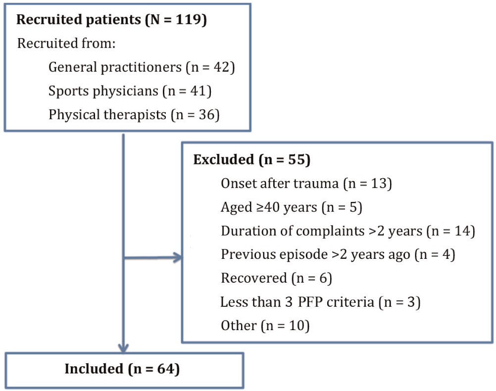
Flowchart of recruited patients. PFP, patellofemoral pain.
Characteristics of Study Participants a
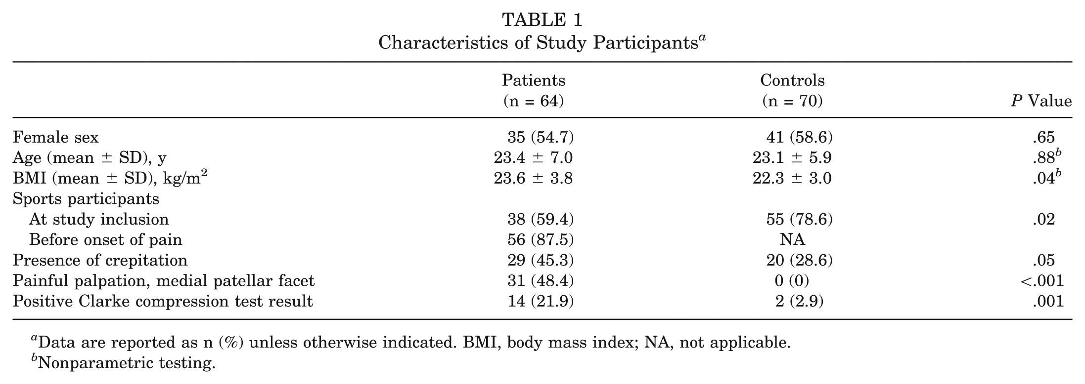
Data are reported as n (%) unless otherwise indicated. BMI, body mass index; NA, not applicable.
Nonparametric testing.
Compared with adult patients, function (AKPS score) was significantly lower in adolescent patients. Bilateral knee pain and painful palpation of the medial patellar facet occurred significantly more in adolescents compared with adult patients (Table 2).
Characteristics of the PFP Patient Population, Subdivided Into Adults and Adolescents a
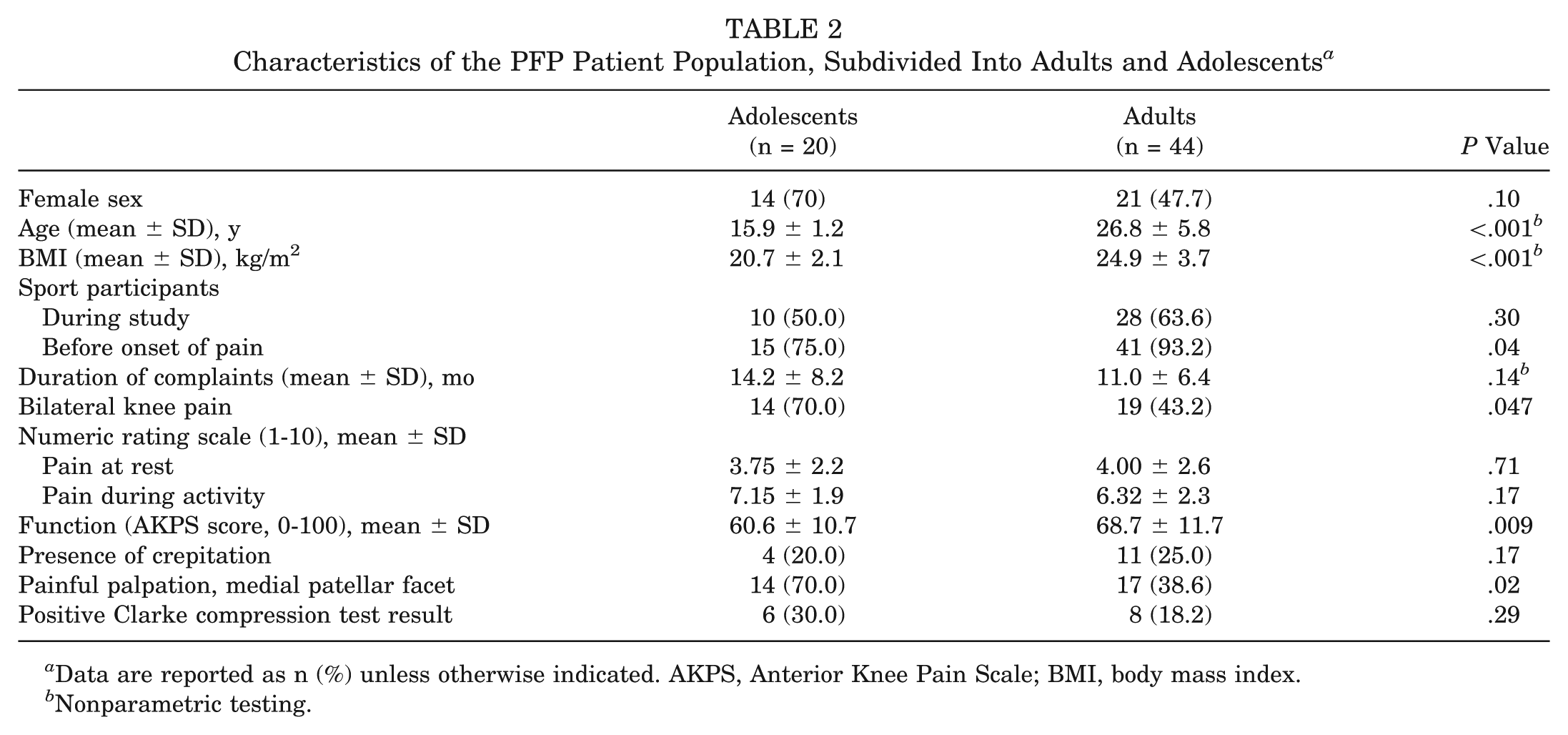
Data are reported as n (%) unless otherwise indicated. AKPS, Anterior Knee Pain Scale; BMI, body mass index.
Nonparametric testing.
Structural Abnormalities on MRI
The results described below are the key outcomes from the regression analyses based on adjusted P values.
The most frequently found abnormalities in patients and control subjects were minor cartilage defects of the patella (high signal or hypertrophy or fraying; 23.4% vs 21.4% for patients and controls), patellar BMLs (53.1% vs 51.4%), patellar osteophytes (70.3% vs 60.0%), high signal intensity of Hoffa fat pad superolaterally (57.8% vs 51.4%), high signal intensity or thickening of the patellar tendon (37.5% vs 30%), high signal intensity of prefemoral and quadriceps fat pad (70.3% vs 65.7% and 37.5% vs 34.3%), ganglion cyst (32.8% vs 34.3%), and popliteal cyst (67.2% vs 71.4%) (Table 3). Full-thickness cartilage loss of the patellofemoral joint was not present in this study population. Partial-thickness cartilage loss of the patella occurred in 9% of patients and 1% of control subjects.
Presence of Structural Abnormalities of the Patellofemoral Joint on MRI a
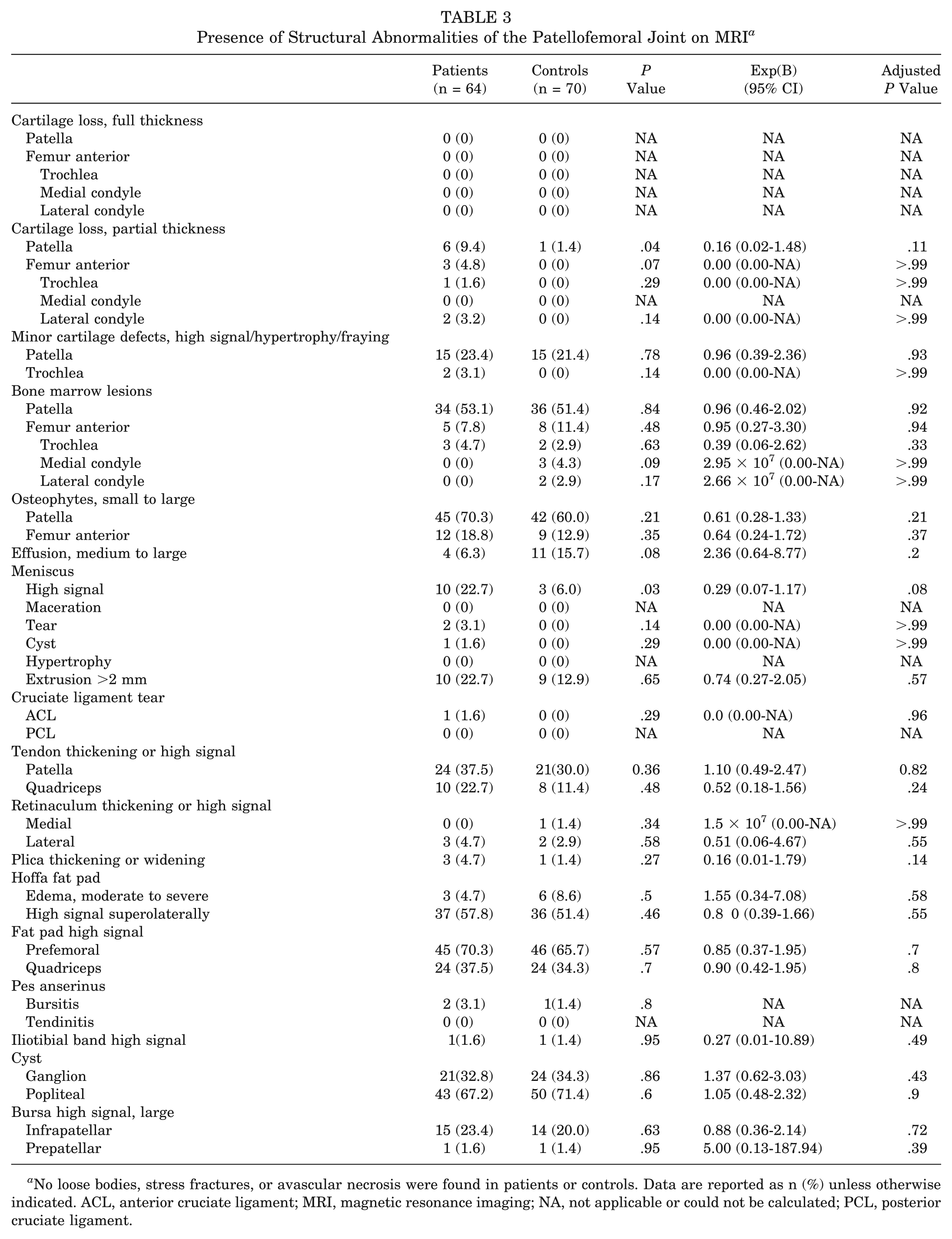
No loose bodies, stress fractures, or avascular necrosis were found in patients or controls. Data are reported as n (%) unless otherwise indicated. ACL, anterior cruciate ligament; MRI, magnetic resonance imaging; NA, not applicable or could not be calculated; PCL, posterior cruciate ligament.
Osteophytes of the anterior femur occurred in 18.8% of patients and 12.9% of control subjects. Other structural abnormalities of the anterior femur occurred less frequently. For instance, partial-thickness cartilage loss occurred in only 3 patients and not in control subjects. BMLs of the anterior femur were present in 5 patients and 8 control subjects. Minor cartilage defects of the trochlea were present in only 2 patients.
High signal intensity of the meniscus occurred in 22.7% of patients and 6.0% of control subjects. Meniscal extrusion occurred in 22.7% of patients and 12.9% of control subjects. Medial retinaculum thickening or high signal occurred in only 1 control subject. Plica thickening or widening occurred in 3 patients and 1 control subject. After adjustment for age, BMI, sex, and sports participation, none of the structural abnormalities seen on MRI were statistically significantly associated with the presence of PFP.
Subgroup analyses of a selection of the abnormalities of the patellofemoral joint on MRI in the patient population comparing adult and adolescent patients revealed no significant differences between these patient groups (Table 4). The most frequently found abnormalities in adult and adolescent patients were patellar BMLs (47.8% vs 65% in adults and adolescents), patellar osteophytes (79.5% vs 50%), high signal intensity of Hoffa fat pad superolaterally (65% vs 40%), and high signal intensity or thickening of the patellar tendon (38.6% vs 35%). Partial-thickness cartilage loss of the patella occurred in 13.6% of adult patients but not in adolescents. Minor cartilage defects of the patella (high signal intensity or hypertrophy or fraying) were present in 31.8% of adult patients and 5% of adolescent patients. Trochlear BMLs, partial-thickness cartilage loss of the trochlea, and minor cartilage defects of the trochlea occurred in adults only.
Presence of Structural Abnormalities of the Patellofemoral Joint on MRI in Patients, Subdivided Into Adults and Adolescents a
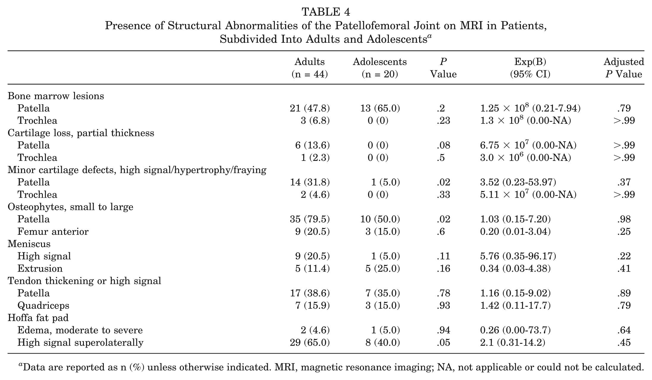
Data are reported as n (%) unless otherwise indicated. MRI, magnetic resonance imaging; NA, not applicable or could not be calculated.
After adjustment for age, BMI, sex, and sports participation, no difference in the presence of structural abnormalities of the patellofemoral joint on MRI was present between adult and adolescent patients.
Discussion
The purpose of this study was to investigate if PFP was associated with structural abnormalities of the patellofemoral joint using high-resolution MRI, which enabled us to detect even small lesions. Full-thickness cartilage loss of the patellofemoral joint was not present in this study population. Minor patellar cartilage defects, patellar BMLs, patellar osteophytes, high signal intensity of Hoffa fat pad, and high signal intensity or thickening of the patellar tendon were frequently seen in both patients and control subjects. Structural abnormalities of the trochlea occurred less frequently. For partial-thickness patellar cartilage loss and meniscal high signal intensity, both present only in small numbers, the difference between patients and control subjects was not statistically significant, but both of them occurred seemingly more in patients. Medial synovial plica and patellar retinaculum abnormalities were rarely seen in this population. Overall, our results indicate that the presence of structural abnormalities of the patellofemoral joint on MRI is not associated with PFP.
Subgroup analyses comparing adult and adolescent patients showed consistently more abnormalities in adult patients. However, after adjustment for sex, BMI, and sports, no statistically significant differences were present between these groups. Additional analyses in the total study population also revealed no differences between adults and adolescents (data not presented).
It could be hypothesized that abnormalities are most likely to occur at the medial part of the patella, since this is the most frequent pain location in PFP patients. Furthermore, pain might actually be located in a specific part of a surrounding structure, like the medial meniscus or superior patellar tendon. Therefore, explorative analyses of specific subregions of the patella (medial, lateral, superior, and inferior), meniscus (medial and lateral), and patellar tendon (superior, middle, and inferior) were performed. These analyses also showed no significant differences between patients with PFP and healthy control subjects (see Appendix 2, available online). Differences in the presence of combinations of abnormalities were not tested due to a lack of power.
Comparison With the Literature
So far, only 1 previous study by Kang et al 16 has investigated MRI findings in patients with PFP. However, theyapplied MRI at 1.5 T, whereas in our study, high-resolution MRI at 3 T was performed. Furthermore, their study consisted of male soldiers with a mean age of 22 years, whereas our study consisted of the general PFP population seeking care in primary care and even included a group of adolescents. Similar to our study, abnormalities of the patellofemoral joint (eg, cysts, effusion, bone marrow signal change, meniscal lesions) were common in both patients and controls in the study by Kang et al. 16 The prevalence of abnormalities on MRI in asymptomatic knees has been frequently reported in the literature.3,5,13,23 This raises the question of whether these features really contribute to the pathogenesis of knee pain and, if they do, which other factors need to be present to induce pain. Kang et al 16 did find a higher prevalence of abnormalities of the patellofemoral joint and the extensor mechanism in patients, and especially a higher prevalence of thick medial plica in the patient group compared with the control group (9% vs 0%). In our study, plica thickening or widening occurred in 2 patients and 1 control subject only. This might be due to a difference in scoring, as they scored medial patellar plicae thickening when 2 mm or more, contrary to the 3 mm or more in our protocol, which we based on the study of Boles et al. 6 Furthermore, it is important to note that their population consisted of soldiers, and consequently, the physical activity level is assumed to be higher compared with that of our population.
It was apparent in this study that sports participation was not associated with the presence of BMLs, cartilage defects, and meniscal lesions of the PFP. For BMLs and cartilage defects, these results are in contrast to previous literature stating that subjects with higher physical activity levels have a higher incidence of these abnormalities.19,24-27,30 This might be due to the fact that we have dichotomized physical activity into sports participation or not, instead of using a continuous scale for physical activity level.
Retropatellar cartilage damage has been implicated as a possible etiological factor for PFP for many years. In the 20th century, arthroscopic studies clarified that PFP was not necessarily related to cartilage defects.1,14,20 This was confirmed in an MRI study by Kannus et al, 18 who found no correlation between cartilage defects and PFP. Similar to these studies, our results indicate that cartilage defects are not associated with PFP. The current high-resolution imaging technique allowed us to look at even minor cartilage defects, including high signal intensity, fraying/fissuring, and hypertrophy. These were also not associated with the presence of PFP. Therefore, it seems that there is conclusive evidence that major or minor patellofemoral cartilage defects are not associated with PFP.
Strengths and Limitations
This is the first case-control study on structural abnormalities of the patellofemoral joint in PFP that includes a large group of PFP patients, including a group of adolescent patients who are often excluded in the literature. To our knowledge, there has been no previous study in PFP using high-resolution MRI, which enables detection of even minimal structural abnormalities.
There are, however, some limitations to our study that need to be addressed. Our intention was to match patients and controls on age, sex, BMI, and sports participation. However, some differences were present concerning BMI and percentage of sports participants; therefore, all analyses were adjusted for these confounders.
We used the MOAKS, a semiquantitative score primarily developed to study osteoarthritis on MRI. To make this score more appropriate for the relatively young population studied, some additional features were added. Therefore, we believe that we covered all potential items.
Finally, a lack of power might be the reason that we did not find an association between certain structural abnormalities and PFP. Although a higher percentage of partial-thickness patellar cartilage loss and more frequent meniscal high signal intensity were present in PFP patients compared with control subjects, these differences were not statistically significant. Although this is one of the largest case-control studies in the PFP research field, we lacked power due to the small numbers of abnormalities found in the study population. However, since these abnormalities are present in such small numbers, their contribution to the pathogenesis of PFP is unlikely.
Conclusion
A large number of structural abnormalities of the patellofemoral joint on MRI were seen in this relatively young population. Our results indicate that structural abnormalities of the patellofemoral joint on MRI are not associated with PFP.
Footnotes
Acknowledgements
The authors acknowledge all participants for their participation and the caregivers for recruiting the participants.
One or more of the authors has declared the following potential conflict of interest or source of funding: Financial support was received from the Dutch Arthritis Foundation and the Erasmus University Rotterdam. G.P.K. is a consultant to Bracco SA. G.P.K. and E.H.G.O. receive research support from General Electric Healthcare.
References
Supplementary Material
Please find the following supplemental material available below.
For Open Access articles published under a Creative Commons License, all supplemental material carries the same license as the article it is associated with.
For non-Open Access articles published, all supplemental material carries a non-exclusive license, and permission requests for re-use of supplemental material or any part of supplemental material shall be sent directly to the copyright owner as specified in the copyright notice associated with the article.