
Abstract
Background:
Subfibular ossicles are frequently found in patients with chronic lateral ankle instability (CLAI). However, there is a lack of consensus about the optimal surgical treatment for CLAI with subfibular ossicle.
Purpose:
To evaluate the clinical and radiographic outcomes of the modified Broström procedure with subfibular ossicle excision compared with the same procedure for CLAI without subfibular ossicle.
Study Design:
Cohort study; Level of evidence, 3.
Methods:
Ninety-six patients (96 ankles) treated with the modified Broström procedure using bone tunnel and suture anchor techniques for CLAI constituted the study cohort. The 96 ankles were divided into 2 groups with and without subfibular ossicles. The ossicle group (42 ankles) and nonossicle group (54 ankles) consisted of patients with a mean age of 26.6 and 30.3 years, respectively, at the time of surgery with a mean follow-up duration of 63.7 and 62.1 months, respectively. Subfibular ossicles were excised in the ossicle group.
Results:
Mean Karlsson scores improved from 55.2 to 95.3 in the ossicle group and from 56.4 to 94.8 in the nonossicle group at final follow-up. Mean American Orthopaedic Foot and Ankle Society (AOFAS) ankle-hindfoot scores also improved from 63.3 to 95.9 in the ossicle group and from 62.8 to 95.1 in the nonossicle group at final follow-up. Mean talar tilt angles were 14.0° in the ossicle group and 12.2° in the nonossicle group preoperatively and 7.6° and 6.8° at the final follow-up, respectively. Mean anterior talar translations in the ossicle group and nonossicle groups improved from 9.3 and 9.4 mm preoperatively to 5.8 and 5.7 mm at final follow-up, respectively. No significant differences were found between the 2 groups in terms of Karlsson score, AOFAS score, talar tilt angle, and anterior talar translation at final follow-up (P > .05).
Conclusion:
The modified Broström procedure with subfibular ossicle excision provided similarly good clinical and radiographic outcomes compared with the same procedure without subfibular ossicle excision. Accordingly, the study results suggest that these procedures appear to be effective and reliable method for the treatment of CLAI with subfibular ossicle.
Ankle sprains are the most common musculoskeletal injury in sports. Recurrent sprains and recurrent episodes of the ankle giving way often cause chronic lateral ankle instability (CLAI). Most patients with ankle instability see improvement with nonsurgical treatment including strengthening of peroneal muscles and proprioception exercise. For patients with persistent lateral ankle instability after nonsurgical treatment, lateral ankle ligament repair or reconstruction is then recommended. Various surgical procedures have been introduced for the treatment of CLAI. Among those procedures, modification of the Broström procedure that repairs the anterior talofibular ligament (ATFL) and calcaneofibular ligament (CFL), including extensor retinaculum augmentation, has brought about satisfactory results and is considered the primary treatment strategy for patients in need of surgical treatment.1,11,14
There are 2 theories describing the precise origin of subfibular ossicles. These small bony fragments of the lateral malleolus may indicate nonunited avulsion fractures by ATFL2,4,24 or accessory bones derived from a secondary ossification center, os subfibulare.26,27 Commonly, the term subfibular ossicle is used as a concept including nonunited avulsion fracture and os subfibulare. The subfibular ossicle has been subjected to ankle inversion injury, resulting in disruption of the fibrous or cartilaginous attachment and causing subfibular pain and recurrent irritation of the surrounding soft tissue by the ossicle. 9 The combination of subfibular ossicle and recurrent ankle sprain is thought to accelerate lateral ankle instability. The subfibular ossicle has various shapes and sizes, and its incidence is reported in 0.2% to 6.7% of the normal population.3,21,27 However, it is frequently found in 10% to 38.5% of patients with CLAI.5,17,30 This higher incidence implies that there is a correlation between subfibular ossicle and CLAI.
There is still debate about the surgical treatment of CLAI with subfibular ossicle. Some have reported inferior clinical outcomes after the modified Broström procedure with subfibular ossicle excision, 16 whereas others have suggested that the modified Broström procedure with subfibular ossicle excision was an equally successful treatment compared with the same procedure without the ossicle. 6
The purpose of the present study was to evaluate the clinical and radiographic outcomes of the modified Broström procedure for CLAI with and without subfibular ossicle. We hypothesized that the modified Broström procedure could provide similar good clinical and radiographic outcomes even if subfibular ossicle excision was carried out simultaneously compared with the same procedure without subfibular ossicle excision.
Methods
We obtained approval from our institutional review board, and all patients granted their informed consent. The study was carried out in accordance with the World Medical Association Declaration of Helsinki. Two hundred six patients (208 ankles) who underwent the modified Broström procedure for CLAI between January 2005 and December 2012 were retrospectively analyzed. The indications for operation were persistent pain, giving-way sensation, limited physical activity for more than 6 months, and failure of nonoperative treatment for a period of at least 3 months. The inclusion criteria for this study were first-time surgery, no history of fracture around the ankle, an uninjured contralateral ankle, no simultaneous surgery for ankle osteoarthritis or anatomic deformity, no peroneal tendon subluxation or tear, no neuromuscular disorders, and no foot malalignment such as cavovarus foot. Ninety-seven patients (99 ankles) who did not satisfy the criteria were excluded. Thirteen patients (13 ankles) with inadequate follow-up duration (<24 months) were also excluded.
Accordingly, the remaining 96 patients (96 ankles) with CLAI constituted the study cohort and were divided into 2 groups: a group with subfibular ossicle (the ossicle group) and a group without subfibular ossicle (nonossicle group). The ossicle group was composed of 42 patients (42 ankles), and the nonossicle group was made up of 54 patients (54 ankles). The ossicle group consisted of 28 men and 14 women with a mean age of 26.6 years at the time of surgery with a mean follow-up duration of 63.7 months. The nonossicle group consisted of 38 men and 16 women with a mean age of 30.3 years with a mean follow-up duration of 62.1 months. Duration of preoperative symptoms and nonoperative treatment were similar between the 2 groups. No significant intergroup differences were observed in patient demographics (P > .05) (Table 1).
Patient Demographics of Chronic Lateral Ankle Instability With and Without Subfibular Ossicle a
Values are expressed as mean ± SD unless otherwise indicated. BMI, body mass index.
Chi-square test or independent t test.
The ossicle group was subdivided into small (≤10 mm; 20 ankles) and large (>10 mm; 22 ankles) ossicles to compare clinical and radiographic outcomes. The prognostic factors that affect clinical outcome were investigated according to age, sex, body mass index (BMI), symptom duration, and combined intra-articular lesion in both groups. Magnetic resonance imaging (MRI) was used to identify ligament, cartilage, and other injuries. If combined intra-articular lesions were observed by MRI and the patient reported ankle joint pain, additional arthroscopic management was performed.
The clinical and radiographic evaluations were made preoperatively and postoperatively at 3 months, 6 months, 1 year, and annually thereafter. Comparison of the results between the 2 groups was performed preoperatively and at the final follow-up by 2 orthopaedic surgeons who were not directly involved in the surgical procedures.
Clinical Evaluations
The Karlsson scoring 15 and American Orthopaedic Foot and Ankle Society (AOFAS) ankle-hindfoot scoring system 17 were administered to the patients for assessment of clinical outcomes. The Karlsson score was described by Karlsson and Peterson 15 to assess subjective symptoms like pain and stiffness, ankle stability, and physical activity. The 100-point AOFAS scoring system evaluates clinical parameters using a combination of subjective and objective data; scores are allocated as follows: pain (40 points), function (45 points), and alignment (15 points).
Radiographic Evaluations
Standing ankle radiographs (anteroposterior, lateral, and mortise views) were taken to evaluate alignment and to rule out fracture. The size of a subfibular ossicle was also measured by the longest diameter on ankle radiographs (Figure 1). Three-dimensional computed tomography was used for observing shape and location of the subfibular ossicle before surgery. For further evaluation for ankle instability, stress radiographs using a Telos stress device were included and used to determined talar tilt angle and anterior translation of the talus before and after surgery. Patients were requested to relax leg muscle to prevent muscle guarding, and then an anterior and varus force of 150 N was placed on the distal tibia via a Telos device. All radiographic measurements were evaluated accurately using a picture archiving and communication system (PACS; Maroview version 5.4; Marotech Inc) by 2 orthopaedic surgeons who were blinded to the surgical treatment group.
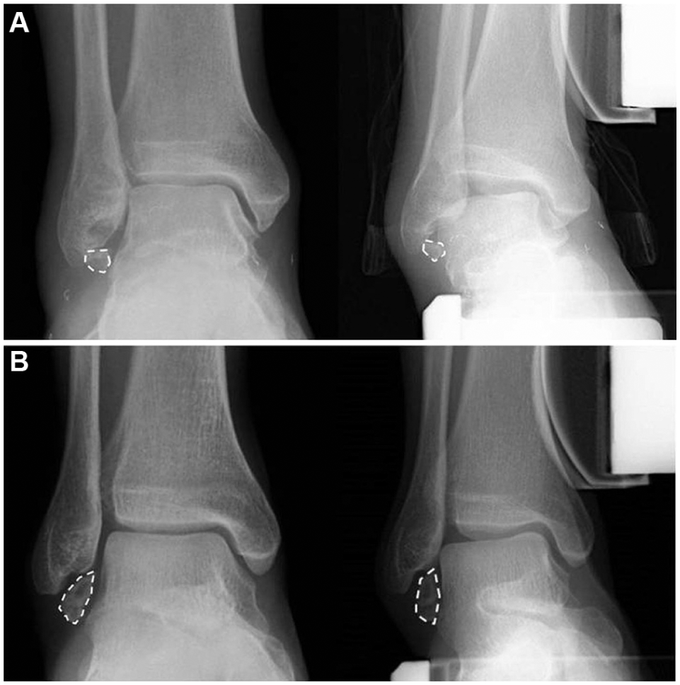
Subfibular ossicle on anteroposterior plain and stress radiographs preoperatively. The size of a subfibular ossicle was also measured by the longest diameter on ankle radiographs. (A) Small (≤10 mm) ossicle and (B) large (>10 mm) ossicle were shown at the inferior tip of the fibula (dashed areas).
Surgical Techniques
All operations were performed by a single orthopaedic surgeon (K-B.L.). Under general anesthesia, the patient was placed in the supine position with a pillow under the ipsilateral buttock, and a thigh pneumatic tourniquet was applied. After induction of anesthesia, all ankles were evaluated for ankle joint stability and laxity. A curvilinear skin incision of about 4 cm was made anteroinferiorly above the tip of the lateral malleolus, avoiding the superficial peroneal nerve and sural nerve. Subcutaneous dissection was performed, carefully identifying the inferior extensor retinaculum. The joint capsule, ATFL, and CFL were dissected from the tip of the lateral malleolus after inferior retraction of the peroneal tendons. In this process, if the ossicle at the tip of the lateral malleolus was observed, it was excised through an open procedure, regardless of size, while care was taken not to injure the ligament (Figure 2). If the ATFL or the CFL was attached to the ossicle, careful dissection and separation from the lateral ligament complex were done.
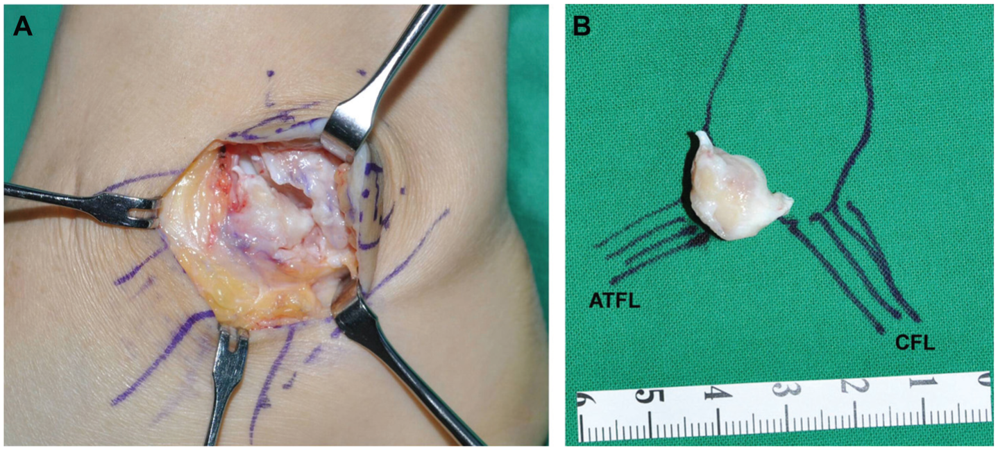
(A) Intraoperative photograph of the modified Broström procedure with subfibular ossicle excision on the left ankle. The exposed subfibular ossicle is shown. (B) Drawing representing the relationship between the removed ossicle and the 2 lateral ankle ligaments (anterior talofibular ligament [ATFL] and calcaneofibular ligament [CFL]).
After determining the anatomic footprint of the ATFL and the CFL, fibular decortication was performed. Then, the modified Broström procedure was performed with the bone tunnel or suture anchor technique. After bone tunnels were made at the anteroinferior portion of the lateral malleolus, the ATFL, CFL, and joint capsule were then reinserted into the roughened surface on the fibula using nonabsorbable suture material (2-0 Ethibond; Ethicon). When the suture anchor technique was performed, a single suture anchor with nonabsorbable 4-strand suture (Fastin RC; DePuy Mitek) was used and inserted into the center of the anatomic footprint of the ATFL and CFL. These procedures were done with the foot in neutral dorsiflexion and 5° eversion. Finally, the extensor retinaculum was reinforced to the periosteum of the distal fibula. The ankle joint stability was reexamined before skin closure.
Postoperatively, a short leg splint was applied and nonweightbearing with crutches was required for 2 weeks. Then, patients were placed in a cam walker and weightbearing was progressed. Gentle active range of motion of the ankle, peroneal strengthening, and proprioception exercises were initiated at 4 weeks after surgery. Patients were instructed to begin straight running and functional activities at 8 weeks postoperatively (see the Appendix, available in the online version of this article and at http://ajsm.sagepub.com/supplemental).
Statistical Analysis
The independent t test was used to determine the significance of intergroup differences in age, BMI, symptom duration, follow-up duration, Karlsson score, AOFAS score, talar tilt angle, and anterior talar translation. Categorical variables such as sex of the patient were compared using the Pearson chi-square test. In addition, univariate analysis was used to identify the prognostic factors in each group separately. Statistical significance was accepted for P < .05, and all components of the statistical analysis were performed and reviewed by 2 statisticians.
Results
Clinical Outcomes
The mean Karlsson score was 55.2 in the ossicle group and 56.4 in the nonossicle group preoperatively. At final follow-up, these scores improved to 95.3 and 94.8, respectively. The mean AOFAS score improved from 63.3 to 95.9 in the ossicle group and from 62.8 to 95.1 in the nonossicle group. There were no significant differences in the mean Karlsson score and AOFAS score at the final follow-up between ossicle and nonossicle groups (P > .05) (Table 2).
Comparison of Clinical Outcomes Between the Ossicle and Nonossicle Groups After Modified Broström Procedure for Chronic Lateral Ankle Instability a
Values are expressed as mean ± SD. AOFAS, American Orthopaedic Foot and Ankle Society.
Independent t test.
To investigate prognostic factors that affect clinical outcome, the ossicle and nonossicle groups were divided according to age, sex, BMI, symptom duration, and combined intra-articular lesions. Median values were used as a standard for dividing the groups. However, there was no significant correlation between these factors and clinical outcome in the 2 groups (P > .05) (Table 3).
Prognostic Factors and Clinical Outcomes Between the Ossicle and Nonossicle Groups After Modified Broström Procedure for Chronic Lateral Ankle Instability a
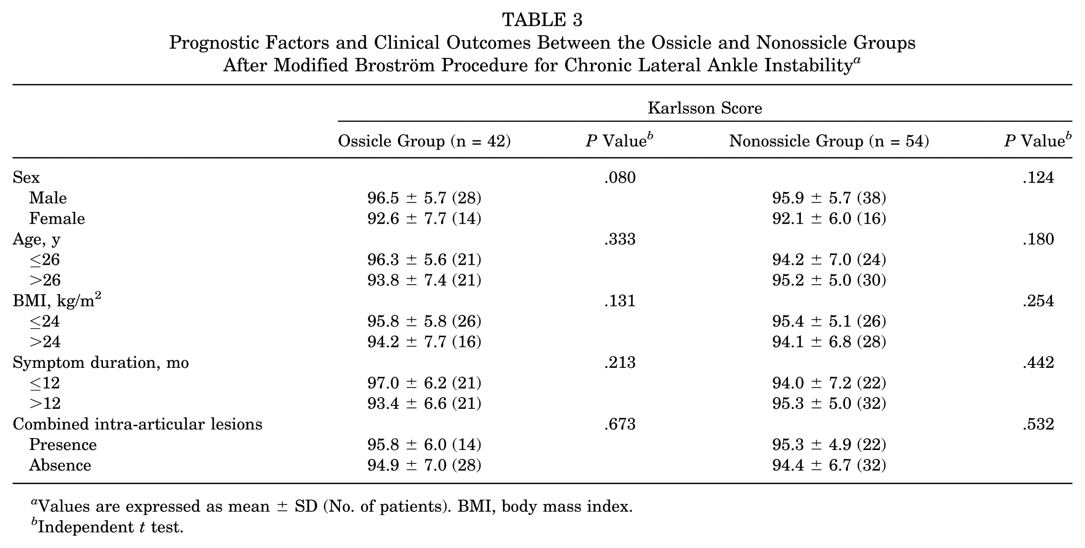
Values are expressed as mean ± SD (No. of patients). BMI, body mass index.
Independent t test.
Radiographic Outcomes
The mean talar tilt angle improved from 14.0° preoperatively to 7.6° at final follow-up in the ossicle group and from 12.2° preoperatively to 6.8° at final follow-up in the nonossicle group. The mean anterior talar translation improved from 9.3 mm preoperatively to 5.8 mm at final follow-up in the ossicle group and from 9.4 mm preoperatively to 5.7 mm at final follow-up in the nonossicle group. The mean talar tilt angle and anterior talar translation at final follow-up showed no significant difference between the ossicle and nonossicle group (P > .05). The mean side-to-side difference also showed no significant difference at final follow-up (P > .05) (Table 4).
Comparison of Radiographic Outcomes Between the Ossicle and Nonossicle Groups After Modified Broström Procedure for Chronic Lateral Ankle Instability a
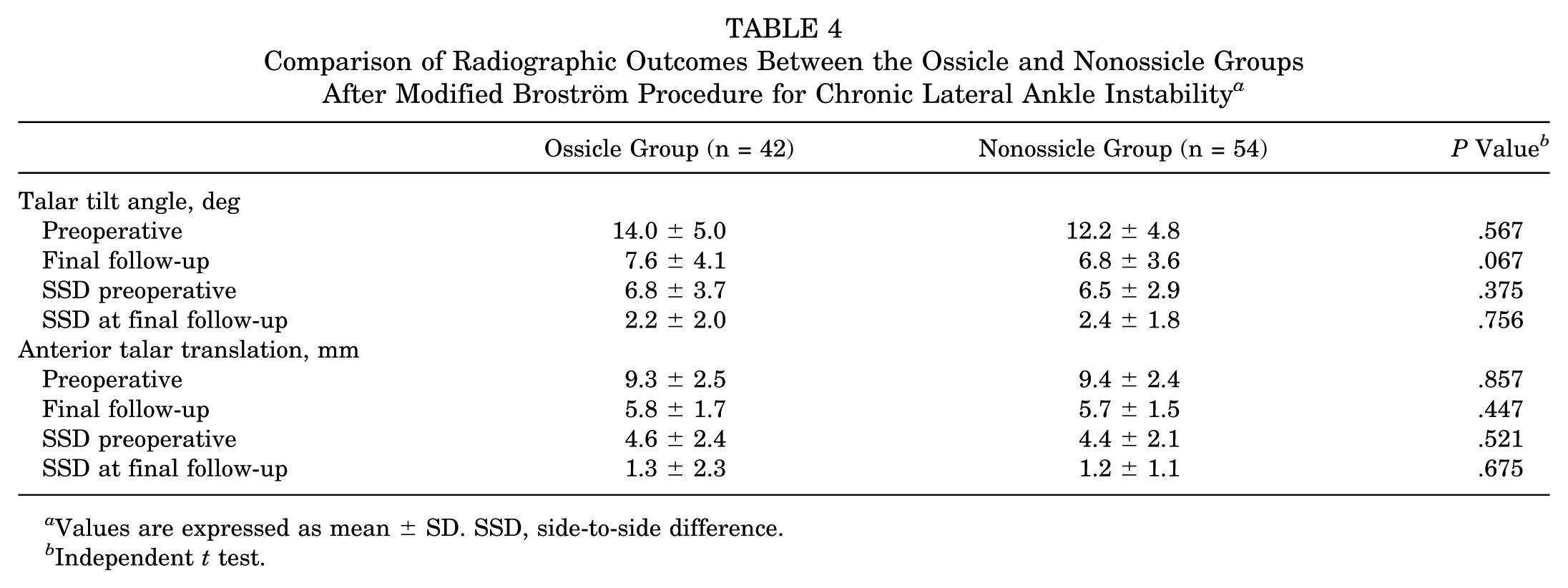
Values are expressed as mean ± SD. SSD, side-to-side difference.
Independent t test.
Ossicle Size and Outcomes
The mean subfibular ossicle size was 8.8 ± 3.7 mm, and the mean sizes of small and large ossicles were 5.8 ± 2.1 mm and 12.0 ± 1.7 mm, respectively. Between small and large ossicle groups, the Karlsson score and the AOFAS score showed no significant differences at final follow-up (P > .05). Similarly, the mean talar tilt angle and anterior talar translation showed no significant difference preoperatively and postoperatively (P > .05) (Table 5).
Comparison of Clinical and Radiographic Outcomes According to the Subfibular Ossicle Size After Modified Broström Procedure for Chronic Lateral Ankle Instability a
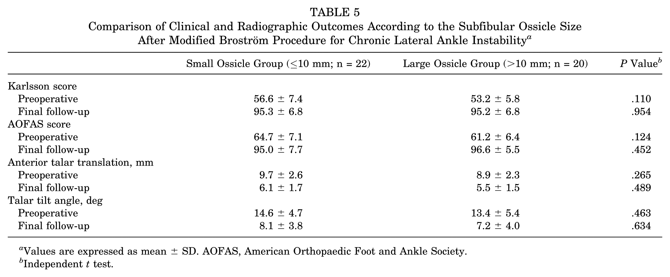
Values are expressed as mean ± SD. AOFAS, American Orthopaedic Foot and Ankle Society.
Independent t test.
Additional Arthroscopic Findings
Arthroscopic procedures were simultaneously performed to treat other accompanying intra-articular pathologic lesions demonstrated by MRI, such as soft tissue impingement with synovial hypertrophy, bony impingement due to osteophyte formation, osteochondral lesion of talus, and loose body. Arthroscopic management for 14 (33%) ankles in the ossicle group and 22 (41%) ankles in the nonossicle group was carried out. Arthroscopic synovectomy or debridement for soft tissue impingement was performed in 6 (14%) ankles in the ossicle group and in 14 (26%) ankles in the nonossicle group. All hypertrophied soft tissue impingement lesions occurred on the anterolateral gutter of the ankle. Arthroscopic multiple drilling or microfracture for osteochondral lesion of the talus was performed in 6 (14%) ankles in the ossicle group and in 6 (11%) ankles in the nonossicle group. Six osteochondral lesions were seen in the ossicle group; 3 on the medial talar dome and 3 on the lateral, with a mean size of 1.2 ± 0.4 cm2. All 6 lesions in the nonossicle group occurred on the medial dome, with a mean size of 1.1 ± 0.3 cm2. Arthroscopic bony spur resection was performed in 3 (7%) ankles in the ossicle group and in 2 (4%) ankles in the nonossicle group. Intra-articular loose body removal was performed in 2 (5%) ankles in the ossicle group and 3 (6%) ankles in the nonossicle group. Statistical analysis using the chi-square test did not show a significant difference regarding proportion of combined intra-articular lesions in both groups (P > .05).
Complications
At 2 months after operation, 1 superficial wound dehiscence was reported in the nonossicle group, and wound closure was then performed. One case of superficial peroneal nerve neurapraxia was reported in the ossicle group, which resolved spontaneously within 4 weeks after operation.
Discussion
This was a comparative study with the largest series of patients and relative long-term follow-up period among the studies investigating treatment for CLAI with subfibular ossicle. We assessed the Karlsson and AOFAS scores and investigated prognostic factors affecting clinical outcomes. In this study, the ossicle and nonossicle groups showed similar results that were good in terms of the Karlsson score, AOFAS score, talar tilt angle, and anterior talar translation on stress radiographs at the final follow-up. These results mean that the excision of subfibular ossicle during the modified Broström procedure does not have a negative effect on clinical results.
There are several surgical methods for subfibular ossicle including ossicle excision, ossicle fixation, modified Broström procedure with the ossicle excision, and anatomic or nonanatomic reconstructions with the ossicle excision. Because these options have both benefits and drawbacks, there are uncertainties about the appropriate surgical treatment for subfibular ossicle. Subfibular ossicle excision has been performed on patients with only tenderness of the subfibular area and showed good clinical results. 10 Ossicle fixation with small screws after resection of the pseudarthrosis has also been performed,10,19 but osteosynthesis of the ossicle has several weaknesses, such as nonunion and delayed rehabilitation. Moreover, small screws can cause soft tissue irritation and persistent pain after surgery. Some surgeons have performed lateral ankle stabilization procedures using the peroneus brevis tendon.2,22 Mancuso et al 22 reported that a lateral ligament reconstruction using a split peroneus brevis was performed after excision of the ossicle because the ATFL was found to be too short to reanastomosis with the malleolus. However, Rosenbaum et al 28 reported that peroneal tenodesis did not restore stability to the ankle joint. In addition, patients undergoing Evans tenodesis with peroneus brevis tendon transfer had significantly more laxity, significantly lower functional outcomes, and more degenerative changes on radiographs compared with patients undergoing anatomic reconstruction at a minimum 15-year follow-up. 20 Besides, some disadvantages have been reported for nonanatomic reconstructions, including abnormal ankle kinematics and limited subtalar motion. 29 Although various anatomic reconstructions using autologous tendon or allograft have been used in patients with weak remnant of the lateral ligament with satisfactory results,7,12,31 these reports are mostly for short-term outcomes, and they have several drawbacks such as longer operative time and incorporation of the graft.
On the other hand, favorable long-term results have been reported for anatomic repair using remnants of the lateral ligaments.1,14 For our study, the modified Broström technique was chosen and the ossicle excision simultaneously carried out. From our results, there were no significant differences between the ossicle and nonossicle groups in clinical and radiographic outcomes. These results were congruent with the surgical techniques. When subfibular ossicle was observed, it was excised while care was taken to avoid injury of the lateral ligament complex. Although the ATFL or CFL was sometimes shortened, the lateral ligament complex was fully advanced and reattached to the fibula. In this study, all ATFL and CFL could be repaired, and augmentation using extensor retinaculum was enough to give additional stability.
Although ossicle excision of large ossicles more than 10 mm was performed, the present study showed good clinical outcomes compared with ossicle excision of small ossicles less than 10 mm. These results can be explained by several intraoperative findings. Subfibular ossicles were usually less than 15 mm, and there were no cases larger than 15 mm that looked like a distal fibular tip fracture. The ossicle size had little effect on clinical outcomes because difference in mean ossicle size between small and large ossicle groups was not substantial. Hasegawa et al 10 reported that the status and quality of the lateral ligament complex were more important than ossicle size in postoperative ankle instability. Furthermore, the subfibular ossicle was not always attached to the ATFL and some attached to the CFL. Kono et al 18 reported that there was no attachment of ligament fibers at the proximal or the distal ends of the ossicle. Because the ATFL plays the most important role in lateral ankle stability, this supports our results.
To the best of our knowledge, there have been few reports on prognostic factors with regard to age, sex, obesity, and symptom duration in patients who have undergone the modified Broström procedure. There have been some reports related to intra-articular lesions and CLAI. Osteochondral lesion of the talus, soft tissue impingement syndrome, loose body, peroneal tendon disorder, and other associated lesions could be the causes of prolonged pain in CLAI.5,9,13 On the other hand, some authors reported that cartilage lesions did not affect the results of the lateral ankle ligament reconstruction or the modified Broström procedure.8,23,25,30 In this study, combined intra-articular lesions did not influence the clinical outcomes at the final follow-up. These results suggest that if an intra-articular lesion is treated appropriately, it will not affect the clinical outcomes. Sex of the patient was also not a clinical factor affecting Karlsson score in the ossicle and nonossicle group (P > .05). Xu et al 32 reported that there are no differences between males and females in terms of clinical and radiographic outcomes after the modified Broström procedure.
This study had some limitations. First, if it were a prospective randomized study, more rigorous information could have been provided. Second, arthroscopic procedure was performed only on patients who had intra-articular symptoms and lesions on MRI, although the intra-articular lesions associated with CLAI occur at a high incidence.
In conclusion, the modified Broström procedure and subfibular ossicle excision provided good clinical and radiographic outcomes comparable with those of the same procedure without subfibular ossicle excision. Therefore, the modified Broström procedure with ossicle excision is recommended as an effective and reliable treatment strategy for CLAI in patients with subfibular ossicle.
Footnotes
The authors declared that they have no conflicts of interest in the authorship and publication of this contribution.
References
Supplementary Material
Please find the following supplemental material available below.
For Open Access articles published under a Creative Commons License, all supplemental material carries the same license as the article it is associated with.
For non-Open Access articles published, all supplemental material carries a non-exclusive license, and permission requests for re-use of supplemental material or any part of supplemental material shall be sent directly to the copyright owner as specified in the copyright notice associated with the article.