
Abstract
Background:
The tibial tubercle to trochlear groove (TT-TG) distance is used for screening patients with a variety of patellofemoral joint disorders to determine who may benefit from patellar medialization using a tibial tubercle osteotomy. Clinically, the TT-TG distance is predominately based on static imaging with the knee in full extension; however, the predictive ability of this measure for dynamic patellar tracking patterns is unknown.
Purpose:
To determine whether the static TT-TG distance can predict dynamic lateral displacement of the patella.
Study Design:
Cohort study (Diagnosis); Level of evidence, 2.
Methods:
The static TT-TG distance was measured at full extension for 70 skeletally mature subjects with (n = 32) and without (n = 38) patellofemoral pain. The dynamic patellar tracking patterns were assessed from approximately 45° to 0° of knee flexion by use of dynamic cine-phase contrast magnetic resonance imaging. For each subject, the value of dynamic lateral tracking corresponding to the exact knee angle measured in the static images for that subject was identified. Linear regression analysis determined the predictive ability of static TT-TG distance for dynamic patellar lateral displacement for each cohort.
Results:
The static TT-TG distance measured with the knee in full extension cannot accurately predict dynamic lateral displacement of the patella. There was weak predictive ability among subjects with patellofemoral pain (r2 = 0.18, P = .02) and no predictive capability among controls. Among subjects with patellofemoral pain and static TT-TG distances 15 mm or more, 8 of 13 subjects (62%) demonstrated neutral or medial patellar tracking patterns.
Conclusion:
The static TT-TG distance cannot accurately predict dynamic lateral displacement of the patella. A large percentage of patients with patellofemoral pain and pathologically large TT-TG distances may have neutral to medial maltracking patterns.
Patellofemoral pain (PFP) is one of the most prevalent knee disorders in young adult and adolescent females.5,44 While PFP is occasionally overlooked in epidemiology studies focused on sports injuries, the reported prevalence of PFP in this population is higher than that of anterior cruciate ligament (ACL) injuries. 14 Young females with PFP report equal loss of function and more persistent symptoms compared with their peers who sustain an ACL injury.34,36,44 This chronic, often debilitating pain limits physical activity and may deprive individuals of the physical, social, and psychological benefits of exercise.22,31,34
PFP has numerous potential causes, including maltracking, chondral lesions, quadriceps and patellar tendon abnormalities, and other possible unknown origins. 33 While the diagnosis of patellar dislocation is usually obvious, patellar maltracking is often more difficult to diagnose and typically requires imaging. 33 For the purposes of this study, PFP is defined as anterior knee pain (AKP) with an insidious onset and potential patellar maltracking absent a history of dislocation.
For patients with PFP, the primary goal of treatment is to reduce pain by restoring more normative patellar tracking patterns during dynamic knee motion with active muscle contraction.17,33 Conservative therapies are the mainstay of current practice; however, surgical interventions are occasionally warranted when nonoperative treatments fail.1,11,17,19 Tibial tubercle (TT) osteotomy, sometimes paired with soft tissue procedures, aims to medialize patellar tracking by reducing the lateral pull of the patellar tendon.17,20,26 When the static distance between the TT and the trochlear groove (TT-TG distance) is used for preoperative screening, TT osteotomy has yielded excellent results for patients with a history of patellar dislocation. 46 While some studies18,24,35,46 investigating TT osteotomy for patients with intractable PFP, without a history of patellar dislocation, have also observed positive outcomes, other studies12,30,37 have reported medial patellar instability, surgical failure, and a less favorable prognosis compared with cohorts with a history of patellar dislocation.
The less favorable surgical outcomes reported for patients with intractable PFP relative to patients with a history of patellar dislocation may be directly related to the degree of difficulty in identifying which subjects actually demonstrate dynamic lateral maltracking in this population. 12 In contrast to cohorts with a history of patellar dislocation, for whom the lateral maltracking is made apparent by the dislocation, patients with isolated PFP lack diagnostic events that clearly establish dynamic lateral maltracking. This is a crucial distinction, as neutral and medial tracking patterns have been documented in patients with isolated PFP.27,28,39 Medialization of the patellae in some patients may account for the mixed outcomes observed in this population. However, it remains unknown whether the primary measure used to screen surgical candidates for TT osteotomy (ie, static measures of TT-TG distance) can accurately identify dynamic lateral tracking patterns.24,46 Thus, the aim of this study is to determine the predictive ability of static TT-TG distance for dynamic lateral displacement of the patella.
Methods
As part of an ongoing protocol approved by an institutional review board, clinical and magnetic resonance (MR) imaging data for 70 subjects were collected for this study. Skeletally mature subjects with (n = 32) and without (n = 38) PFP were included (Table 1). Controls consisted of volunteers with no history of pain, injury, or surgery in either knee. For both cohorts, if both knees met criteria and if time permitted, bilateral scans were obtained (11 subjects with PFP and 4 controls). For these subjects, data from the knee that demonstrated the greater TT-TG distance were used for the analysis. All subjects were screened for exclusion criteria, which included prior knee trauma (eg, ligament, cartilage, or meniscus injury), knee surgery, clinically or radiologically diagnosed rheumatoid or osteoarthritis, history of patellar dislocation, generalized joint laxity (eg, diagnosed Ehlers-Danlos syndrome or Beighton score 43 >5), or contraindication to MR scanning. Inclusion in the cohort with PFP required a clinical diagnosis of PFP. Only individuals with symptoms occurring for more than 6 months before scanning were included in this cohort. All subjects with PFP complained of diffuse pain localized to the anterior knee that affected their sports participation and/or activities of daily living. On examination, all subjects with PFP demonstrated discomfort to palpation in the peripatellar region.
Participant Characteristics a
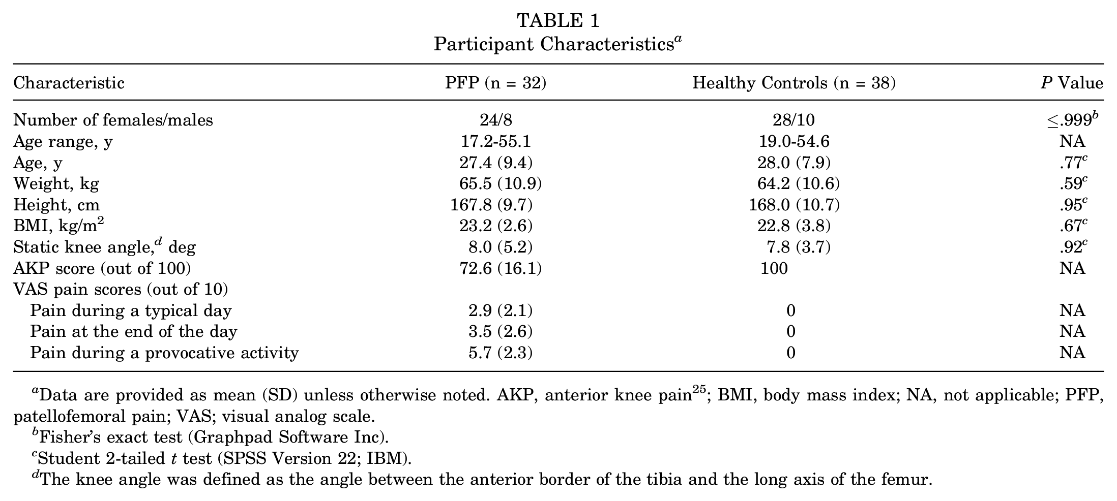
Data are provided as mean (SD) unless otherwise noted. AKP, anterior knee pain 25 ; BMI, body mass index; NA, not applicable; PFP, patellofemoral pain; VAS; visual analog scale.
Fisher’s exact test (Graphpad Software Inc).
Student 2-tailed t test (SPSS Version 22; IBM).
The knee angle was defined as the angle between the anterior border of the tibia and the long axis of the femur.
All clinical and radiologic data were acquired during a single visit. MR data were obtained in a 3-T MR scanner (Phillips Electronics). Before MR scanning, all subjects underwent a history and physical examination. Clinical intake focused on the knee and included determination of the AKP score and visual analog scale (VAS) scores for pain during a typical day, pain at the end of the day, and pain during provocative activities. 25
For static imaging, subjects were situated in a supine position with the lower extremity in an anatomically neutral position, supported by a cushioned heel holder. The fully extended knee was positioned within an 8-channel knee coil for enhanced image quality. The static MR series included sagittal 3D gradient recalled echo (GRE), 3D GRE with fat saturation (GRE-FS), and proton density weighted images. The 2 GRE images had a pixel resolution of 0.27 × 0.27 × 1.0 mm (512 × 512 pixels). All 3 image sets were converted, using the raw MR data, into 3D axial and coronal images. A musculoskeletal radiologist read all images to screen for underlying osteoarthritis and other knee injuries. Any cases demonstrating potential cartilage defects were referred to the senior musculoskeletal radiologist for grading. All subjects (n = 10) demonstrating greater than grade 2 patellofemoral or tibiofemoral cartilage defects on the International Cartilage Repair Society scoring system were excluded (these 10 subjects are not included in Table 1 or in the final study population). 21
The static TT-TG distance and knee angle were measured from the axial and sagittal GRE series. The static TT-TG distance was measured according to the current gold standard technique (Figure 1).8,13 The knee angle (Figure 2) was defined as the angle between the anterior border of the tibia (Ty) and the long axis of the femur (Fy). Due to the inherent bowing of the femur and angulation of the anterior border of the tibia, this measure of knee flexion overestimates the clinical measure (ie, hip, knee, and ankle) by 10°. 16 Thus, full knee extension, as measured using the clinical measure, corresponds to 10° using the current MR-based method.
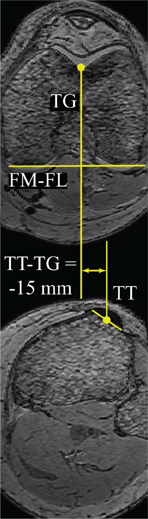
Static axial magnetic resonance images used to calculate static distance from the tibial tubercle to the trochlear groove (TT-TG distance). The tangent to the posterior aspect of the femoral condyles (FM-FL) was drawn on the femoral slice that depicted the widest distance between the femoral epicondyles and the deepest TG (top). A line perpendicular to the tangent bisecting the deepest aspect of the TG was drawn and transferred to the most proximal tibial slice depicting complete insertion of the patellar tendon onto the TT (bottom). The difference between the transferred line and a parallel line bisecting the patellar tendon determined static TT-TG distance.
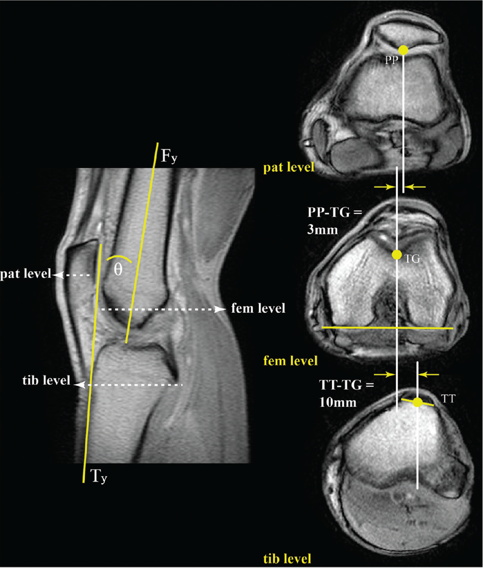
Key landmarks on dynamic cine-phase contrast (CPC) magnetic resonance images. The left image is the full extension image from the CPC dataset for this subject. The knee angle (θ) was defined as the angle between the anterior border of the tibia (Ty) and the long axis of the femur (Fy). The 3 cine axial planes (pat, fem, and tib) were selected based on this image and are shown on the right (top, middle, and bottom, respectively). The locations of most posterior point of the patella (PP) in the image at level of the midpatella (top), the deepest aspect of femoral sulcus (TG) in the image representing the widest aspect of the femoral epicondyles (middle), and the midpoint of the patellar tendon in the image containing complete insertion onto the TT (bottom) were identified. Tracking data from the CPC analysis were used to track the knee angle and these points throughout the motion cycle. These data, in turn, were used to quantify the TT-TG distance and the distance from TG to PP throughout the motion cycle.
For dynamic imaging, subjects were situated in a supine position with their knee flexed and supported by a cushioned block. Lateral stabilization was provided by a custom-built coil holder, which supported a pair of large flex-coils medial and lateral to the knee. An additional pair of medium flex-coils were placed directly anterior to the knee. Subjects were taught to flex and extend the knee, touching the top and bottom of the MR bore, at 30 cycles per minute to the beat of an auditory metronome. The typical range of motion was from 45° of flexion to full extension. Sagittal dynamic cine-phase contrast (CPC) MR images (anatomic and 3D velocity) were obtained throughout the motion. In addition, 4 anatomic, cine, axial-plane images were acquired during the same motion. Through use of the full extension image from the sagittal CPC image set, these images were selected at the level of the midpatella, the femoral epicondylar width, and the TT (Figure 2).
The CPC dataset provided the ability to accurately (<0.3 mm; Behnam et al 3 ) track the motion (ie, translation and rotation) of the patella, femur, and tibia throughout the extension-flexion movement. On the cine images, the anatomic sites used to calculate the static TT-TG distance (ie, the midpoint of the patellar tendon at the level of complete insertion onto the TT and the deepest point of the TG at the widest aspect of the femoral epicondyles) were identified (Figure 2). The most posterior point of the patella (PP) at the level of the midpatella was also located. Based on the movement profiles obtained with the CPC data, the PP, TG, TT, and knee angle were tracked in even 80-millisecond increments throughout the movement. All dynamic data were interpolated to single knee angle increments for further analyses. For a complete description of the CPC imaging technique and how it is used to derive the kinematic data, see Seisler and Sheehan. 38
The dynamic lateral patellar displacement (LPD) and dynamic TT-TG distances (Figure 2) were defined as the distance from PP and TT, respectively, to TG:
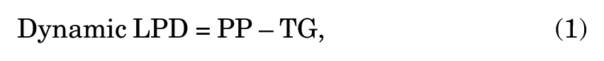
where PP is the most posterior point on the patella, defined in the axial image at the level of the midpatella and tracked by use of the dynamic data; and TG is the deepest point in the femoral groove, defined in the axial image at the level of the femoral epicondyles and tracked by use of the dynamic data:

where TT is the midpoint of the patellar tendon insertion onto the tibia, as defined and tracked by use of dynamic MR data.
Linear regression analysis was used to determine whether the static TT-TG distance could predict dynamic LPD. The dynamic LPD measured at the dynamic knee angle (Figure 2) corresponding to the static knee angle for each subject was used for the correlation analysis. Using thresholds recommended for interpreting Pearson’s correlation coefficients, we defined r 2 values <0.09 as negligible, 0.09-0.25 as weak, >0.25-0.64 as moderate, and >0.64 as strong. 29 As males represented a small percentage of the total population, all regressions were calculated for 2 groups, one that combined males and female subjects and the other that included only females.
To further explore the relationship between the static TT-TG distance and dynamic LPD, we determined the prevalence of subjects with PFP and pathological static TT-TG distances demonstrating neutral or medial patellar tracking patterns. As recommended by prior studies using MR images, 15 mm was defined as the threshold for pathological TT-TG distance.2,24,46 Individuals in the cohort with PFP who had a dynamic LPD value that was within 1 standard deviation and >1 standard deviation medial to the control group’s average were considered to have a neutral and medial tracking pattern, respectively. This comparison was done for a single knee angle (10°).
To calculate the number of subjects required to achieve adequate power, we conducted an a priori power analysis using G-Power. 15 The analysis was based on the assumption that the static TT-TG distance to dynamic LPD regression would be similar to the dynamic TT-TG distance to dynamic LPD regression (r 2 = 0.45) observed in a prior study. 48 Alpha and power were assumed to equal 0.05 and 0.95, respectively. This led to the conclusion that cohort sizes of 15 subjects or more were required to determine significant regression between static TT-TG distance and dynamic LPD. Sample sizes beyond the required number were included if the additional data were available. Intraobserver variability for static TT-TG distance was assessed for 30 randomly selected subjects using the Bland-Altman 95% limits of agreement (LOA) and a 2-way mixed-effects model intraclass correlation coefficient (ICC).4,42
Results
No demographic differences were observed between the subjects with PFP and the asymptomatic controls (Table 1). However, the cohort with PFP demonstrated a greater average (±SD) static TT-TG distance relative to the control cohort (13.6 ± 3.8 mm vs 10.3 ± 3.7 mm, P < .001). In addition, the cohort with PFP demonstrated greater lateral shift (−1.8 mm, P = .048) and greater lateral tilt (6.3°, P = .01) than the control cohort. The kinematic comparison was done at a single dynamic knee angle (10°).
The static TT-TG distance demonstrated weak predictive ability for dynamic LPD (Table 2) for subjects with PFP (Figure 3). The static TT-TG distance could predict LPD with 95% confidence to 9.0 mm for this cohort (Table 2). The static TT-TG distance could not predict the dynamic LDP for healthy controls.
Regression Coefficients for Static TT-TG Distances With Dynamic LPD a

Insignificant regressions and SEs are represented with dashes. LPD, lateral patellar displacement; PFP, patellofemoral pain; SE, standard error of the estimate; TT-TG, tibial tubercle and trochlear groove.
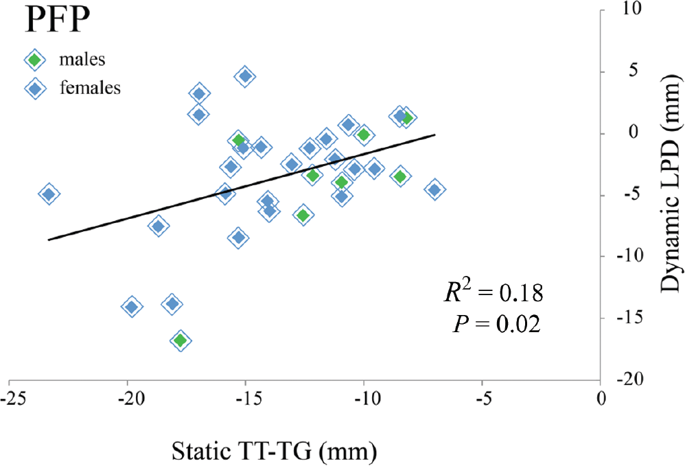
Linear regression analysis for static distance between the tibial tubercle and the trochlear groove (TT-TG distance) and dynamic lateral patellar displacement (LPD) for the cohort with patellofemoral pain (PFP). Although all subjects were placed in “full knee extension” for static imaging, the static knee angle varied slightly across subjects. Thus, the dynamic LPD was defined at the dynamic knee angle corresponding to the knee angle in which the static TT-TG measures were acquired.
Within the cohort with PFP, 13 of 32 (41%) subjects demonstrated a pathological static TT-TG distance of >15 mm. Of these, 6 of 13 (46%) and 2 of 13 (15%) demonstrated neutral and medial maltracking, respectively (Figure 4). The subjects with the most extreme lateral tracking (–17.7 mm) and the second most extreme medial tracking (+2.3 mm) both had TT-TG values between 17 and 18 mm (Figure 5).
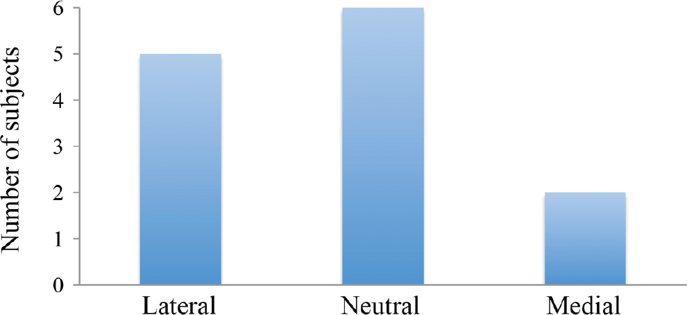
Subjects with patellofemoral pain and pathological distance between the tibial tubercle and the trochlear groove (TT-TG distance) >15 mm demonstrating lateral, neutral, and medial patellar tracking patterns.
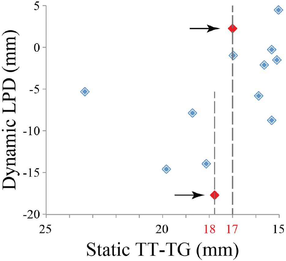
Individual lateral patellar displacements (LPD) among subjects with patellofemoral pain (PFP) and with pathological values for the static distance between the tibial tubercle and the trochlear groove (TT-TG distance). Thirteen subjects with PFP demonstrated a pathological TT-TG distance (>15 mm).
The mean difference in the Bland-Altman analysis for intraobserver variability was 0.27 mm, with LOA of –1.30 to 1.99 mm. The ICC for intraobserver variability (0.991; 95% confidence interval, 0.98-0.99) was above the threshold for clinical reliability. The average knee flexion angles observed on static imaging for each cohort were similar and demonstrated slight hyperextension (Table 1). 16
Discussion
This study demonstrates that static measures of TT-TG distance cannot accurately predict dynamic lateral tracking of the patella with both measurements acquired at full knee extension. This is in direct contrast to previous studies which stated that TT-TG distances are radiological measures of patellofemoral alignment in the axial plane.41,45,48 Due to the difficulty of determining lateral patellar maltracking on clinical examination, in the absence of a history of patellar dislocation, it is critical to obtain additional screening measures before recommending TT osteotomy for patients with PFP. In the current study, 41% of all subjects with PFP were potential candidates for TT osteotomy, based on an isolated static TT-TG distance >15 mm.24,46 Yet 61% of these individuals demonstrated neutral or medial dynamic patellar tracking patterns. TT medialization in these patients may increase contact pressures between the medial patellar and trochlear facets, thereby exacerbating symptoms or causing iatrogenic medial instability, particularly if the osteotomy is coupled with a lateral release.6,37 This potential overcorrection may account for the less favorable outcomes reported in populations with isolated PFP (ie, no history of dislocation) compared with patients who have pathologic TT-TG distances and a history of patellar dislocation. 12 Thus, for patients with intractable PFP and a lateralized patellar tendon force (static TT-TG distance >15 mm), the decision to perform corrective TT osteotomy should not be based on isolated measures of the static TT-TG distance. Additional steps to identify dynamic lateral patellar tracking patterns are critical to avoid excessive medialization of dynamically neutral or medial patellae.
A previous study 16 assessed the ability of static markers, other than static TT-TG distance, to predict dynamic lateral patellar tracking. This prior study found that static measures of LPD predicted 47% of the variability for dynamic LPD, which is clearly superior to the static TT-TG distance (r2 = 0.18). Further, including the static MR Q-angle in the regression analysis increased the predictive capacity to 62%. Thus, when dynamic images (computed tomography, 48 cine MR, 7 CPC MR 40 ) cannot be acquired to precisely identify dynamic patellar tracking patterns, static measures of LPD with or without the static MR Q-angle are preferable to isolated static measures of TT-TG distance. Further research is necessary to determine whether other static measures can more accurately predict dynamic lateral patellar tracking.
Greater bony and soft tissue constraints in healthy controls, relative to individuals with PFP, likely account for the reduced ability of static TT-TG distances to predict dynamic LPD among controls. A prior study 16 evaluating static alignment to dynamic tracking also found reduced or absent regression among healthy controls, whereas such relationships existed for cohorts with PFP. Subjects with PFP often have other predisposing factors to PFP (eg, alta, ligament laxity), which permit increased patellar mobility due to decreased bony restraint.23,32 As a result, force imbalances surrounding the knee are more likely to present as maltracking in subjects with PFP relative to controls. It follows that the factors responsible for force imbalances (eg, vastus muscle weakness,47,50 pathological TT-TG distance9,10,51) present in cohorts with PFP would demonstrate stronger regression for the patellar tracking patterns.
The closest comparison to the current analysis is a recent pair of studies45,48 focused on the correlation between the dynamic TT-TG distance and the dynamic LPD. If the current analysis was changed such that the dynamic TT-TG distance replaced the static TT-TG distance in the regression analysis, then our results would concur with the prior findings. Specifically, the dynamic TT-TG distance demonstrated a stronger ability to predict lateral patellar tracking, relative to the static TT-TG distance. However, the predictive ability only achieved 45% in the cohort with PFP and 13% among controls. This was similar to the regression values observed for dynamic TT-TG distance and dynamic LPD in the prior studies45,48 (r2 = 0.45 and 0.49) focused on individuals with a history of patellar dislocation, suggesting that the dynamic relationship between dynamic TT-TG distance and dynamic LPD is similar across these 2 pathologic conditions. Yet in all prior studies, the regression analysis was performed by use of dynamic TT-TG distance, not static TT-TG distance. This distinction is crucial, as TT-TG distance is most commonly obtained clinically by use of static imaging techniques, and thresholds for surgical candidacy have been established only through use of static TT-TG distance.20,24,46
The free knee extension exercise without axial loading of the tibiofemoral joint was specifically designed to emphasize potential relationships between TT-TG distances and patellar tracking. This exercise requires a strong quadriceps load in terminal extension, which pulls the patella to the superior aspect of the femoral groove, limiting the bony constraint on the patella. At this position, the primary forces on the patella are from the soft tissues. Thus, if a relationship does exist between the TT-TG distance and patellar tracking, it would be expected to be strongest at full extension during the exercise. In comparison, a loaded activity such as squatting, which is often used to evaluate patellofemoral tracking in vivo, does not necessitate a high quadriceps load at full extension. As a result, the relationship may be masked by the forces exerted on the patella from the femur. 49
The primary limitation of this study is the lack of surgical outcomes to support the proposed screening. Validation of these recommendations is encouraged through performance of a prospective clinical trial. In addition, other factors beyond LPD and pathological TT-TG distance may influence outcomes after TT osteotomy. A comprehensive study investigating the potential effects of patella alta, patellar tilt, and femoral rotation among other factors is needed.
In conclusion, the static TT-TG distance offers limited insight into dynamic lateral patellar tracking patterns in patients with isolated PFP. Specifically, the static TT-TG distance cannot identify which patents with isolated PFP may or may not have lateral maltracking. Before performing a TT osteotomy in this population, clinicians should conduct additional screening aimed at identifying lateral displacement of the patella. In patients found to have dynamically neutral or medial patellar tracking patterns, caution is recommended to avoid excessive medialization of the patella in the trochlear groove.
Footnotes
Acknowledgements
The authors thank Judith Welsh, MLS, and Katharine Alter, MD, for their help and support. In addition, they thank the NIH Clinical Center Radiology Department, headed by David Bluemke, MD, for supporting this work.
One or more of the authors has declared the following potential conflict of interest or source of funding: This work was funded by the Intramural Research Program of the National Institutes of Health (NIH) Clinical Center, Bethesda, MD, USA.