






Abstract
Background:
The majority of distal biceps tendon injuries can be repaired in a single procedure. In contrast, complete chronic tears with severe tendon substance deficiency and retraction often require tendon graft augmentation. In cases with extensive partial tears of the distal biceps, a human dermal allograft may be used as an alternative to restore tendon thickness and biomechanical integrity.
Hypothesis:
Dermal graft augmentation will improve load to failure compared with nonaugmented repair in a tendon-deficient model.
Study Design:
Controlled laboratory study.
Methods:
Thirty-six matched specimens were organized into 1 of 4 groups: native tendon, native tendon with dermal graft augmentation, tendon with an attritional defect, and tendon with an attritional defect repaired with a graft. To mimic a chronic attritional biceps lesion, a defect was created by a complete tear, leaving 30% of the tendon’s width intact. The repair technique in all groups consisted of cortical button and interference screw fixation. All specimens underwent cyclical loading for 3000 cycles and were then tested to failure; gap formation and peak load at failure were documented.
Results:
The mean (±SD) load to failure (320.9 ± 49.1 N vs 348.8 ± 77.6 N, respectively; P = .38) and gap formation (displacement) (1.8 ± 1.4 mm vs 1.6 ± 1.1 mm, respectively; P = .38) did not differ between the native tendon groups with and without graft augmentation. In the tendon-deficient model, the mean load to failure was significantly improved with graft augmentation compared with no graft augmentation (282.1 ± 83.8 N vs 199.7 ± 45.5 N, respectively; P = .04), while the mean gap formation was significantly reduced (1.2 ± 1.0 mm vs 2.7 ± 1.4 mm, respectively; P = .04). The mean load to failure of the deficient tendon with graft augmentation (282.1 N) compared with the native tendon (348.8 N) was not significantly different (P = .12). This indicates that the native tendon did not perform differently from the grafted deficient tendon.
Conclusion:
In a tendon-deficient, complete distal biceps rupture model, acellular dermal allograft augmentation restored the native tendon’s biomechanical properties at time zero. The grafted tissue-deficient model demonstrated no significant differences in the load to failure and gap formation compared with the native tendon. As expected, dermal augmentation of attritional tendon repair increased the load to failure and stiffness as well as decreased displacement compared with the ungrafted tissue-deficient model. Tendons with their native width showed no statistical difference or negative biomechanical consequences of dermal augmentation.
Clinical Relevance:
Dermal augmentation of the distal biceps is a biomechanically feasible option for patients with an attritionally thinned-out tendon.
Ruptures of the distal biceps brachii tendon have received significant attention as a result of an increased awareness of injuries, numerous surgical fixation techniques, and debate regarding 1- versus 2-incision surgery.2,8,12,13,18,19,23 The incidence of complete ruptures of the biceps tendon is 1.2 per 100,000 people per year, with men predominating between the ages of 40 and 60 years. A spontaneous tendon rupture may result from smoking or anabolic steroids. 22 The biomechanics of fixation have improved progressively with modern implants. A recent meta-analysis suggested that cortical button fixation might be the safest fixation technique. 23
While the majority of distal biceps tendons may be repaired in a single procedure with only a 1% rerupture rate, delayed presentation and rapid atrophy of the tendon may result in the need to use allograft tissue. 22 While longitudinal gaps or defects may be best repaired with autograft or allograft augmentation, 15 a thinned or atrophic tendon can potentially be treated with a human dermal allograft to strengthen the repair construct. 14 Tendon augmentation and reconstruction using dermal allografts have been described with success for various orthopaedic applications.4,17,21 However, the biomechanics of dermal graft augmentation of the distal biceps tendon has not been evaluated.
The purpose of this study was to assess the biomechanical properties of distal biceps tendon repair with and without acellular dermal allograft augmentation in a tendon with its native width and a tendon-deficient (attritional) model. Every tendon had a complete tear of the radial tuberosity. Our hypothesis was that in the attritional model, dermal graft augmentation would improve load to failure and gap formation (displacement) compared with nonaugmented repair.
Methods
Institutional review board approval was not required for this study. Eighteen matched pairs were randomly separated into 2 cohorts. In one cohort, the native tendon (n = 9) was tested alongside the native tendon with dermal allograft augmentation (n = 9); in the other cohort, the tendon with an attritional defect (n = 9) was tested alongside the tendon with an attritional defect repaired with dermal allograft augmentation (n = 9). Tendons with an attritional defect were tendons with a decreased width. All tendons were completely cut from the radial tuberosity.
For the tendon-deficient model, the tendon was step cut; specifically, a sharp demarcation between the intact tendon and the deficient tendon was created to 30% of the native tendon’s width over a longitudinal distance of 20 mm. This defect model was chosen during pilot studies. We examined 30%, 40%, and 50% residual tendon stumps (specifically, the percentage of the maximum width of the normal tendon that was left intact). Repair of the 50% defect was technically feasible and performed well in biomechanical testing. The 40% defect model had a wide range of loads to failure, and the 30% model was challenging to repair, which consistently failed at low loads. We wanted to emulate a clinical situation in which surgeons would intraoperatively choose to enhance the repair, and as such, the 30% residual tendon model was selected.
Dual-fixation distal biceps repair with a cortical button and interference screw was performed in all specimens as described by Sethi et al.18,20 The distal end of the tendon was debrided and whipstitched, and these sutures were threaded through a cortical button. Once the biceps button was passed through the radial tuberosity using the “tension-slide technique,” an interference screw was inserted on the radial side, and suture limbs were tied over the screw. 18
A modified version of this technique was performed for all repairs augmented with a dermal allograft. 14 A 2.0-mm acellular human dermal graft (ArthroFlex; LifeNet Health) was cut into a trapezoidal shape measuring 25 mm in length, 15 mm across the top, and 12 mm across the bottom. The graft was wrapped around the free distal biceps tendon, with the narrow (12 mm) part placed distally and the wider base (15 mm) proximally. The graft was then secured to the native tendon using a No. 0 Vicryl suture (Ethicon) and to facilitate the whipstitch. Once secured, a No. 2 high-strength nonabsorbable suture (FiberWire; Arthrex) was used to place Krackow stitches through the graft and tendon, incorporating the graft into the repair construct.18,20
Specimens were tested using a servohydraulic mechanical testing system (MTS Systems Corp). Specimens were cut in the midhumerus and distal third of the forearm before being mounted anatomically with the humerus held in a rigid clamp in line with the actuator. The biceps reconstruction construct was secured to the actuator with a custom dry freeze–activated sinusoidal clamp attached to the biceps tendon at the musculotendinous junction. Once the specimen was mounted, the forearm was positioned at 90° of flexion, and the biceps was preloaded with 50 N by threading a rod into the radius and loading it with adjustable weights. The force of 50 N was based on the expected passive contraction force of the biceps tendon in the early postoperative period. This load was registered through a 2500-N load cell.
A 5-mm × 5-mm block of foam bone substitute was glued to the radial tuberosity just distal to the reconstruction construct. A 9-mm differential variable reluctance transducer (DVRT; MicroStrain) was placed with one end inserted into the foam bone substitute and the other end inserted into the biceps tendon reconstruction construct, perfectly aligned to enable linear motion of the DVRT throughout the whole range of motion. Once the load was properly configured, the specimen was cycled from full extension to 90° of flexion at a rate of 0.25 Hz. This method of unrestricted loading allowed the forearm to move from pronation to supination as the arm was flexed. Displacement was measured through 3000 cycles 19 (Figure 1).
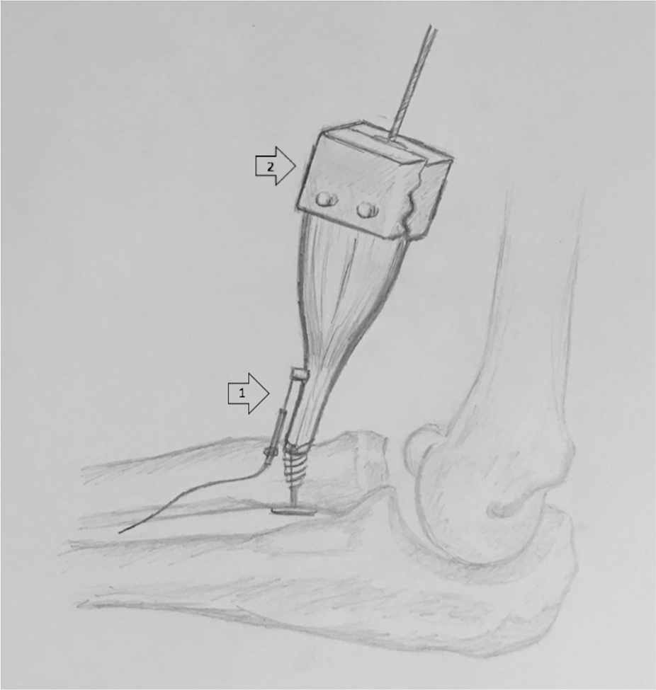
Specimens were tested in an anatomic position for cyclic loading, gap formation, and load to failure. The repair side was loaded with 50 N in neutral position. Weights and rod lengths were adapted accordingly. The tendon was gripped with an appropriately sized soft tissue clamp and secured by dry-ice freezing. (1) Differential variable reluctance transducer (DVRT) and (2) sinusoidal clamp to biceps.
After cyclic loading was complete, specimens were loaded to failure. This was accomplished by restraining the arm at 90° of flexion and loading at a rate of 120 mm/min until reconstruction failure. Displacement and load-to-failure data were recorded with an accuracy of 0.5 mm and 0.5 N, respectively. The mechanism of failure was recorded as well as the final load and maximum displacement over 3000 cycles. Displacement and load-to-failure data were compared and analyzed. 19
Displacement was measured as elongation of the overall construct. This measured length is a combination of gap formation between the bone and the graft, gap formation between the graft and the tendon, elongation and slippage of sutures, and structural tendon elongation.
Descriptive statistics to characterize the study groups were calculated using means ± SDs. Differences in the load to failure, stiffness, and displacement between the groups were analyzed with 1-way analysis of variance. When statistically significant, pairwise differences between the groups were analyzed by independent t tests with a Bonferroni adjustment. All statistical analyses were performed using Stata 12 (StataCorp LLC).
A priori power analysis was carried out. The addition of a graft was presumed to increase the load to failure by 10% to 15% (35-53 N), whereas a critical defect was presumed to diminish the load to failure by approximately 30% (100 N). An SD of 80 N was assumed across all groups. 19 A sample size of 6 per group would provide 80% power to detect a difference in the load to failure at an alpha level of .05.
Results
The mean load to failure (Table 1) was 75% greater (P < .001) for the native tendon group compared with the deficient tendon group. The mean load to failure was 41% greater (P = .04) for the grafted deficient tendon group compared with the deficient tendon group. The mean load to failure was not significantly different for the native tendon compared with the grafted deficient tendon (P = .12), suggesting that the tendon with its native width did not perform differently than the grafted deficient tendon.
Comparisons of Load to Failure for Augmentation of the Distal Biceps: Native Tendon Versus Tendon-Deficient Model
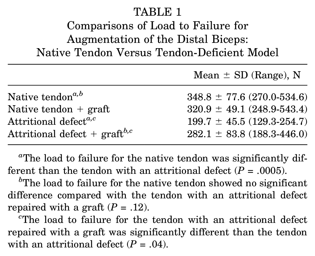
The load to failure for the native tendon was significantly different than the tendon with an attritional defect (P = .0005).
The load to failure for the native tendon showed no significant difference compared with the tendon with an attritional defect repaired with a graft (P = .12).
The load to failure for the tendon with an attritional defect repaired with a graft was significantly different than the tendon with an attritional defect (P = .04).
The mean stiffness (Table 2) was 155% greater (P < .001) for the native tendon group compared with the deficient tendon group. The mean stiffness was 53% greater (P = .04) for the grafted deficient tendon group compared with the deficient tendon group.
Comparisons of Stiffness for Augmentation of the Distal Biceps: Native Tendon Versus Tendon-Deficient Model

The stiffness for the native tendon was significantly different than the tendon with an attritional defect (P = .0006).
The stiffness for the native tendon was significantly different compared with the tendon with an attritional defect repaired with a graft (P = .012).
The stiffness for the tendon with an attritional defect repaired with a graft was significantly different than the tendon with an attritional defect (P = .04).
The mean displacement (Table 3) was not significantly different for the native tendon group compared with the deficient tendon group (P = .11). The mean displacement for the deficient tendon group was 125% greater (P = .04) when compared with the grafted deficient tendon group. The mean displacement for the native tendon group was not significantly different compared with the grafted deficient tendon group (P = .47), indicating that the tendon with its native width did not perform differently than the grafted deficient tendon.
Comparisons of Displacement for Augmentation of the Distal Biceps: Native Tendon Versus Tendon-Deficient Model
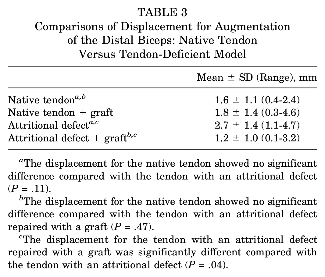
The displacement for the native tendon showed no significant difference compared with the tendon with an attritional defect (P = .11).
The displacement for the native tendon showed no significant difference compared with the tendon with an attritional defect repaired with a graft (P = .47).
The displacement for the tendon with an attritional defect repaired with a graft was significantly different compared with the tendon with an attritional defect (P = .04).
Each of the 4 groups had different modes of failure. The augmented native tendon group had 3 early failures including 2 knot slippages and 1 suture knot failure, none of which disrupted tendon quality. The native tendon group had 3 early failures including 1 bone fracture and 2 suture slippages. In the grafted deficient tendon group, 5 of the specimens failed from tendon pullout from the socket, whereas in the deficient tendon group, 4 specimens failed from suture pulling through the tendon, which disrupted tendon quality.
Discussion
This study demonstrated that dermal allograft augmentation of the repair of complete attritional lesions of the biceps tendon restored the load to failure and stiffness and reduced gap formation under cyclical loading back to levels comparable with the repair of injuries in tendons with their native width. Dermal allograft–augmented repair improved these same parameters when statistically compared with a nonaugmented tendon-deficient matched pair. In contrast, augmented and nonaugmented repairs of native tendons did not demonstrate any statistically significant biomechanical differences.
The impetus to study this technique has come from our clinical experience. Despite a low rate of postoperative reruptures, we have encountered cases with significant thinning, loss of elasticity, and atrophy of the distal tendon in the acute setting of a distal biceps rupture (<4 weeks after injury). 10 When the patient has reasonable tendon quality, the tendon can be successfully repaired to the tuberosity under tension or at high flexion angles. 15 However, when the ruptured tendon has attritional changes or volumetric loss, we believe that the rate of reruptures increases.
This tendon attrition may be further exacerbated by fixation techniques. Intramedullary fixation, with a cortical button, interference screw, or both, may place the repair site under stress at the tendon–bone tunnel interface. One concern is that with forearm rotation, the biceps tendon’s angle of insertion into the tunnel changes from 0° in full supination (a straight line pull) to over 90° in full pronation. This potentially causes edge loading of the tendon at the bone-tendon interface, which may make it more susceptible to retearing. We believed that the addition of biological tissue between the tendon–bone tunnel interface could act as a buffer to reduce this effect and restore native biomechanical tendon properties, particularly in an attritionally thinned tendon.
The matched specimens with a native tendon, as expected, showed no statistical difference or negative biomechanical consequence of dermal allograft augmentation. With the dermal graft, there was no decrease in load to failure, and there was no increase in gap formation. Importantly, the graft did not cause any tendon slippage and showed no compromise in fixation. Normal specimens were tested to see if there were any biomechanically deleterious effects of adding a graft, specifically, to ensure that there was no compromise in fixation strength or gap formation as well as ensure that the native tendon did not slide past or piston within the graft, causing potential concern for clinical applications.
The second part of this study examined a tendon-deficient distal biceps. There is no predicate model for studying tendon deficiency in the distal biceps. A model for an attritional biceps tendon was elaborated in pilot testing. This model was created and determined in pilot studies examining 30%, 40%, and 50% residual tendon stumps. The 50% residual tendon retained a significant load to failure, whereas the 30% residual tendon was significantly weaker and emulated a condition in which a surgeon would want to enhance the surgical repair. In this study, as predicted by our pilot testing, there was a significant difference in the mean load to failure between the native tendon (348.8 N) and the deficient tendon (199.7 N) (P < .001).
The relevant findings of this study were that augmentation of deficient tendons with a dermal allograft significantly increased the load to failure and stiffness as well as decreased gap formation when compared with an ungrafted attritional tendon model (Figure 2).
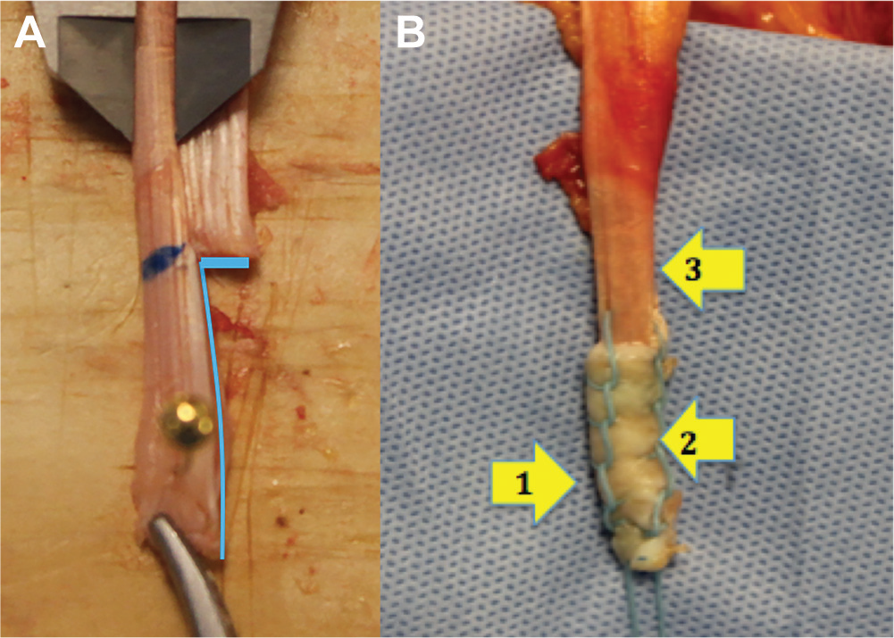
(A) A tendon that has been “step cut” and (B) an attritionally thinned-out tendon repaired with a dermal allograft. The deficient tendon with a graft showed a significant increase in load to failure and stiffness as well as a decrease in displacement compared with the deficient tendon alone (P = .04). Furthermore, these same parameters were not different between the native tendon and the grafted attritional tendon, suggesting a restoration of biomechanical behavior with graft augmentation. (1) Locked running suture, (2) deficient tendon, and (3) native tendon.
Furthermore, the grafted tissue-deficient model performed similar to the native tendon, with no significant differences in load to failure (P = .12) and displacement (P = .47). These data were derived from an appropriately powered study, suggesting that acellular dermal graft augmentation in an atrophic tissue situation can potentially restore its native biomechanical properties.
On the basis of the findings of this study, the authors consider dermal augmentation of distal biceps repair when there is substantial distal tendon thinning. This typically reflects a greater than 50% narrowing or tapering of the distal tendon and is often manifested with sutures cutting into or through the tendon during placement of the whipstitch. This study did not evaluate the graft as bridge tissue or a method to gain tendon length.9,11
While modes of failure were not dissimilar between the groups, the quality of the residual tendon was compromised in all ungrafted specimens. Repair of the native tendon failed, with the suture tearing through the residual tendon with significant shearing and tendon damage. The grafted tendon more often had suture breakage, and the residual tendon could be easily re-repaired as the graft shielded the tendon from significant intrinsic damage (Figure 3).
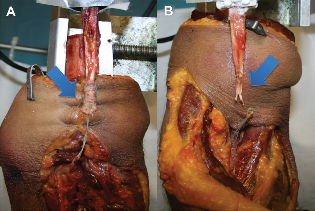
(A, B) The biceps tendon with and without a dermal allograft after cyclically loading for 3000 cycles and then tested to failure. The native tendon failed, with the suture tearing through the residual tendon with significant shearing and tendon damage. The grafted tendon more often had suture failure, and the residual tendon could be easily repaired as the graft shielded the tendon from significant intrinsic damage. The left arrow indicates a strong tendon with the graft, while the right arrow shows a shredded tendon without the graft.
The use of dermal graft augmentation is well described in both orthopaedic basic science and clinical orthopaedic applications.1,3,5-7 There are several commercially available acellular dermal matrices on the market. These are biological scaffolds with very high suture pullout strengths, 4 which allow for vascularization, host cell ingrowth, and proliferation. Omae et al 16 demonstrated that an acellular dermal graft may increase the maximum load to failure of an augmented rotator cuff. A histological examination showed evidence of native cell infiltration and neotendon formation when used as an interposition graft in a canine model. 16 Barber et al 4 reported on improved outcomes in humans with dermal augmentation of massive rotator cuff tears. The authors found intact repairs in 85% of the augmented group and 40% of the nonaugmented group of massive tears as well. Similar clinical improvements have been reported in foot and ankle applications, including bridging or “gap jumping” applications.4,17
This study has several weaknesses. This is a biomechanical and not a clinical study. As such, these data only reflect the time-zero application of the dermal graft and do not address the biological processes that occur after surgery. Also, the measured length of displacement is a combination of gap formation between the bone and the graft, gap formation between the graft and the tendon, elongation and slippage of sutures, and structural tendon elongation. However, there are sufficient data suggesting the appropriate ingrowth and vascularity of dermal grafts, leading us to believe that the construct may become stronger rather than weaker with time. 21 Furthermore, the mean age of the specimens used in this study was slightly higher than the age at which a distal biceps tendon rupture is clinically observed. It is conceivable that if specimens were obtained from middle-aged donors, a higher load to failure would have been observed. The native variability of tendon sizes as well as final cross-section diameters of the reconstruction construct differed. Further studies are warranted to evaluate biceps function and graft incorporation in a longitudinal clinical study.
Conclusion
In a tendon-deficient model, acellular dermal allograft augmentation of the distal biceps restored the native tendon’s biomechanical properties at the time of surgery. Dermal allograft augmentation of attritional tendon repair increased the load to failure and stiffness as well as decreased displacement compared with the ungrafted model. The native tendon showed no statistical difference or negative biomechanical consequences of dermal augmentation.
Footnotes
Acknowledgements
The authors gratefully acknowledge the assistance of William Grant Franco in drafting and editing the article.
Presented as a poster at the 43rd annual meeting of the American Orthopaedic Society for Sports Medicine, Toronto, Ontario, Canada, July 2017.
One or more of the authors has declared the following potential conflict of interest or source of funding: LifeNet Health provided dermal allograft patches. Arthrex (Science Care) provided cadaveric specimens and the BicepsButton, No. 2 FiberLoop with a straight needle, PEEK Tenodesis Screw, Drill Pin, Button Inserter, Headed Reamer, No. 2 FiberWire, and Driver for the Bio-Tenodesis Screw. P.S. has received royalties and honoraria from Arthrex. R.M. has received research grants from Arthrex, Joint Restoration Foundation, and BioD and owns stock in Cayenne Medical, ITS Implant, and AlignMed. A.D.M. is a consultant for Arthrex and is involved with Arthrex Research Support.