
Abstract
Background:
Bone bruises are frequently found on magnetic resonance imaging (MRI) after anterior cruciate ligament (ACL) injury and have been related to the force associated with the trauma. Yet, little is known about the bone bruise distribution pattern of skeletally immature (SI) patients, as the presence of an open physis may play a role in energy dissipation given its unique structure.
Purpose:
To describe and compare the location and distribution of tibial and femoral bone bruises, observed on MRI, between 2 groups of ACL-injured knees: the first group with an open physis and the second with a closed physis. Additionally, based on the bone bruise distribution pattern, the secondary aim of the study was to propose a new classification of bone bruise in SI patients.
Study Design:
Cross-sectional study; Level of evidence, 3.
Methods:
A retrospective review was conducted to identify all cases of primary ACL tears in patients ≤16 years old, with MRI within 6 weeks of injury between January 2012 and December 2016. Overall, 106 patients were identified: 53 with open physis (skeletally immature [SI] group) and 53 with closed physis as control (skeletally mature [SM] group). MRI scans were reviewed to assess for the presence and location of bone bruises. Longitudinal bone bruise distribution was described as epiphyseal and metaphyseal in both femur and tibia. The proposed classification for tibia and femur has 2 parts: the location of the bone bruise in the (I) lateral, (II) medial, or (III) medial and lateral parts of the bone; and if the bone bruise (a) does not or (b) does cross the physis. For the tibia, if the bone bruise is also present in the central portion, a letter C is added.
Results:
The SI group had significantly fewer bone bruises cross the physis and extend into the metaphysis than did the SM group for both the tibia (25% vs 85%, respectively; P < .0001) and the femur (4% vs 42%; P < .0001). The most common patterns observed in the SI group were type IIICa in the tibia (medial/lateral and central, not extending into the metaphysis: 42%) and type Ia in the femur (lateral, not extending into the metaphysis: 59%).
Conclusion:
The data from this study shows that patients with an open physis at the occurrence of an acute ACL rupture have unique bone bruise patterns as compared with those with a closed physis. In the SI patients, the bone bruise pattern is significantly less frequently observed in the tibial and femoral metaphysis.
Keywords
Bone bruises are frequent findings on magnetic resonance imaging (MRI) after an acute anterior cruciate ligament (ACL) injury.4,5,14,17,22,23 Owing to the physical impact between the femoral and tibial articular surfaces during injury, bone contusions are often seen on MRI and have been associated with osseous edema and/or trabecular microfracture.4,9,15,22,29,30 Previous studies have correlated the bone bruise location and volume to the mechanism of injury1,13,18,19 and concomitant knee injuries, 10 respectively. Furthermore, the degree of bone bruising has been associated with the amount of energy involved at the time of injury10,11,27 and with cartilage damage adjacent to the initial subchondral lesion at early7,24,25 and intermediate 5 follow-up. Meanwhile, sex-specific injury mechanisms and differences in bone bruise patterns have yet to be clearly established in the literature.6,16,28
Despite previous studies reporting the high prevalence of bone bruising in adult patients, little is known about the bone bruise pattern in skeletally immature (SI) patients. In these patients, the presence of an open physis may play a role in the bone bruise pattern as energy dissipates across the physis because of its unique cartilaginous and corticocancellous structure. Furthermore, as the presence and location of a bone bruise have been shown to be clinically relevant, based on concomitant injuries and clinical outcomes, a classification of the bone bruise distribution pattern in the SI population is warranted.
The purpose of this study was to describe and compare the location and distribution of tibial and femoral bone bruises, observed on MRI, between 2 groups of ACL-injured knees: the first group with an open physis and the second with a closed physis. Additionally, based on the bone bruise distribution pattern, the secondary aim of the study was to propose a new classification of bone bruise among SI patients. It was hypothesized that the bone bruise patterns would differ between SI and skeletally mature (SM) patients after an acute ACL rupture owing to the presence of an open physis. Instead of the common distribution pattern directed toward the metaphysis as seen on SM patients, the bone bruise would not extend into the metaphysis in SI patients and instead be concentrated along the epiphysis.
Methods
Institutional review board approval was obtained before this study was conducted. A retrospective review of the electronic medical database of our institution was conducted to identify all cases of complete primary ACL tears between January 2012 and December 2016. All patients were ≤16 years old at time of surgery and had MRI performed within 6 weeks of the injury. The 6-week threshold was selected according to a previous study that demonstrated decreased MRI bone bruise incidence after this period. 9 Clinical notes, operative reports, and MRIs were reviewed. Patients were excluded if they had previous knee trauma or surgery, partially open physis, tibial avulsion, or unavailable injury date data and MRI. Overall, 106 patients were identified. First, 53 cases that met the inclusion criteria were selected for the SI group. Subsequently, 53 consecutive patients aged 20 to 30 years with MRI performed within 6 weeks of the injury and with a completely closed physis were selected as the control group.
MRI scans were reviewed by 2 fellowship-trained sports medicine orthopaedic surgeons to evaluate the presence and location of bone bruises. MRI was considered adequate for evaluation if there were T2 or proton density fat-suppressed sequences in the coronal and sagittal planes, with slice thickness ≤5 mm. The interobserver agreement was expressed in terms of the κ statistic, with extremity MRI interpreted as positive or negative for bone bruises. Interobserver agreement was defined as almost perfect (κ > 0.80), good (κ = 0.80-0.61), moderate (κ = 0.60-0.41), fair (κ = 0.40-0.21), or poor (κ < 0.21). Overall interobserver reliability in this study was good (κ = 0.74). A bone bruise was defined as geographic and nonlinear areas of high signal intensity adjacent to the joint surfaces.2,3,8 The longitudinal bone bruise distribution pattern was classified in the femur and in the tibia as epiphyseal and metaphyseal, based on whether the bone bruise was clearly observed crossing the physis or the physeal scar in SI and SM patients, respectively.
A classification of bone bruise distribution pattern for the pediatric population was proposed for the tibia and the femur. For the tibia, the classification has 3 parts: (1) the location of the bone bruise in the (I) lateral, (II) medial, or (III) medial and lateral parts of the bone; (2) if the bone bruise (a) does not or (b) does cross the physis; and (3) if the bone bruise is also present in the central portion of the tibia, for which a letter C is added (Figure 1A). For the femur, the classification has 2 parts: (1) the location of the bone bruise in the (I) lateral, (II) medial, or (III) medial and lateral parts of the bone; (2) if the bone bruise (a) does not or (b) does cross the physis (Figure 1B). The bone bruises were classified according to the location observed in the MRI analysis.
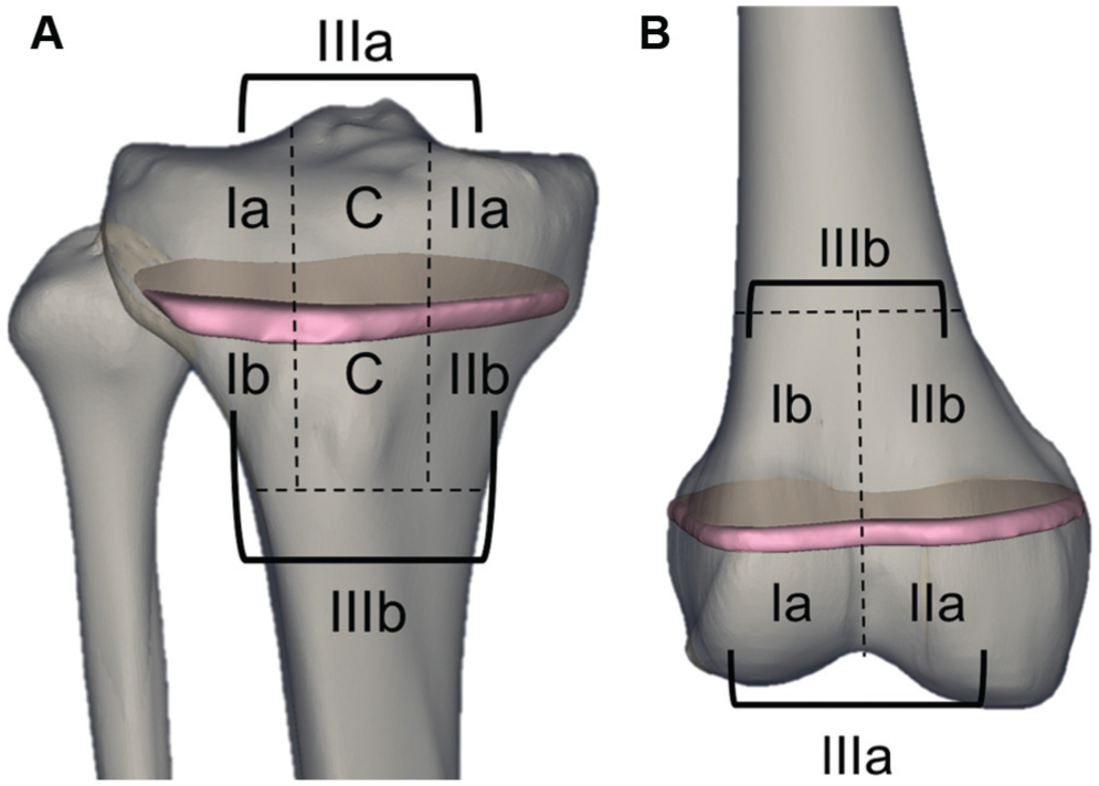
Proposed classification of bone bruise distribution for the pediatric population: (A) tibia and (B) femur. I, lateral; II, medial; III, medial and lateral. a, does not cross physis; b, crosses physis. C, present in the central portion (tibial classification only).
Descriptive statistics—including means, SDs, frequencies, and ratios—were calculated for all variables. The Student t test was used to analyze continuous variables. Differences in bone bruise pattern distribution on MRI were compared with chi-square tests between the SI and SM groups. Comparisons in bone bruise locations between males and females and meniscal injuries between groups were also made via chi-square tests. The alpha level for statistical significance was set at P < .05.
Results
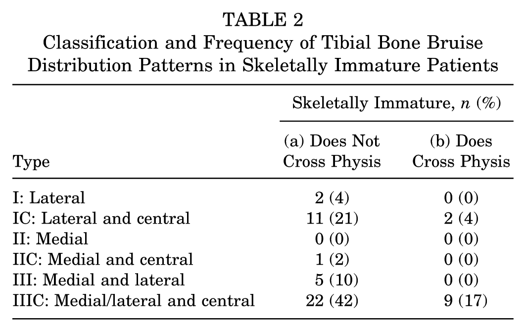
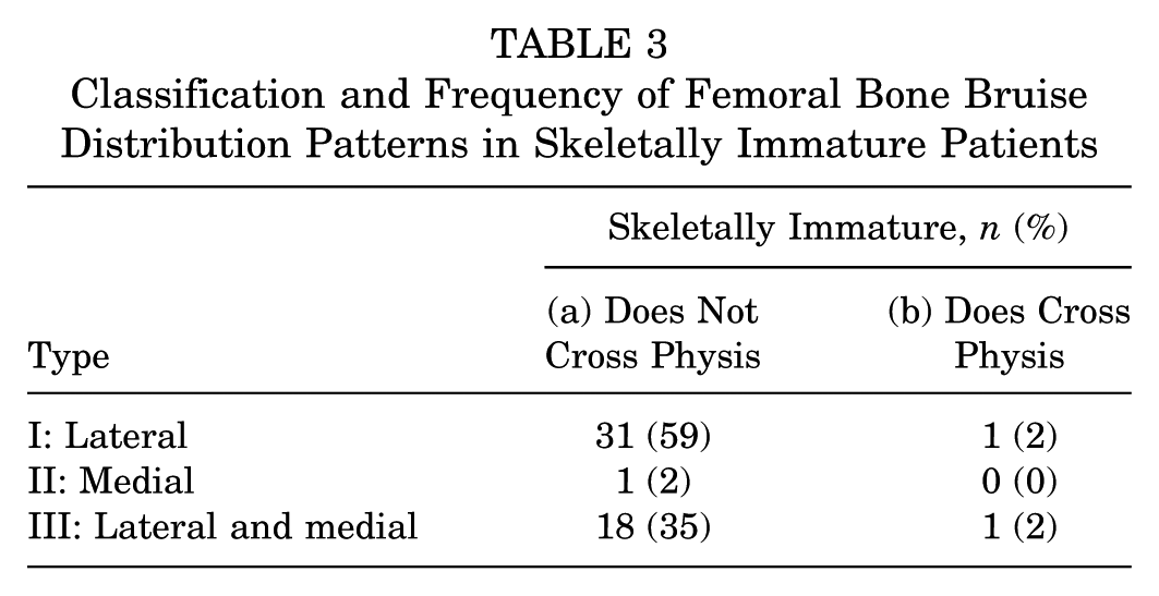
Table 1 displays the characteristics of the included patients. The SI group had significantly fewer bone bruises cross the physis and extend into the metaphysis than did the SM group for both the tibia (25% vs 85%, respectively; P < .0001) and the femur (4% vs 42%; P < .0001) (Figure 2). Tables 2 and 3 present the distribution of tibial and femoral bone bruises in our proposed classification system for the pediatric population. The most common patterns observed among the SI patients were type IIICa for the tibia (medial/lateral and central, not extending into the metaphysis: 42%) and type Ia for the femur (lateral, not extending into the metaphysis: 59%).
Patient Data a
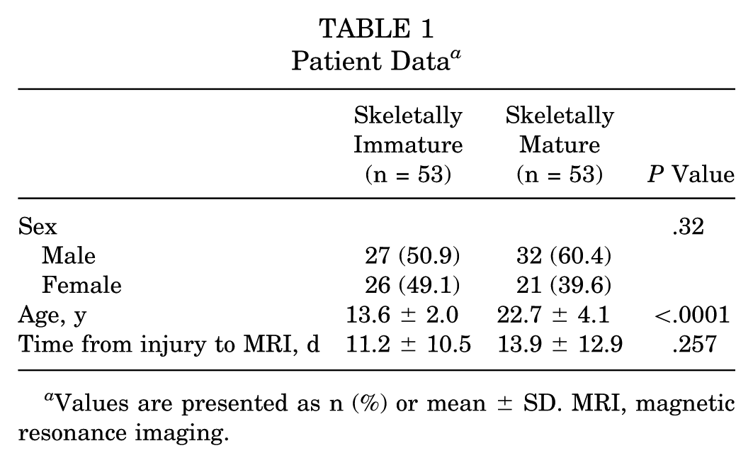
Values are presented as n (%) or mean ± SD. MRI, magnetic resonance imaging.
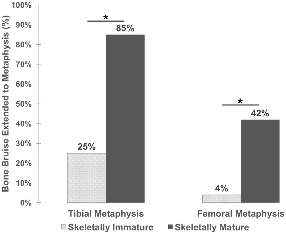
Comparison of bone bruise percentages extending to the metaphysis in the tibia and femur for the skeletally immature and mature groups. *P < .05.
Classification and Frequency of Tibial Bone Bruise Distribution Patterns in Skeletally Immature Patients
Classification and Frequency of Femoral Bone Bruise Distribution Patterns in Skeletally Immature Patients
Meniscal tears were present in 47.2% of the SI patients and 54.7% of the SM patients, with no significant difference between groups (P = .44). There were 10 medial and 19 lateral meniscal tears in the SI group, as opposed to 17 medial and 24 lateral meniscal tears in the SM group. In both groups, the most frequent location of meniscal injury was the posterior horn of the lateral meniscus, with no significant difference between groups (84.2% in the SI group and 91.2% in the SM group; P = .45). Regarding differences by sex in bone bruise location, no sex differences were observed in the SI and SM groups (Appendix Tables A1 and A2, available in the online version of this article).
Discussion
The main findings of this study were that the bone bruise pattern of SI patients with an acute ACL rupture was significantly less likely to cross the tibial and femoral physes when compared with that of the SM patients. Since the physis is a unique structure with different zones, it has biomechanical properties that are unique versus the rest of the bone. The decreased frequency of bone bruises found in the metaphysis of the SI group, in comparison with the SM group (Figure 2), may be explained by a shock-absorbing function of the physis. As such, the energy from the injury would be absorbed in the physis, minimizing the energy that reaches the metaphysis and thus preventing metaphyseal microfractures (Figures 3 and 4). The unique properties of the physis may have prevented the dissipation of the traumatic energy across the physis; thus, energy may have been dissipated toward other areas within the epiphysis. In the SM group, the lack of an energy-absorbing function of the physis may be responsible for the increased incidence of bone bruises that extend from the epiphysis to the metaphysis of both the femur and the tibia.
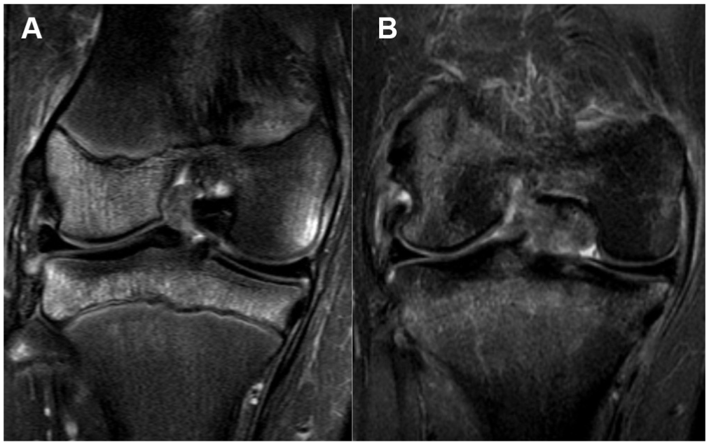
Different bone bruise distribution patterns between the skeletally immature and mature groups in magnetic resonance imaging of the coronal plane. (A) Bone bruise in a skeletally immature patient distributed along the tibia epiphysis but not crossing the physis. (B) Tibial bone bruise in a skeletally mature patient distributed toward the metaphysis.
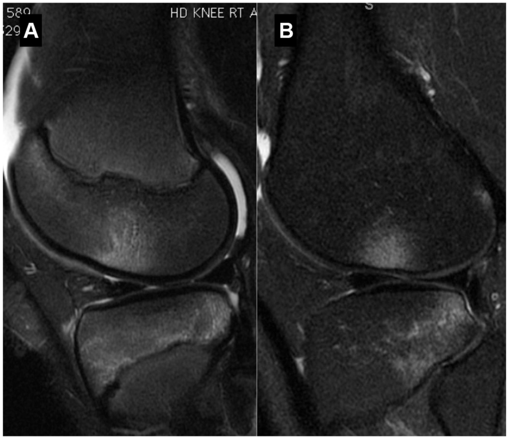
Different bone bruise distribution patterns between the skeletally immature and mature groups in magnetic resonance imaging of the sagittal plane. (A) Bone bruise in a skeletally immature patient distributed along the tibia epiphysis but not crossing the physis. (B) Tibial bone bruise in a skeletally mature patient distributed toward the metaphysis.
Bone bruises have been associated with concomitant injuries and poor clinical outcomes. A recent study 21 found that, after an acute noncontact ACL injury, bone bruises in the lateral femoral condyle and lateral tibial plateau were associated with a high-grade pivot shift and concomitant lateral meniscal lesions. Furthermore, 56% of patients with a bone bruise had concomitant lateral meniscal tears 23 and the presence of a large femoral bone bruise related to medial and lateral meniscal tears. 10 Regarding clinical outcomes, a prior study reported 11 that patients with a bone bruise after an ACL injury had larger effusions, which required a longer length of time to dissipate; they needed more time to achieve not just nonantalgic gait without external aids but also symmetric and equal range of motion; and they had worse visual analog scale pain scores. Meanwhile, Vincken et al 26 demonstrated that whereas patients with a bone bruise initially had poorer function and decreased activity as compared with patients without bone bruises, after 6 months there were no significant differences.
Currently, there is a lack of literature that clearly defines the anatomic distribution pattern of a bone bruise in the pediatric ACL reconstruction population. The goal of our proposed classification is to aid in the standardization of reporting bone bruises in the tibia and the femur in the pediatric population for clinical practice and across the literature. Future studies should evaluate whether bone bruises extending to the central area of the tibia (subtype C) and the ones crossing the physis (type b) have deleterious effects on patient outcomes, which might demonstrate prognosis relevance of the classification.
The results in this study are supported by prior literature. Viskontas et al 27 found, in a population of a wide age range (13-61 years old), that tibial bone bruising was mainly located in the lateral tibial plateau. The higher frequency of bone bruises in the lateral tibial plateau has also been reported in other studies evaluating SM patients.17,23,25 Kaplan et al, 12 also with a wide-ranging population age (15-70 years; mean, 32 years), observed that bone bruises more frequently reached the metaphysis in the tibia than in the femur—the same distribution observed in this study for both the SI and the SM groups. This phenomenon was attributed to the less dense bone in the tibia versus the femur, making the tibia more susceptible to trabecular microfractures. A study evaluating bone bruises in SI patients with tibial eminence fractures indicated that 90% of its cohort had bone contusions on MRI. 20 Its results were also in agreement with the current study, as bone bruises were more commonly seen in the lateral femoral condyle (80%) and the lateral tibial plateau (75%) as compared with the medial femoral condyles (60%) and the medial tibial plateau (30%).
In the current study, 47.2% of the SI patients and 54.7% of the SM patients had meniscal tears. The observed prevalence is higher than the 40% of the patients with meniscal tears reported by Shea et al 20 and lower than the 56% of patients with bone bruise and concomitant lateral meniscal tears reported by Spindler et al. 23 Additionally, lateral meniscal tears were more frequently found than medial meniscal tears in both groups, while Shea et al 20 reported an equal distribution between medial and lateral injuries. Wittstein et al 28 reported rates of 46.6% for medial meniscal tears and 39.7% for lateral tears in their study, which evaluated bone contusions in patients with noncontact ACL ruptures.
In this study, no sex differences were observed in the SI and SM groups. Therefore, with the results from our study, we cannot state that there is a difference by sex on bone bruise distribution patterns in SI and SM patients. These findings are similar to those of a previous study that evaluated patients with noncontact ACL ruptures and compared the location of tibial and femoral contusions on MRI between sexes. 28 Wittstein and colleagues 28 also did not observe significant differences between males and females in bone bruise distribution patterns.
This study investigated only patients who had ACL reconstruction based on retrospective review of electronic medical records. It is possible that patients who did not go on to have ACL surgery may have had less clinical instability and different patterns of bone contusion.
Conclusion
The data from this study shows that patients with an open physis at the occurrence of an acute ACL rupture have unique bone bruise patterns as compared with those with a closed physis. In SI patients, the bone bruise pattern is significantly less frequently observed in the tibial and femoral metaphysis.
Supplemental Material
DS_10.1177_0363546518777247 – Supplemental material for Bone Bruise Patterns in Skeletally Immature Patients With Anterior Cruciate Ligament Injury: Shock-Absorbing Function of the Physis
Supplemental material, DS_10.1177_0363546518777247 for Bone Bruise Patterns in Skeletally Immature Patients With Anterior Cruciate Ligament Injury: Shock-Absorbing Function of the Physis by João V. Novaretti, Jason J. Shin, Marcio Albers, Monique C. Chambers, Moises Cohen, Volker Musahl and Freddie H. Fu in The American Journal of Sports Medicine
Footnotes
One or more of the authors has declared the following potential conflict of interest or source of funding: J.V.N. has been awarded a scholarship from the CAPES Foundation—Programa de Doutorado Sanduíche no Exterior (88881.135480/2016-01; Brazil). V.M. is a consultant for Smith & Nephew and has received payments from Arthrex for education and from Mid-Atlantic Surgical Systems in the form of a grant. J.J.S. has received payments from Smith & Nephew for education.
References
Supplementary Material
Please find the following supplemental material available below.
For Open Access articles published under a Creative Commons License, all supplemental material carries the same license as the article it is associated with.
For non-Open Access articles published, all supplemental material carries a non-exclusive license, and permission requests for re-use of supplemental material or any part of supplemental material shall be sent directly to the copyright owner as specified in the copyright notice associated with the article.