






Abstract
Background:
Engraftment and longevity of transplanted cells are crucial for stem cell–based cartilage treatment.
Purpose:
To determine whether cultured spherical cell masses of human bone marrow–derived mesenchymal stem cells (hBM-MSCs) could improve engraftment at defect sites and to examine their corresponding effects on osteochondral regeneration.
Study Design:
Controlled laboratory study.
Methods:
A cylindrical osteochondral defect (5 mm wide × 5 mm deep) was created in trochlear grooves of rabbit knees. The single-cell type of hBM-MSCs with fibrin glue, the spherical type of hBM-MSCs with fibrin glue, and cell-free fibrin glue (control) were each implanted into osteochondral defect sites. A total of 18 rabbit knees were randomly assigned to 1 of the 3 groups (3 rabbits per group). Animals were sacrificed at 6 and 12 weeks after transplantation. Repaired tissues were evaluated via gross examination, histologic examination, and immunofluorescence analysis.
Results:
Transplantation with spherical hBM-MSCs exhibited superior overall osteochondral restoration when compared with the single-type group, as evidenced by well-ordered mature collagen fibrils produced during subchondral bone formation in the zonation phenomenon. Immunofluorescence analysis of osteochondral defect areas with human-specific antigen revealed a larger number of mesenchymal stem cells in the spherical-type group than the single cell–type group.
Conclusion:
Transplantation of spherical hBM-MSCs was better than single cells from monolayer culture in improving osteochondral regeneration.
Clinical Relevance:
The findings demonstrate a simple strategy for enhancing the potency of stem cells required for restoration of osteochondral defects. Furthermore, this strategy may be implemented with other types of stem/progenitor cell–based therapies.
Tissue repair of an osteochondral defect or cartilage lesion is difficult owing to limited movement of progenitor cells from bone marrow and blood vessels into the defect site. In addition, continuous flow of joint fluid as a result of physical movement can lead to constant washing out of cartilage matrix created by adjacent and ectal stem cells around the defect region following bone marrow stimulation procedures.9,27,32 Therefore, surgical techniques with mesenchymal stem cells (MSCs) with multidifferentiation potential have been developed for cartilage treatment. 17
However, engraftment and longevity of cells after transplantation are limited. In particular, the avascular environment of joint space usually leads to cell death within several days after transplantation. Therefore, tissue engineering techniques with MSCs have been explored for regeneration of osteochondral defect tissues.4,7,12,20,24 Efforts to enhance early engraftments of implanted cells by preventing cell death via maintenance of cell-to-cell interactions are especially important for treating osteochondral defects. However, maintaining the purity and multipotency of stem cells with currently available 2-dimensional (2D) culture systems remains a challenging task. The structure of cell membrane is easily influenced by interactions with surrounding tissues and cells.10,25,27 Stem cells with successful anchorage might better sustain early regeneration via paracrine and autocrine signaling. 28
MSCs can be cultured and isolated via adherent culture based on their ability to survive through adhesion to a flat flask. However, culturing reduces the differentiation and reproductive potential of MSCs and makes them vulnerable to cell damage caused by interactions between implanted stem cells and the surrounding extracellular matrix. 2 Lee et al 15 employed a 3-dimensional (3D) culture system instead of the conventional 2D system to support the spherical cell shape, which also induces and maintains the generation of spherical cell masses.
The 3D suspension culture method is currently being developed to overcome the limitations of 2D culture. It also increases space efficiency and maintains desired stem cell properties. Generating embryonic bodies by inducing aggregation of embryonic stem cells is one of the most commonly applied culture methods for producing MSCs with spherical shapes. 22 Cell aggregation during 3D culture facilitates active and efficient delivery of signals between cells. Thus, the 3D culture method can provide an in vitro environment that is more similar to in vivo conditions of the 2D culture method, which in turn enhances the therapeutic potential of cells.1,15 However, lower survivorship of stem cells after implantation owing to harsh conditions of diseased target tissues, such as hypoxia, poor nutrient supply, inflammation, and immunologic attacks from host cells, is a principal limitation in stem cell therapy. Therefore, the objective of this study was to determine whether spherical cell masses cultured with a high-activation culture system were effective for osteochondral regeneration by inducing early engraftment and preventing early cell death. Specifically, we determined whether cultured spherical-shaped masses of human bone marrow–derived MSCs (hBM-MSCs) in a monolayer culture state could improve engraftment rates of stem cells through improved longevity of MSCs, and we examined their corresponding effects on osteochondral regeneration. We hypothesized that transplantation of hBM-MSCs as aggregated spheres could enhance the efficiency of engraftment and result in improved regeneration at osteochondral defect sites.
Methods
Preparation of Single-Cell and Aggregated Spherical hBM-MSCs
hBM-MSCs
15
were purchased from Lonza. Cells were cultured in MSC basal medium (Lonza) supplemented with MSC growth medium, a hMSC SingleQuots kit (Lonza), 5% fetal bovine serum, 1%
For sphere formation after trypsinization (0.025% trypsin; HyClone) of hBM-MSCs in a monolayer culture state, cells were cultured in suspension with a bacterial dish containing sphere medium—namely, bovine serum-free Dulbecco’s modified Eagle’s medium/nutrient mixture F-12 (Invitrogen) supplemented with 20% serum replacement (Gibco BRL) at 37°C in a humidified cell incubator with 5% CO2. After 24 hours, spheres sized 40 to 100 μm were sorted out with 100- and 40-μm mesh strainers. These sorted spheres were then transplanted into the defect site with a 31-gauge syringe (nominal inner diameter: 133 μm) (Figure 1). To adjust the cell number of transplanted spheres, half were dissociated with trypsin and counted, while the other half were used for in vivo experiment.
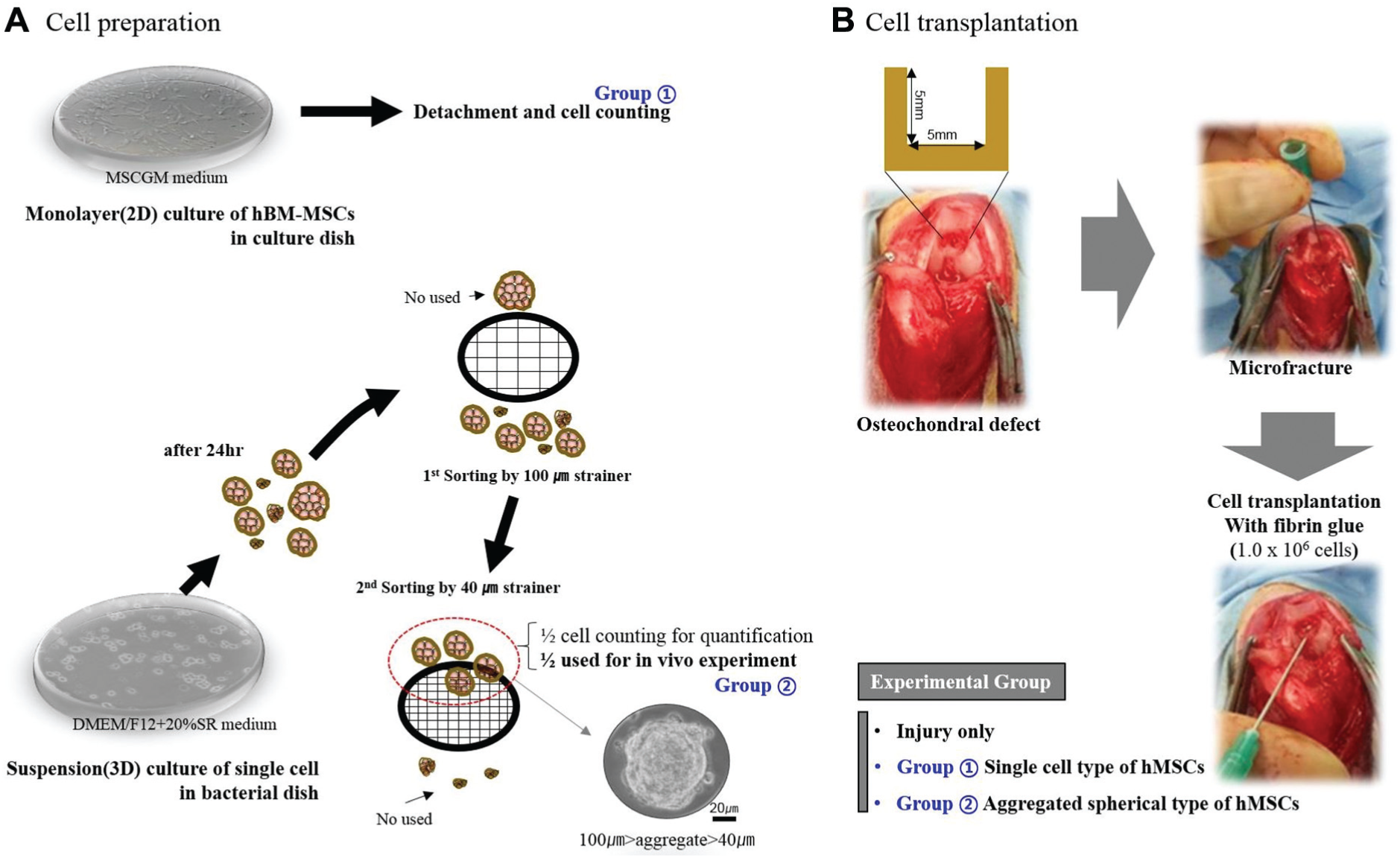
Experimental scheme and protocol for cultivation of single-cell and aggregated spherical human bone marrow–derived mesenchymal stem cells (hBM-MSCs). 2D, 2-dimensional; 3D, 3-dimensional.
Cells were periodically observed with phase-contrast microscopy to identify any morphologic changes associated with cell growth, multiplication, or cell structure. hBM-MSCs in monolayer and aggregated sphere suspensions showed significant differences in cell morphology (Figure 2).
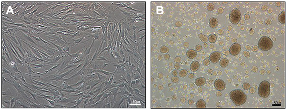
Differences in cell morphology between common and aggregated spherical human bone marrow–derived mesenchymal stem cells: (A) monolayer cells at 4 days after seeding; (B) aggregated cells at 24 hours after anchorage deprivation.
Single-cell hBM-MSCs (1.0 × 10⁶) mixed with fibrin glue, aggregated spherical hBM-MSCs (1.0 × 10⁶) mixed with fibrin glue, and cell-free fibrin glue only were implanted into osteochondral defect sites. Rabbit knees were randomly assigned to 1 of the 3 groups (6 animals per group): fibrin glue only (control), fibrin glue with single-cell hBM-MSCs (single MSCs), or fibrin glue with aggregated spherical hBM-MSCs (aggregated MSCs) (Table 1).
Treatment Groups for Stem Cell Implantation in Rabbit Osteochondral Defect Model
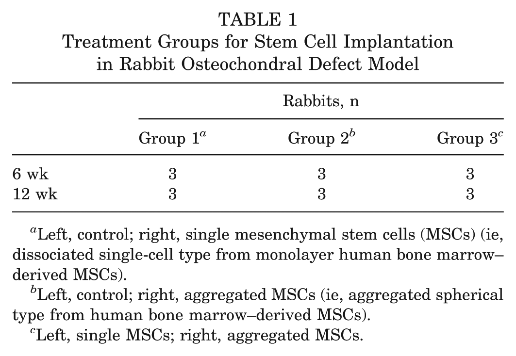
Left, control; right, single mesenchymal stem cells (MSCs) (ie, dissociated single-cell type from monolayer human bone marrow–derived MSCs).
Left, control; right, aggregated MSCs (ie, aggregated spherical type from human bone marrow–derived MSCs).
Left, single MSCs; right, aggregated MSCs.
Animals and In Vivo Transplantation
Animal selection and care, surgical protocol, 11 and preparation procedures were approved by the Institutional Animal Care and Use Committee of Samsung Medical Center (approval 20140805001). Eighteen skeletally mature male New Zealand White rabbits (5-6 months old) weighing ~3 kg (range, 2.9-3.4 kg) were used in this study. After a 1-week acclimation period, rabbits were anaesthetized via enflurane (Geroran) inhalation combined with an intramuscular injection of xylazine (Rompun) at 5 mg/kg and ketamine (Ketalar) at 35 mg/kg. For each rabbit, both knee joint areas were shaved, cleaned with 10% betadine solution, and sterilely draped. Surgical procedures were performed by an orthopaedic surgeon (B.H.L.). Both knee joints were opened with a medial parapatellar approach. The patella was everted laterally, and intra-articular structures were thoroughly inspected for any abnormal conditions, such as infection and deformity. After confirmation of normal intra-articular structure, the knee joint was fully flexed, and a cylindrical osteochondral defect (5 mm in diameter, 5 mm in depth) was created in the trochlear groove with a customized surgical drill bit with scale marks.
The surgical field was continuously flushed with sterile saline solution to minimize heat generated by the drilling process. Thereafter, 5 microfracture holes (1 mm in diameter, 3 mm in depth, 2 mm apart) were then created with a syringe needle. Visible bleeding was observed in all rabbits. Sealed samples of 3 groups were transplanted into the defect site of both knees with the 3 groups of 3 pairs. To prevent leakage of transplants, the patella retinaculum and overlying skin were carefully closed.
Intramuscular antibiotics (cefazolin, 11 mg/kg) were administered daily for 7 days. Rabbits were provided tap water and food. They were housed in separate cages and allowed to move freely. Rabbits were sacrificed by injection of excess ketamine hydrochloride (35 mg/kg) at 6 and 12 weeks postoperatively (9 rabbits for each time point).
Gross and Histologic Evaluation
Arthrotomy was performed postmortem in the same manner as during transplantation for gross and histologic examinations of the intra-articular structure. Samples were observed for evidence of abnormal responses suggesting rejection or infection, such as severe inflammation and extensive fibrosis. The degree of osteochondral regeneration was evaluated via gross examination. Coloration, luster, irregularity, presence of any depression or bulging of repaired tissue in the defect area, and the state of the border surrounding the normal cartilage tissue were carefully examined. 23 Representative sections of each sample were independently scored by 2 blinded observers according to the modified International Cartilage Repair Society (ICRS) gross grading scale (Wayne scoring system) 29 (see Appendix Table A1, available in the online version of this article).
For histologic analysis, full-thickness samples (cartilage and bone) were obtained from both knee joints of each rabbit at 6 and 12 weeks posttransplantation. Specimens were fixed in 10% formaldehyde, decalcified in 10% nitric acid for 3 days, dehydrated in a graded ethanol series, and embedded in paraffin wax. Samples were cut in the sagittal plane into at least thirty 5-µm-thick sections through the center of the defect. Paraffin sections were subjected to immunofluorescence analysis and hematoxylin and eosin staining. For detection of osteochondral repair, sections were stained with 0.1% safranin O solution.
Sections were analyzed semiquantitatively with the ICRS grade scoring system. 19 The surface, matrix, cell distribution, cell population viability, subchondral bone, and cartilage mineralization were evaluated 19 (see Appendix Table A2, available online). Two independent researchers blinded to treatment assignments completed the scoring and averaged their results. The scale consisted of 6 categories. Total score ranged from 0 to 18.
Cell Tracking With Anti-human Nuclei Antibody
To successfully evaluate engrafted cells after transplantation as aggregated spherical or single-cell type, we used anti-human nuclei antibody (MAB1281; Merck Millipore) for staining for in vivo tracking. At 6 and 12 weeks after transplantation, we performed phase-contrast microscopy of the osteochondral defect area on the distal femoral condyle of knee joints to determine the number of stained hBM-MSCs.
Rabbit joint tissue was fixed in 10% formalin solution, dehydrated through a graded ethanol series, cleared in xylene, and processed for embedding in paraffin according to routine protocols. Paraffin sections were then incubated with primary antibodies against cell nuclei (1:1000 dilution) at room temperature for 1 hour. For detection, goat anti-guinea pig Immunoglobulin G (IgG) (H+L) highly cross-adsorbed secondary antibody, Alexa Fluor 594 conjugate (A-11076; Thermo Fisher Scientific), goat anti-mouse IgG (H++L) cross-adsorbed secondary antibody, and DyLight 488 conjugate (35505; Thermo Fisher Scientific) were used at 1:200 dilution according to the manufacturer’s instructions. Slides were counterstained with liquid VECTASHIELD antifade mounting medium containing DAPI (H-1200; VECTOR) and then visualized with a ZEISS LSM 880 confocal laser scanning microscope (Carl Zeiss Microscopy). Images were captured via bright field microscopy at 20× magnification. Five images from randomly selected visual fields were captured from each slide for quantification. Human nuclei positive cells were counted with image processing and analysis software Image J (v 1.45; http://rsb.info.nih.gov/ij/) by 3 observers blinded to these samples.
Statistical Analysis
Differences in sums of ICRS gross and histologic grading scores were compared between samples at 6 and 12 weeks after transplantation with 2-tailed Mann-Whitney U test for both sides among the 3 groups (control, single cells, and spheres). A nonparametric Kruskal-Wallis test with Bonferroni correction was performed to compare the 3 groups. P < .05 was considered statistically significant. All statistical analyses was conducted with SAS (v 9.4; SAS Institute).
Results
Macroscopic Findings
There were no abnormal findings suggesting rejection or infection, such as severe inflammation or extensive fibrosis, in all 18 rabbits at 6 and 12 weeks after transplantation. Regeneration of osteochondral defects in the articular cartilage was evaluated with the modified ICRS gross grading scale (Wayne scoring system) at 6 and 12 weeks.
Osteochondral defect sites in control knees were filled with granulated tissue, whereas defects in MSC-transplanted knees had firm regenerated tissue with pearly white appearance. This newly formed tissue resembled articular cartilage. It adhered to the adjacent cartilage and restored the contour of femoral condyles (i.e., the smooth articular surface without depression required for articulation with the tibia). The best gross appearance was observed in rabbits transplanted with aggregated spherical hBM-MSCs at 12 weeks after treatment. When compared with control knees, these MSC-transplanted knees exhibited superior osteochondral restoration. They showed relatively smooth surface and coverage, similar to surrounding normal cartilage (Figure 3). Total score of the modified ICRS gross grading scale (Wayne scoring system) for the spherical hBM-MSC group (mean, 10.2 of 16 points) showed better improvement than that of the other 2 groups (8.8 in the single-cell group, 6.8 in the control group) at 6 weeks after treatment. Total score at 12 weeks also showed better improvement in the spherical hBM-MSC group (mean, 11.3) than in the other 2 groups (10.5 in the single-cell group, 9.3 in the control group) (Figure 3C).
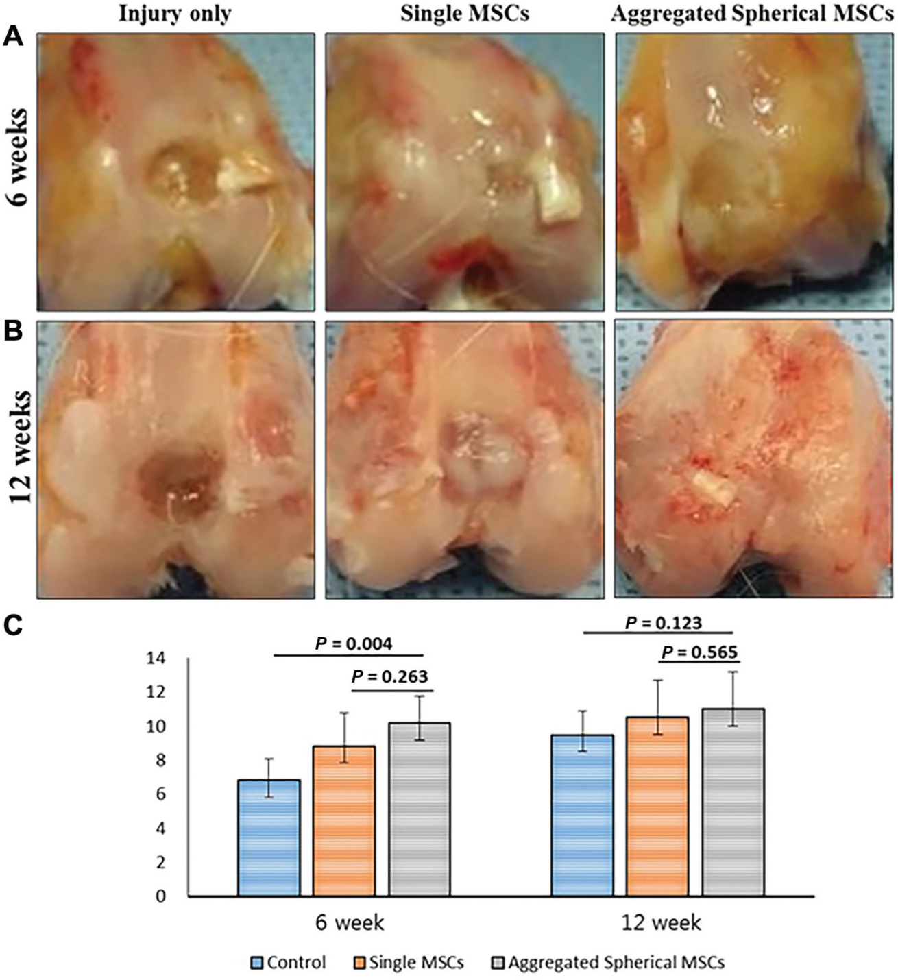
Representative gross appearance of the distal femoral cartilage in the control group, single mesenchymal stem cell group, and aggregated spherical mesenchymal stem cell group at (A) 6 weeks and (B) 12 weeks after treatment. (C) Macroscopic cartilage repair assessment for the 3 groups. Error bars represent 95% CI. y-axis: sum of modified ICRS gross grading scale (Wayne scoring system).
In the aggregated spherical hBM-MSC group, transplant showed significantly improved coverage and surface at 6 and 12 weeks postoperatively (coverage: from 2.8 ± 0.3 at 6 weeks to 3.3 ± 0.8 at 12 weeks, P = .05; surface: from 2.4 ± 1.2 at 6 weeks to 3.7 ± 1.2 at 12 weeks, P = .034) (Table 2).

Differences among the 3 groups were compared with the Kruskal-Wallis test with Bonferroni corrections. Differences with P < .05 are displayed in bold. ICC, intraclass correlation coefficient; ICRS, International Cartilage Repair Society; MSC, mesenchymal stem cell.
Interrater reliability for gross measures.
Differences were compared between 6 and 12 weeks with the Student t test and Mann-Whitney U test.
Osteochondral defects in knees implanted with single hBM-MSCs were occupied with partially restored, new, and white tissue, whereas defect sites transplanted with aggregated spherical hBM-MSCs were completely covered with neotissue that was undistinguishable from the surrounding host tissue. Aggregated spherical hBM-MSCs achieved better restoration of osteochondral defects, and the regenerating cartilage also showed good integration with adjacent host tissues (Figure 3).
Microscopic Findings
Histologic analysis of osteochondral regeneration in the 3 groups was carried out via hematoxylin and eosin staining and safranin O staining at 6 and 12 weeks postimplantation. Osteochondral defects in the control group were not reconstructed, showing eburnated bone at the joint surface or replaced with yellowish inflammatory tissues. In addition, a gap was obvious in the defect. Only a small portion of the regenerative tissue present was primarily fibrocartilage (Figure 4).
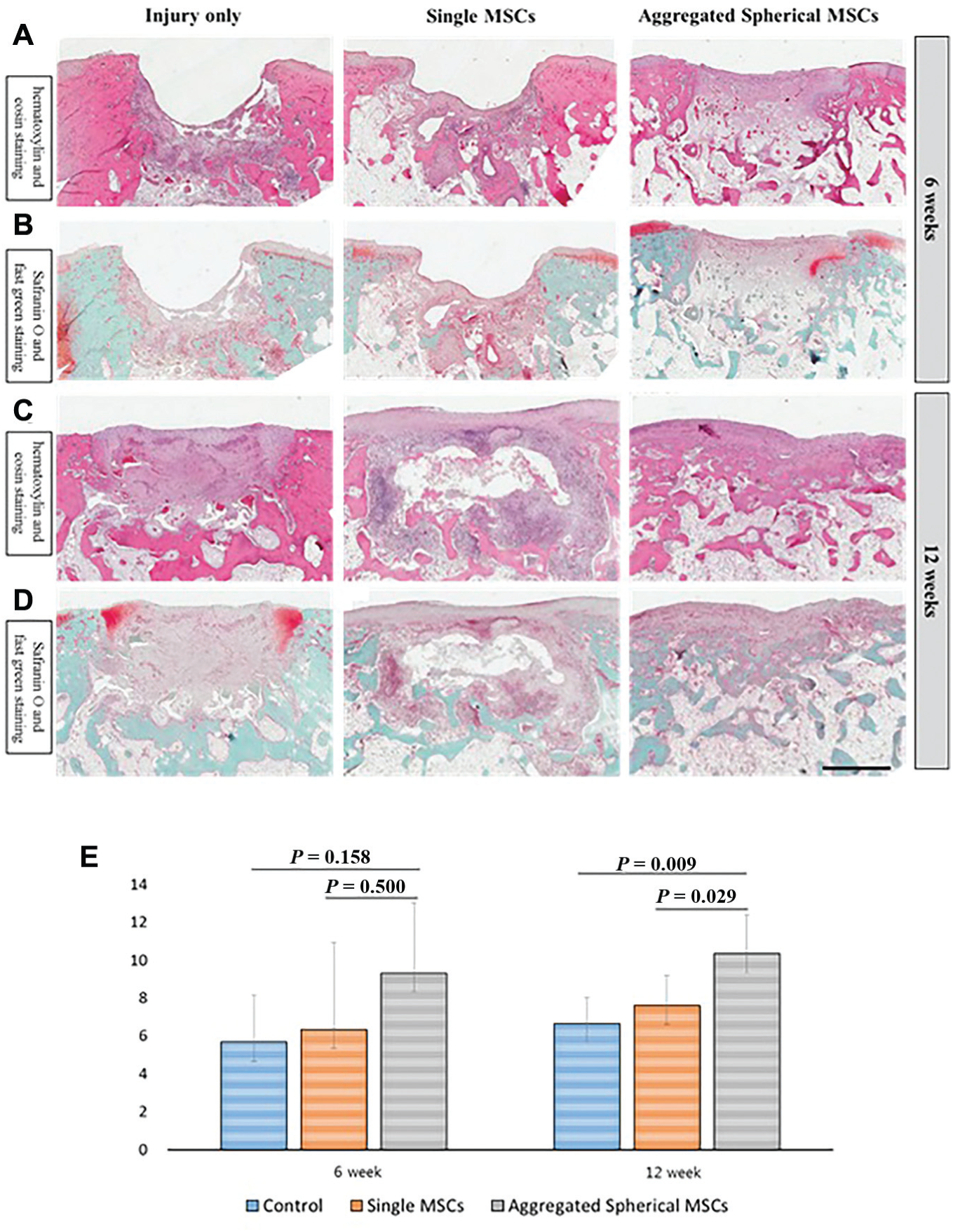
Microscopic findings of regenerating osteochondral defects on articular cartilage in the control group, single mesenchymal stem cell (MSC) group, and aggregated spherical MSC group at 6 and 12 weeks after transplantation (9 knees/group). (A, C) Hematoxylin and eosin staining, 12.5× magnification. (B, D) Safranin O and fast green staining, 12.5× magnification. Scale bars: 2 mm. (E) Semiquantitative analysis of tissue repair at articular cartilage defect sites in rabbit knees at 6 and 12 weeks. Sections were histologically evaluated per a modified O’Driscoll score. Error bars represent 95% CIs. y-axis: sum of International Cartilage Repair Society Visual Histologic Assessment Scale score.
Defect sites in the single MSC group showed formation of a superior osteochondral junction, whereas control knees showed minimal reconstruction at the defect site (predominantly occupied by nonspecific inflammatory cells). However, knees transplanted with aggregated spherical MSCs displayed better bony structural restoration potential at 6 weeks than the other 2 groups (Figure 4, A and B).
At 12 weeks, rabbits transplanted with spherical aggregated hBM-MSCs showed reformation of the subchondral bone. Formation of a good osteochondral junction was observed throughout the defects. Four of the 12 specimens in the spherical aggregated group exhibited normal cartilage mineralization. All 4 specimens showed hyaline cartilage repair with excellent structural integrity of the matrix (Figure 4C). In particular, osteochondral defects after implantation of spherical aggregated spherical hBM-MSCs were restored by well-ordered mature collagen fibrils during subchondral bone formation in the zonation phenomenon. Subchondral bone formation was observed at stem cell implant sites. However, deeply recessed surface and limited bone formation were observed in controls. Moreover, granulated tissue caused by inflammatory reactions and predominantly nonchondrocytic tissue occupied defect sites (Figure 4D). Safranin O staining revealed chondral differentiation in repaired tissues. Denser and more pronounced staining without a remarkable gap was observed in the spherical aggregated hBM-MSC group, whereas weak staining and smaller regions of repaired tissue were observed in the single MSC group (Figure 4D).
Semiquantitative analysis of sections with the ICRS Visual Histologic Assessment Scale revealed histologically superior repaired tissue in transplanted knees versus control knees (Figure 4E). Stem cell implantation stimulated tissue regeneration in osteochondral defects according to macroscopic analysis and histologic scoring of the regenerated tissues. Total score of the ICRS Visual Histologic Assessment Scale for the spherical hBM-MSC group (mean, 9.3 of 18 points) showed better improvement than that of the other 2 groups (6.3 for the single-cell group, 5.7 for the control group) at 6 weeks. Total score at 12 weeks also showed better improvement in the spherical hBM-MSC group (mean, 10.3) as opposed to the other 2 groups (7.6 in single-cell group, 6.7 in the control group) (Figure 4E).
The 3 treatment groups showed significant differences in the sum of ICRS histologic score at 12 weeks postimplantation (P = .009, P = .029) (Figure 4E). Further comparisons among the 3 groups revealed significant differences in matrix characteristics (P = .016 at 6 weeks), cell distribution (P = .009 at 6 weeks, P = .026 at 12 weeks), cell population viability (P = .015 at 6 weeks, P = .049 at 12 weeks), and subchondral bone formation (P = .019 at 6 weeks) (Table 3).
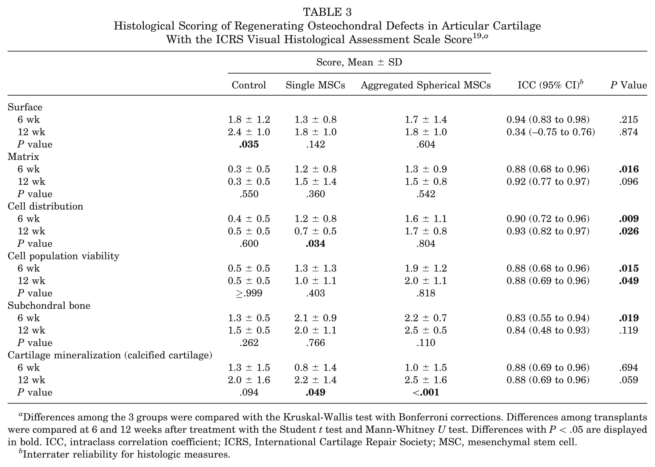
Differences among the 3 groups were compared with the Kruskal-Wallis test with Bonferroni corrections. Differences among transplants were compared at 6 and 12 weeks after treatment with the Student t test and Mann-Whitney U test. Differences with P < .05 are displayed in bold. ICC, intraclass correlation coefficient; ICRS, International Cartilage Repair Society; MSC, mesenchymal stem cell.
Interrater reliability for histologic measures.
Cell Tracking With Anti-human Nuclei Antibody
Tracking of grafted cells as either aggregated spherical MSCs or single MSCs was performed by immunofluorescence staining with an anti-human nuclei antibody after transplanting these cells in the articular cartilage defect region. Phase-contrast microscopy for the osteochondral defect area in the knees at 6 weeks showed a larger number of human nucleus–stained MSCs in the spherical aggregated hBM-MSC transplantation group than in the single hBM-MSC group (Figure 5).
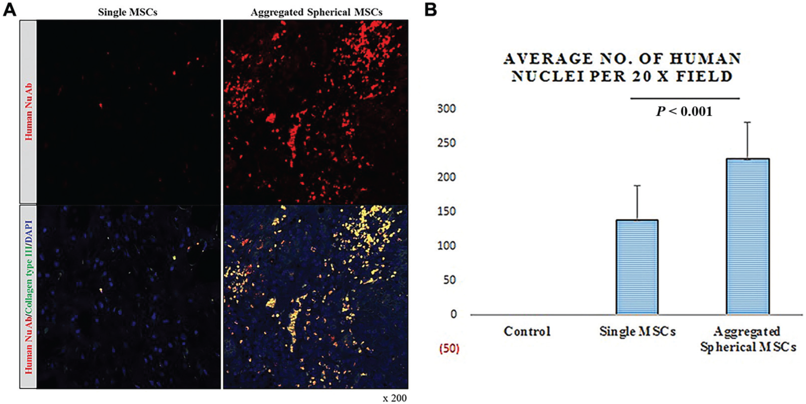
Osteochondral defects at 6 weeks after cell transplantation as observed by confocal laser scanning microscopy. (A) Cell tracking with anti-human nuclei antibody. (B) Human nucleus–positive cells were more frequently observed in the spherical aggregated mesenchymal stem cell group. Error bars represent 95% CIs.
Anti-human nuclei antibody signals decreased gradually at 6 and 12 weeks posttransplantation (Figure 6). No signals were detected in the negative control tissue.
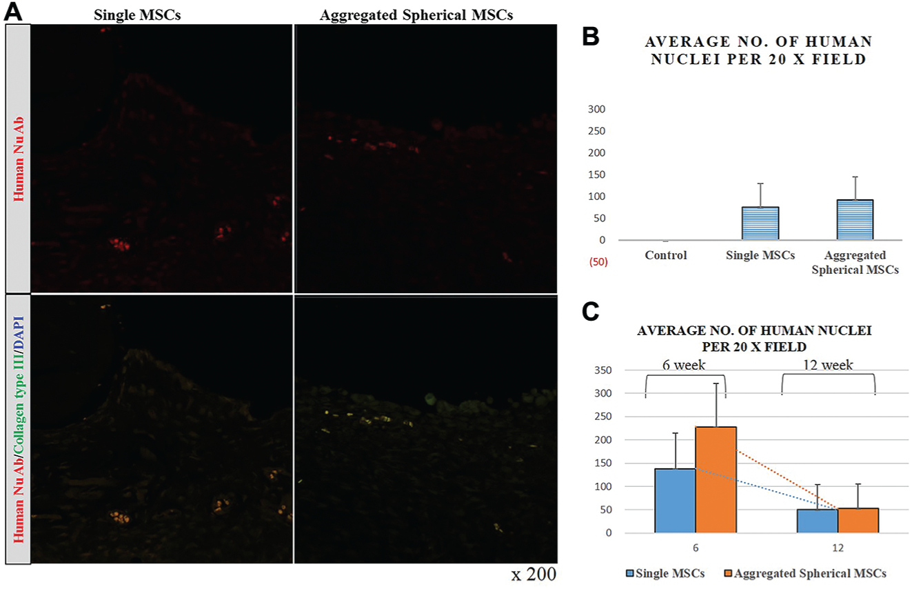
(A) Osteochondral defects at 12 weeks after cell transplantation were observed with a confocal laser scanning microscope. (B) Engraftment of human bone marrow–derived mesenchymal stem cells (MSCs) was improved after transplantation with spherical aggregated cells than with single cells. (C) Anti-human nuclei antibody signals gradually decreased at 6 and 12 weeks posttransplantation. Error bars represent 95% CIs.
Discussion
Stem cell transplantation has recently emerged as an attractive therapeutic approach for osteochondral defects or cartilage lesions. However, there are several challenges and considerations regarding the clinical application of stem cell treatment for articular cartilage lesions. It has been reported that most transplanted stem cells will die within 1 week after implantation owing to harsh conditions of diseased target tissues, such as hypoxia, poor nutrient supply, inflammation, and immunologic attack from host cells. 15 To improve the survival of grafted cells, some researchers explored the optimal tissue source of MSCs and optimal time point for stem cell delivery. 26
The low therapeutic efficacy of stem cells is a major obstacle that prevents full-scale clinical applications of stem cells.16,21 Several approaches have been introduced to improve the therapeutic effect of MSCs.16,24 The most important factor influencing adequate therapeutic efficacy of stem cell therapy is a stable supply of cells with optimal engraftment efficiency. In cell transplantation, an important limitation that reduces efficacy of treatment is poor engraftment rate after transplantation. Therefore, we developed a culture strategy to produce 3D spheres of hBM-MSCs that could preserve cell-cell interactions without needing additional cytokines, serum, or matrices. It was previously demonstrated that delivery of cells without disrupting the extracellular matrix can result in higher engraftment efficiency and therapeutic efficacy. 14
Self-assembled spherical hBM-MSCs used in the present study have several advantages. First, the cultivation of spherical cells was induced under anchorage-deprived conditions that resulted in the expansion of the extracellular matrix via cell-cell interactions until a sufficient physical size was reached (ie, 40-100 μm) as described previously. 15 Second, these self-assembled spherical MSCs do not require an exogenous matrix or cytokines. They are also useful for developing biomimetic 3D organoids that could be used as an in vitro model for studying differentiation, organogenesis, migration, and tumor biology. 15 Third, the currently proposed technique for self-assembled spherical hBM-MSCs is safer and more economic with higher efficacy than preexisting methods.
To demonstrate these benefits, spherical aggregated cell colonies were transplanted onto osteochondral defects to determine whether transplantation could enhance engraftment and consequently improve cartilage repair. We previously reported that primary sphere formation and subsequent generation of secondary spheres can enhance proliferative, paracrine, and immune modulatory activity within an in vitro experiment. 15 Proliferation and differentiation potentials of stem/progenitor cells in secondary spheres have been demonstrated for neural stem cells and cardiac cells.3,8,18
Paracrine secretions (eg, transforming growth factor ß secretion) and immune-suppressive properties are considered the most important benefits of MSCs in regenerative medicine. Secretion power is highly correlated with the number of engrafting cells. Therefore, counting the number of engrafting cells without early apoptosis prior to engraftment is important in determining the secretion power. To evaluate whether transplanted hBM-MSCs in the current study retained their stem cell potential after spherical formation, osteochondral defects of the knee joints were observed at 6 and 12 weeks after cell transplantation with phase-contrast microscopy. Results revealed enhanced engraftment after transplantation with spherical aggregated hBM-MSCs.
Transplantation with spherical aggregated hBM-MSCs resulted in more favorable cartilage repair based on gross and histologic examination when compared with single hBM-MSC transplantation or no treatment in a rabbit model. In particular, repaired tissues after transplantation were more similar to surrounding normal articular cartilages than repaired tissues in controls. Additionally, repaired tissues induced by transplanted aggregated spherical hBM-MSCs contained considerably higher amounts of type II collagen, and the deep portion of the repaired tissue was replaced by subchondral bone, as found in the zonation phenomenon.
Several studies reported that hBM-MSCs show multilineage differentiation potential and retain chondrogenic differentiation potential in vitro.13,30,31 As previously reported, these findings suggest that persistent paracrine action of engrafted hBM-MSCs that survive transplantation might have stimulated ordered regeneration of bone and cartilage as well as chondrogenic differentiation and cartilage-specific extracellular matrix synthesis.5,26
The present study has several limitations. First, this study might not be confirmative in that the stem cell potential was determined on the basis of the superior survival rate of engrafting cells or the enhanced efficacy of the stem cell itself. It is well known that experiments with primitive animals have limitations to detect the origin of cartilage matrix and products, such as type II collagen created by injected stem cells or innate adjacent stem cells because of their cross-reactivity of antibody. Consistent with our hypothesis, transplantation of hBM-MSCs as spherical masses was significantly more effective in regenerating osteochondral defects than transplantation of hBM-MSCs as a single-cell suspension. We inferred that the improved therapeutic efficacy was associated with a superior engraftment rate and higher paracrine activity. However, further studies are required to determine whether transplanted hBM-MSCs can directly restore cartilaginous tissue via chondrogenic differentiation or whether they support cartilage restoration by host cells. Nonetheless, current results highlight the potential therapeutic use of spherical hBM-MSCs as secondary cell engineering technology. Second, this study demonstrated that xenogeneic MSC-based treatment could be applied to repair osteochondral defects. However, xenografting in an animal model can create a delayed immune response, which may influence the validation of results. Osteochondral tissue is relatively avascular. Therefore, host immune responses might be reduced. In addition, >6 months of follow-up might be needed to rule out the delayed immune response. Immunofluorescence analysis showed markedly decreased cells in both groups at 12 weeks posttransplantation. Therefore, the follow-up period used in our study might have been short to observe this delayed immune response.
Conclusion
Transplantation of spherical hBM-MSCs was better than single cells from monolayer culture in improving osteochondral regeneration. These results demonstrate a simple strategy for enhancing the potency of stem cells, which is required for restoration of osteochondral defects. Furthermore, this strategy can be implemented with other types of stem/progenitor cell–based therapies.
Supplemental Material
DS_10.1177_0363546518780991 – Supplemental material for Therapeutic Efficacy of Spherical Aggregated Human Bone Marrow–Derived Mesenchymal Stem Cells Cultured for Osteochondral Defects of Rabbit Knee Joints
Supplemental material, DS_10.1177_0363546518780991 for Therapeutic Efficacy of Spherical Aggregated Human Bone Marrow–Derived Mesenchymal Stem Cells Cultured for Osteochondral Defects of Rabbit Knee Joints by Byung Hoon Lee, Jong Nam Park, Eun Ju Lee, Young Wan Moon and Joon Ho Wang in The American Journal of Sports Medicine
Footnotes
Acknowledgements
The authors are grateful to the registered veterinary technicians at the Laboratory Animal Research Center of the Samsung Biomedical Research Institute for their support during the animal procedures. The authors also thank Da-Hye Ko for histologic staining and ABION Inc for its expert technical assistance.
One or more of the authors has declared the following potential conflict of interest or source of funding: This research was supported by a grant of the Korea Health Technology R&D Project through the Korea Health Industry Development Institute, funded by the Ministry of Health and Welfare, Republic of Korea (HI15C2424), and supported by the Basic Science Research Program through the National Research Foundation of Korea, funded by the Ministry of Science and ICT (NRF-2015R1A2A1A15054779). The funding sources were not involved in the study design, collection, data analysis or interpretation, writing of the manuscript, or in the decision to submit the manuscript for publication.
References
Supplementary Material
Please find the following supplemental material available below.
For Open Access articles published under a Creative Commons License, all supplemental material carries the same license as the article it is associated with.
For non-Open Access articles published, all supplemental material carries a non-exclusive license, and permission requests for re-use of supplemental material or any part of supplemental material shall be sent directly to the copyright owner as specified in the copyright notice associated with the article.
