






Abstract
Background:
Although the cause of rotational instability after revision anterior cruciate ligament reconstruction (ACLR) is multifactorial, the rationale of adding an extra-articular procedure is based on its ability to restrict rotational instability.
Purpose:
To assess the effect of anterolateral ligament (ALL) reconstruction on revision ACLR.
Study Design:
Cohort study; Level of evidence, 3.
Methods:
A total of 87 patients who underwent revision ACLR between March 2011 and July 2014 with a follow-up of more than 3 years were included in this retrospective study. Patients were divided into the isolated revision ACLR group (group I, n = 45, from March 2011 to January 2013) or the revision ACLR in combination with ALL reconstruction group (group C, n = 42, from February 2013 to July 2014). Subjective knee assessments including the subjective International Knee Documentation Committee (IKDC) form, Lysholm score, Tegner activity scale, and Anterior Cruciate Ligament–Return to Sport after Injury (ACL-RSI) scale were used. Clinical and functional tests were performed before surgery and at ≥6 months after surgery. All tests were usually completed at 36 months of follow-up.
Results:
The mean follow-up duration for groups I and C were 41.5 ± 8.2 and 38.2 ± 6.9 months, respectively (P = .451). The subjective IKDC score, Tegner score, and ACL-RSI score were significantly better in group C compared with those in group I at the last follow-up (84.3 ± 18.5 vs 75.9 ± 19.2, 7.0 ± 0.8 vs 6.3 ± 0.7, and 69.5 ± 25.4 vs 51.9 ± 23.1, respectively), although they were not significantly different between the 2 groups at 12 months after surgery (79.2 ± 18.8 vs 76.7 ± 17.2, 6.7 ± 0.7 vs 6.5 ± 0.9, and 50.2 ± 24.6 vs 49.9 ± 25.1, respectively). There were no significant differences in KT-2000 arthrometer, isokinetic extensor strength, single-legged hop for distance, co-contraction test, or carioca test results between the 2 groups at the last follow-up (P = .304, .068, .125, .056, and .066, respectively). Preoperatively, 43 (95.6%) patients in group I and 40 (95.2%) patients in group C had a grade 2 or 3 pivot shift (P = .387). Postoperatively, 23 (53.5%) patients in group I and 38 (90.5%) patients in group C had a negative pivot shift (P < .001). Group C showed a higher rate of return to the same level of sports activity than group I (57.1% vs 25.6%, respectively; P = .008), although there was no significant difference in the rate of return to any sports activity at the last follow-up (88.4% in group I vs 88.1% in group C; P = .713).
Conclusion:
Revision ACLR in combination with ALL reconstruction significantly reduced rotational laxity and showed a higher rate of return to the same level of sports activity than revision ACLR alone, although there were no significant differences in anterior laxity or functional test results between the 2 groups.
Keywords
Except for new trauma, the most common cause for failure of primary anterior cruciate ligament (ACL) reconstruction (ACLR) is a nonanatomic tunnel position, which produces recurrent rotatory instability and graft reruptures.2,24 Untreated instability resulting from concomitant injuries to the posterolateral corner or anterolateral ligament (ALL) at the time of primary reconstruction is also a key factor that contributes to failure. 2 Although acceptable clinical results have been reported after revision ACLR, 24 numerous studies have reported that revision ACLR has less favorable results than primary ACLR.2,32,62 Hence, we need to carefully evaluate the reasons for the failure of primary reconstruction and try to minimize residual rotational instability related to functional outcomes.3,21
While the reasons for rotatory instability are multifactorial, the effect of the anterolateral complex including the ALL, with the capsule, lateral collateral ligament, and lateral meniscus as secondary restraints to anterior displacement and internal rotation of the tibia, has been receiving interest.22,44,46 Several biomechanical studies have demonstrated that ALL injuries can affect anterolateral rotatory instability related to the pivot-shift phenomenon.44,46,60 Recurrent and residual abnormal pivot shifts can result in permanent anterolateral complex injuries. 61 In this regard, extra-articular tenodesis or anatomic ALL reconstruction can be a meaningful attempt in case of revision ACLR to improve rotatory stability.15,46,48,55
The load-sharing effect of extra-articular procedures can reduce reinjuries on return to sports after revision ACLR, as insufficient biological graft healing is another cause of ACLR failure. 2 If the ACL graft is not able to share stress with another native structure appropriately, higher tension loads on it would lead to delayed healing and subsequent graft failure.10-12 Improved control of internal tibial rotation by additional extra-articular tenodesis or ALL reconstruction could protect the ACL graft from excessive stress during the postoperative period and return to sports.6,10,41,53,54,58
A multicenter study has shown that combined revision ACLR and extra-articular lateral tenodesis could reduce the failure rate and increase knee stability, although it could not significantly alter the International Knee Documentation Committee (IKDC) score. 57 However, the study group or surgical technique was not homogeneous in that study. Recently, another multicenter study has also demonstrated that additional ALL stabilization could improve rotational stability and the rerupture rate compared with previous isolated revision ACLR. 34 Unfortunately, there was no control group in that study. Studies that compare isolated revision ACLR versus revision ACLR in combination with an extra-articular procedure are lacking. Therefore, the purpose of the current study was to assess the effect of ALL reconstruction on revision ACLR. We compared subjective and objective outcomes of isolated revision ACLR versus revision ACLR in combination with ALL reconstruction in a single surgeon’s series for a large number of consecutive patients. We hypothesized that revision ACLR combined with ALL reconstruction would be more effective in reducing rotatory instability and result in a more satisfactory rate of return to sports than isolated revision ACLR.
Methods
Patients
A total of 108 patients who underwent revision ACLR between March 2011 and July 2014 by an experienced single surgeon (J.G.K.) were enrolled. The inclusion criteria for this study were (1) a rerupture after primary ACLR, (2) revision ACLR using an anatomic outside-in technique, (3) a minimum 36 months of follow-up, and (4) age less than 45 years. Exclusion criteria were (1) multiple ligament injuries including high-grade partial ruptures of the medial collateral ligament and posterolateral complex that could affect the pivot-shift test, (2) contralateral injuries, (3) a subtotal or total meniscectomized state, and (4) cartilage lesions with modified Outerbridge grade ≥3. Revision ACLR usually faces numerous problems such as tunnel widening, tunnel malposition, and preexisting implants. Tunnel widening of more than 16 mm and a well-placed femoral tunnel that could make it difficult to produce a new trajectory were also excluded to render patients as homogeneous as possible. On the basis of these selections, 87 patients were divided into the isolated revision ACLR group (group I, n = 45) and the revision ACLR in combination with ALL reconstruction group (group C, n = 42) (Figure 1). The combination of ALL reconstruction and intra-articular ACLR has been performed since 2013 based on biomechanical backgrounds and clinical suspicion that it could be beneficial for eliminating rotational laxity.7,8,10,12,57 Isolated revision ACLR was performed for 45 patients (group I) from March 2011 to January 2013, while revision ACLR in combination with ALL reconstruction was performed for 42 patients (group C) since February 2013 in a consecutive manner. The same inclusion and exclusion criteria were used for group C and group I. Retrospective analysis of preoperative demographics revealed that these 2 groups did not differ significantly (Table 1). This retrospective comparative study was performed after obtaining approval from the ethics committee of Konkuk University Medical Center (approval No. KUH1060155). Signed informed consent was obtained from all patients.
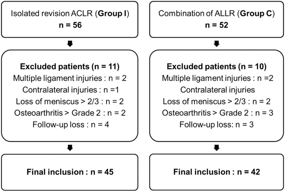
Flowchart showing the selection of participants. ACLR, anterior cruciate ligament reconstruction; ALLR, anterolateral ligament reconstruction.
Preoperative Demographic Data and Clinical Characteristics a

Data are presented as mean ± SD unless otherwise indicated. ACLR, anterior cruciate ligament reconstruction; C, combined revision anterior cruciate ligament reconstruction and anterolateral ligament reconstruction; I, isolated revision anterior cruciate ligament reconstruction; IKDC, International Knee Documentation Committee.
Surgical Techniques
Revision ACLR
After confirmation of an ACL graft rupture by arthroscopic surgery, a tibialis anterior tendon allograft (fresh-frozen) that was irradiated with 50 kGy and controlled by a tissue preservation technique (Clearant Process; Korea Bone Bank) was selected. A graft with a diameter of 9 mm was prepared. The torn graft and residual soft tissue were removed using electrocautery (ArthroCare; Smith & Nephew) to gain better visibility. Through the anteromedial portal, a microfracture awl was placed at a more lateral and posterior position than the previous vertical direction. The FlipCutter (Arthrex) guide tip was located at the center of a new femoral tunnel made using a microfracture awl, and the depth was measured. A stab incision was made at about 3 cm proximal to the lateral epicondyle, and the guide pin was drilled to the intra-articular FlipCutter guide tip using the outside-in method. Following the guide pin, the FlipCutter was engaged into the joint and retrodrilled around 25 to 30 mm to make a femoral socket, the diameter of which was the same as the thickness of the graft (Figure 2). In group C, a new femoral tunnel was made for revision ACLR, followed by another femoral tunnel for ALL reconstruction. The guide pin was drilled 6 mm before reaming for a femoral tunnel for ALL reconstruction. Whether the direction of the guide pin was blended with the femoral tunnel for ACLR was checked. If so, we redirected the guide pin to avoid the overlapping of tunnels (Figure 3).
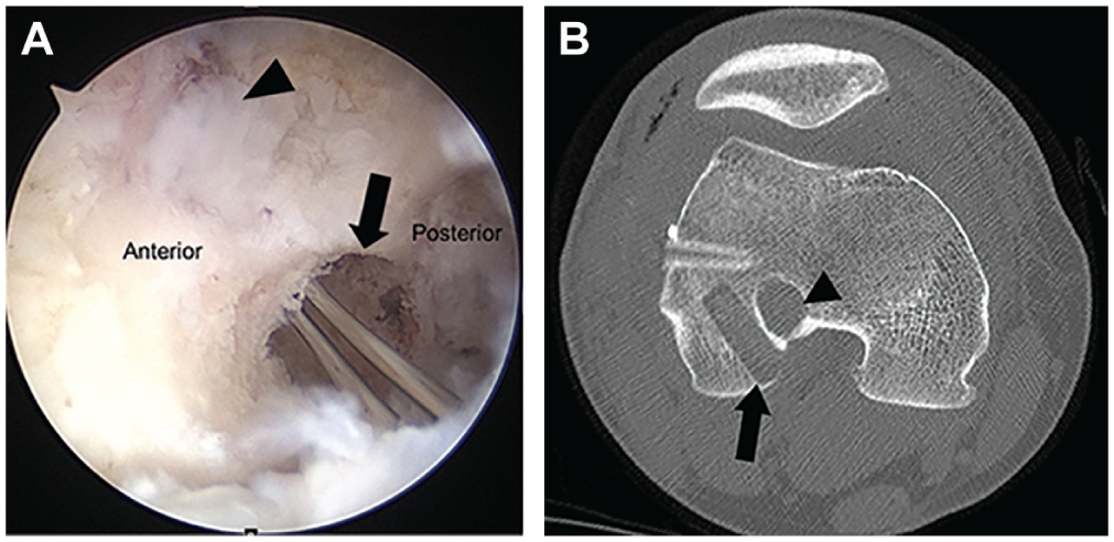
(A) Arthroscopic image and (B) axial computed tomographic image in the right knee show that an entirely new femoral tunnel (arrow) was made using the outside-in technique, and it was not blended with the too vertical and previous anterior femoral tunnel (arrowhead).
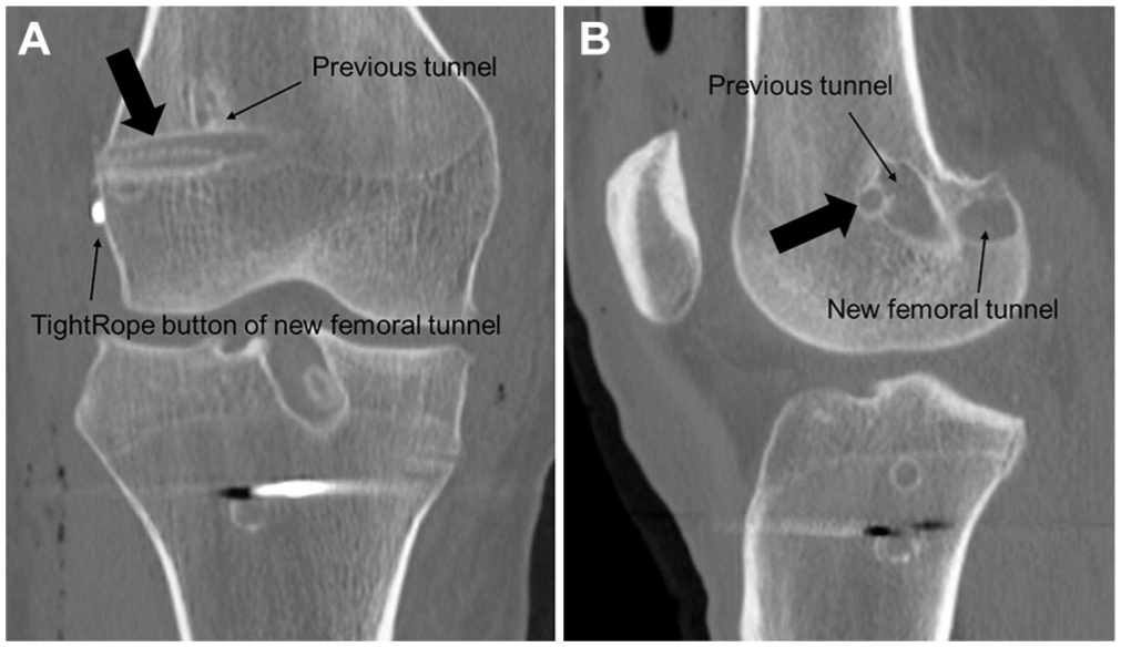
(A) Coronal and (B) sagittal computed tomographic images in the right knee. A new femoral tunnel for anterior cruciate ligament reconstruction was not blended with the femoral tunnel for anterolateral reconstruction (arrow).
After the graft was passed through the tibial tunnel, the ACL TightRope (Arthrex) was used for flipping and compressing the lateral cortex of the femur. Once complete flipping was done, distal pulling was performed for tensioning. A biointerference screw (Matryx; Conmed Linvatec) and additional staple were used for tibial fixation.
ALL Reconstruction
A gracilis tendon allograft (fresh-frozen) irradiated with 50 kGy and controlled by a tissue preservation technique (Clearant Process) with a diameter of 6 mm was prepared. Three bony landmarks (lateral femoral epicondyle, fibular head, and Gerdy tubercle) were identified based on previous ALL anatomy research.7,44 A curvilinear skin incision was then made in the anterolateral aspect of the knee, extending from the lateral epicondyle to the tibial insertion between the Gerdy tubercle and fibular head (Figure 4A). A guide pin was positioned just proximal and posterior to the lateral femoral epicondyle. The femoral socket was made using a 6 mm–diameter cannulated reamer with a depth of 25 mm. This femoral socket preparation was performed before revision ACLR to avoid femoral tunnel communication. A guide pin was then positioned between the fibular head and Gerdy tubercle at approximately 10 mm below the joint line. The tibial socket was made using a 6 mm–diameter cannulated reamer with a depth of 25 mm. After femoral graft fixation was performed with a 7-mm biointerference screw (Matryx), tibial graft fixation was performed with a 7-mm biointerference screw at 30° of knee flexion and neutral rotation (Figure 4B). The placement of the femoral tunnel (proximal and posterior to the lateral epicondyle) and tibial tunnel (the center between the Gerdy tubercle and fibular head) was checked using 64–detector row 3-dimensional computed tomography scans (LightSpeed VCT XT; GE Medical Systems) that were obtained on the day of surgery (Figure 4C).
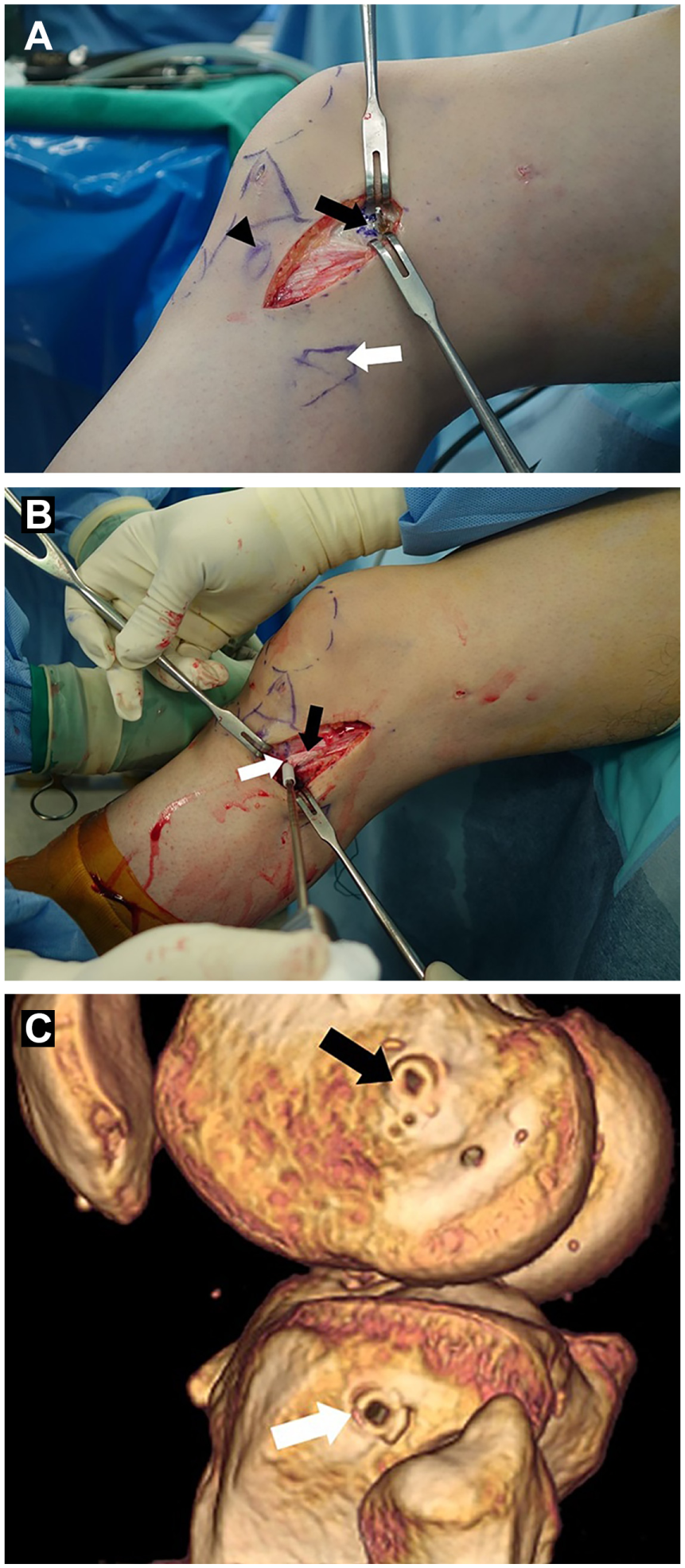
(A) A skin incision was made in the anterolateral aspect of the knee, extending from the lateral epicondyle (black arrow) to the tibial insertion between the Gerdy tubercle (black arrowhead) and fibular head (white arrow). (B) After femoral graft fixation was performed, tibial graft (black arrow) fixation was performed with a biointerference screw (white arrow) at 30° of knee flexion and neutral rotation. (C) The placement of the femoral tunnel (black arrow) and tibial tunnel (white arrow) was evaluated using 3-dimensional computed tomography scans obtained on the day of surgery.
Postoperative Rehabilitation
Rehabilitation was performed with the same method for both groups. Patients were permitted to bear weight with an ACL-supporting brace (Legend; DonJoy) locked in full extension as tolerated immediately after surgery. When meniscus repair was performed, partial weightbearing with crutches was allowed for 6 weeks. Range of motion exercise was started with the brace unlocked from 0° to 90° at 3 weeks postoperatively. Progressive range of motion exercise was maintained, aiming for full flexion by week 6. The brace was completely removed at 2 months postoperatively. Open kinetic chain exercises and a perturbation training program were started at 2 months after surgery. After the course of home-based rehabilitation, running was allowed 6 months after surgery according to the recovery of muscle power and knee function. Competitive sports activity was allowed after 9 months postoperatively.
Clinical Assessment
Clinical and functional tests were administered usually before surgery and at 6, 12, 24, and 36 months postoperatively. They were usually completed at 36 months of follow-up. For subjective knee function assessments, the Lysholm score, subjective IKDC form, and Tegner activity scale were used. The mean duration from revision ACLR to return to sports and level of participation in sports were also assessed. The Anterior Cruciate Ligament–Return to Sport after Injury (ACL-RSI) scale was used to measure psychological readiness for return to sports at 12 months and the last follow-up.4,26
As objective assessments, anterior laxity, manual pivot shift, isokinetic extensor muscle strength, and functional performance tests were administered. Anterior laxity was evaluated by a side-to-side difference on the KT-2000 arthrometer (MEDmetric) with maximal manual stress. The pivot-shift test was performed by the senior author (J.G.K.), who has more than 20 years of ACLR experience. Results were classified as grade 0 = normal, grade 1 = gliding, grade 2 = clunk, and grade 3 = gross shifting as in previous studies.5,16,38,49,50
Isokinetic extensor muscle strength measurements were performed using the Biodex System 3 dynamometer (Biodex Medical Systems) at an angular velocity of 60 deg/s. Measurements were taken initially on the unaffected side, followed by the affected side. The deficit (%) was then calculated as follows: ([peak muscle torque of the uninvolved side – peak muscle torque of the involved side]/peak muscle torque of the uninvolved side) × 100.
To assess functional performance, the single-legged hop for distance reported by Noyes et al, 42 co-contraction test, and carioca test were performed.15,19 For the single-legged hop for distance, patients were asked to hop forward as far as possible, jumping and landing with the same foot. It was performed 3 times, and the longest distances for the involved and uninvolved limbs were measured in centimeters using a ruler on the ground. The limb symmetry index (%) was measured. The co-contraction test was conducted by securing a Velcro belt around the patient’s waist. The belt was attached to a 48-inch length of rubber tubing with a diameter of 1 inch. The tube was anchored to a metal loop that was secured on a wall 60 inches above the floor. A semicircle with a radius of 96 inches from the metal loop was painted on the floor. Patients standing with their toes on the line were asked to run wall to wall along the semicircular line 5 times, and the shortest time was measured. The carioca test was performed by requiring the patient to run laterally 2 lengths of a 12-m distance with a crossover step. The patient ran the course from left to right and then in the reverse direction. The fastest time was recorded.
Statistical Analysis
Statistical analysis was performed using SPSS for Windows version 20.0 (SPSS). An independent t test or Mann-Whitney U test was used to compare parametric or nonparametric variables between the 2 groups. Preoperative and postoperative parametric or nonparametric variables were compared with a paired t test or Wilcoxon signed-rank test in each group. The chi-square test was used to compare categorical data. If more than 20% of expected frequencies were >5, the Fisher exact test was applied. For the chi-square test for trends, linear by linear association was used. Statistical significance was defined at P < .05. To detect a difference of 10 points on the subjective IKDC form between the 2 groups with a level of significance of 5% and a power of 80%, the required sample size was determined to be 36 patients per group. Assuming a dropout rate of 15%, 42 patients per group were required.
Results
The mean follow-up duration for groups I and C were 41.5 ± 8.2 and 38.2 ± 6.9 months (P = .451), respectively. In group I, there were 2 graft ruptures after 12 months. Thus, 43 patients were evaluated at the last follow-up.
Subjective Outcomes
At the last follow-up, both groups showed a significantly improved subjective IKDC score, Lysholm score, and Tegner score compared with preoperative data (all P < .001). However, the subjective IKDC score, Tegner score, and ACL-RSI score were significantly better in group C compared with those in group I at the last follow-up, although they were not significantly different between the 2 groups at 12 months after surgery (Table 2).
Subjective Outcomes a

Data are presented as mean ± SD. ACL-RSI, Anterior Cruciate Ligament–Return to Sport after Injury; C, combined revision anterior cruciate ligament reconstruction and anterolateral ligament reconstruction; I, isolated revision anterior cruciate ligament reconstruction; IKDC, International Knee Documentation Committee.
Objective Outcomes
There were no significant differences in KT-2000 arthrometer, isokinetic extensor strength, single-legged hop for distance, co-contraction test, or carioca test results between the 2 groups at the last follow-up (Table 3).
Preoperatively, 43 (95.6%) patients in group I and 40 (95.2%) patients in group C had a grade 2 or 3 pivot shift (P = .387). Postoperatively, 38 (90.5%) patients in group C and 23 (53.5%) patients in group I had a negative pivot shift (P < .001).
Objective Outcomes at the Last Follow-up a

Data are presented as mean ± SD unless otherwise indicated. C, combined revision anterior cruciate ligament reconstruction and anterolateral ligament reconstruction; I, isolated revision anterior cruciate ligament reconstruction; IKDC, International Knee Documentation Committee.
Return to Sports
The mean time to return to running or sports activity for groups I and C were 6.6 ± 2.5 and 6.1 ± 3.2 months (P = .417) or 11.5 ± 3.7 and 9.6 ± 3.4 months (P = .015), respectively. The average score of preinjury Tegner activity scale was 7.4 ± 0.9 in group I and 7.5 ± 1.0 in group C (P = 0.697). At the last follow-up, the mean Tegner score was 7.0 ± 0.8 in group C and 6.3 ± 0.7 in group I (P < .001) (Figure 5). Group C showed a higher rate of return to the same level of sports activity than group I (57.1% vs 25.6%, respectively; P = .008), although there was no significant difference in the rate of return to any sports activity (88.4% in group I vs 88.1% in group C; P = .713) (Figure 6). Reasons for not returning to the same level of sports are described in Table 4.
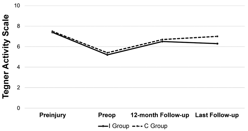
Graphs of Tegner score progression. P < .05 comparing preinjury with preoperatively, 12-month follow-up, and the last follow-up in both groups. C, combined revision anterior cruciate ligament reconstruction and anterolateral ligament reconstruction; I, isolated revision anterior cruciate ligament reconstruction.

Rate of return to sports in both groups. *P < .05. C, combined revision anterior cruciate ligament reconstruction and anterolateral ligament reconstruction; I, isolated revision anterior cruciate ligament reconstruction.
Reasons for Not Returning to the Same Level of Sports at the Last Follow-up a

Data are presented as n (%). C, combined revision anterior cruciate ligament reconstruction and anterolateral ligament reconstruction; I, isolated revision anterior cruciate ligament reconstruction.
Complications
A graft rerupture that needed re-revision surgery was found in 2 (4.4%) patients in group I, while no patient in group C showed a rerupture. The mean time from return to sports to a rerupture was 15.1 ± 3.9 months in the 2 patients. In the current study, 7 patients with a grade 2 pivot shift in group I did not have subjective knee instability. Therefore, they were not recommended to undergo another operative procedure. A complication related to the extra-articular procedure (n = 1) was only femoral interference screw protrusion that required removal. There was no stiffness in either group.
Discussion
The main finding of the present study was that the addition of anatomic ALL reconstruction to revision ACLR was effective in reducing rotational laxity assessed by the pivot-shift test. Anatomic ALL reconstruction plus revision ACLR showed significantly more reduction in residual pivot shift with a higher rate of return to the same level of sports activity compared with isolated revision ACLR. The subjective IKDC score, Tegner score, and ACL-RSI score were significantly better in group C compared with those in group I at the last follow-up, although there were no significant differences in the Lysholm score, anterior laxity, or functional test results between the 2 groups. There is concern that the ceiling effect of the Lysholm score is greater than that of the subjective IKDC score. It should be addressed when assessing knee function postoperatively. 45 However, the subjective IKDC form has merits, such as its point-based calculation system, which means that scoring is not affected by patient factors such as age or sex because it takes various knee-related problems into account.37,43,45 To date, few studies have compared the results of revision ACLR versus a combination of anatomic ALL reconstruction with revision ACLR. There are no such comparative studies in a single surgeon’s series. Moreover, research about revision ACLR has difficulty in selecting a homogeneous group of patients. We tried to carefully decide the reasons for primary reconstruction failure and the inclusion criteria with an aim to have homogeneous patients.
A recent multicenter cohort study by the Multicenter Arthroscopic Knee Surgery (MAKS) group has reported that 4.2% (2/48) in the positive residual pivot-shift group and 13% (42/320) in the negative residual pivot-shift group had a grade ≤2 preoperative pivot shift (grade 1 is subtle gliding and grade 2 is gliding according to their classification). 58 It also investigated risk factors for residual pivot shift after primary ACLR based on a prospective cohort study. Melugin et al 38 revealed that 90.0% (18/20) of patients who had a combined ACL tear with Segond fracture and 82.5% (33/40) of patients who had an ACL tear without Segond fracture showed a grade ≥2 (clunk) preoperative pivot shift, respectively. Similarly, Lee et al 27 showed that 64% (176/275) of patients with acute ACL ruptures had ALL injuries and that 84% of 275 patients had a high-grade pivot shift. Another multicenter study by the French Arthroscopy Society reported that 77% (268/349) showed a grade ≥2 preoperative pivot shift. 34 It also evaluated outcomes of combined intra- and extra-articular grafting during revision surgery after previous ACLR. Several studies have investigated the risk factors for a high-grade pivot shift after an ACL injury and concluded that anterolateral structure disruptions are associated with increased knee rotatory laxity.40,50 We hypothesized that a rerupture after primary ACLR would result in more frequent anterolateral structure injuries and a positive pivot shift. Although most ACLR failures were secondary to technical errors, a subset of patients could also have residual rotatory laxity, despite a well-positioned graft. 39 Such an abnormal pivot shift could result in permanent anterolateral complex injuries. 61 In the current study, 95.4% showed a high-grade (grade ≥2) pivot shift.
Sonnery-Cottet et al 51 have suggested that ACLR failures are caused by persistent rotational instability during sports activity. Hence, in patients with objective excessive rotatory laxity after ACLR, additional tenodesis or reconstruction of anterolateral structures could be considered when performing revision ACLR. Trojani et al 57 have reported that the proportion of negative pivot shifts was 80% with lateral tenodesis plus revision ACLR (n = 84) and 63% with isolated revision ACLR (P = .03), although additional lateral tenodesis did not influence the IKDC score in a multicenter study of 163 revision ACLRs. Louis et al 34 have demonstrated that 99% of 349 patients who underwent combined ALL stabilization and revision ACLR showed a negative pivot shift. Their negative pivot-shift prevalence was lower compared with about 10% in previous reports.24,29,57 In addition, the rerupture rate was 1.2% in their multicenter study. In our study, 90.5% in group C showed a negative pivot shift, while 53.5% in group I had a negative pivot shift (P < .001). There was no rerupture after ALL stabilization in the present study. One prospective study, which was the first clinical series to show that anatomic ALL reconstruction was associated with a reduction in the graft rerupture rate, reported that the rate of graft failure with a combination of ALL reconstruction and ACLR was more than 2 times less than that with isolated ACLR. 54 Therefore, in revision ACLR, an additional extra-articular procedure could be considered as a strategy to improve rotational stability and reduce the rerupture rate. This is in accordance with the concept of “belt and suspenders” and “backup for ACL graft” in biomechanical studies.12,14,17,35,36,53
Few studies have evaluated the rate of return to sports after a combined extra-articular procedure and revision ACLR. Our results showed a higher rate of return to the same sports level compared with 43% of 1167 patients who underwent revision ACLR in a meta-analysis. 1 Lefevre et al 28 reported that 23.7% of 497 patients in the primary ACLR group returned to the same or better sports level at 1 year compared with 12.7% of 55 patients in the revision ACLR group (P = .04). The involved knee was the main reason for changing or stopping the preinjury sports level. In the current study, 57.1% of patients in group C returned to the same level of sports activity at the last follow-up compared with 25.6% of patients in group I (P = .008), although there was no significant difference in the rate of return to a lower level of sports activity (groups I and C: 88.4% vs 88.1%, respectively; P = .713). One multicenter study reported that the mean time to return to sports was less than 8 months in 86% of 349 patients after ALL stabilization plus revision ACLR. 34 Sonnery-Cottet et al 54 showed that the addition of anatomic ALL reconstruction was associated with a greater odds of returning to the preinjury sports level compared with isolated ACLR.
Trojani et al 57 showed that there was no difference in the objective IKDC score between isolated revision ACLR and lateral tenodesis plus revision ACLR at a minimum 2-year follow-up. Vadala et al 59 reported that clinical outcomes other than the pivot shift did not show significant differences between patients with an extra-articular procedure and those without an extra-articular procedure. Similarly, there were no significant differences in KT-2000 arthrometer, isokinetic extensor strength, single-legged hop for distance, co-contraction test, or carioca test results between the 2 groups at the last follow-up in our study. However, ALL reconstruction showed a trend to have better results on the co-contraction and carioca tests in the current study. Lephart et al 31 suggested that the co-contraction, carioca, and shuttle run tests were quantitative measurements of functional capabilities after ACLR. They provide a basis for determining patients’ readiness to return to sports. The co-contraction and carioca tests were designed to reproduce rotational forces, necessitating the control of tibial translation and the pivot-shift phenomenon.30,31 Jang et al 18 reported that the co-contraction and carioca tests, mainly for assessing rotational stability, showed statistically significant differences between return to sports and non–return to sports at a minimum 2-year follow-up.
Numerous recent studies about ALL anatomy and biomechanics have led to a better understanding of them. Subsequently, various anatomic ALL reconstruction techniques have emerged.9,19,23,60 However, regarding extra-articular procedures, no consensus is available on the graft type, location of fixation, or fixation angle. In particular, there have been various fixation angles from full extension to 90°. 23 Schon et al 47 have reported that anatomic ALL reconstruction in conjunction with ACLR can significantly reduce rotatory laxity beyond 30° of flexion. However, it resulted in mild overconstraining, regardless of the fixation angle. However, Thaunat et al 56 demonstrated that the high rates of stiffness and reoperation reported in historical series of nonanatomic extra-articular lateral tenodesis were not found in anatomic ALL reconstruction with a cohort of 548 patients. Our suggestion is that ALL is less isometric structure, therefore, anatomic ligament reconstruction to restore anatomy as close as possible is important.13,20 Furthermore, combined ACLR and anatomic ALL reconstruction using 1 femoral tunnel in revision cases could be a simple and safe strategy to avoid the overlapping of 2 new femoral tunnels. 52 Further research is necessary to fully understand the biomechanics and clinical effects of anterolateral structures.
The present study has some limitations. First, this study was a retrospective comparative study. However, there has been no prospective randomized study about ALL reconstruction and revision ACLR. Second, there was no equipment to evaluate pivot shift quantitatively.25,33 Fixation angle and tension of the graft should be individually adapted according to the amount of rotatory laxity. Hence, equipment that can assess pivot shift needs to be developed. Third, we could not evaluate the evidence of osteoarthritis related to lateral overconstraint at the last follow-up. A 3-year follow-up period is not long enough to identify the progression of osteoarthritis. Thus, a larger sample size and longer follow-up period are required. Finally, the quality of return to sports or psychological readiness of patients to return to sports needs to be more specific. Additional psychological and performance measurements are needed to complement patient-reported outcome scores.
Conclusion
Revision ACLR with ALL reconstruction significantly reduced rotational laxity and showed a higher rate of return to the same level of sports activity than isolated revision ACLR, although there were no significant differences in anterior laxity or functional test results between the 2 groups.
Footnotes
The authors declared that they have no conflicts of interest in the authorship and publication of this contribution. AOSSM checks author disclosures against the Open Payments Database (OPD). AOSSM has not conducted an independent investigation on the OPD and disclaims any liability or responsibility relating thereto.