
Abstract
Background:
Corticosteroid injections in or around tendons for the treatment of athletic injuries are a common practice among orthopaedic surgeons and are apparently efficacious in the short term, although controversies persist related to local complications.
Purpose:
This study evaluated short-term (48 hours) biomechanical, biochemical, and histological alterations after a single injection of betamethasone into the normal tendons of rabbits.
Study Design:
Controlled laboratory study.
Methods:
A total of 72 New Zealand White rabbits were randomly divided into 2 groups: the test group—in which 36 animals underwent 1 intratendinous injection of betamethasone (1.4 mg / 0.2 mL) in the right calcaneal tendon; the control group—in which the right calcaneal tendon of 36 animals was injected with saline (placebo control group) and the left calcaneal tendon was left untreated for normal standards (normal control). Forty-eight hours later, animals were euthanized and tendons were harvested. Metalloproteinase (MMP1 and MMP2) and interleukin (IL1 and IL6) expression levels, biomechanical resistance (load × elongation parameters), and histomorphometry (hematoxylin and eosin and picrosirius red stains for collagen fibers, tenocytes, and inflammatory cells) were analyzed in the tendons.
Results:
The test group had a significant reduction in MMP2 expression as compared with the control groups (P = .027). Regarding the other parameters, there were no additional significant differences between the groups.
Conclusion:
A single injection of corticosteroid into normal calcaneal tendons did not trigger acute local morphological, structural, or biomechanical injuries at 48 hours, but it did promote a significant decrease in MMP2 levels. Additional studies are needed with increased duration of follow-up, various doses, and multiple injections and in tendinopathic models.
Clinical Relevance:
Some previous studies demonstrated early structural changes in tendons after a single corticosteroid injection, which was not corroborated by the present study. Metalloproteinase decrease is usually associated with a reduction in collagen degradation, which would be protective for the healing process. More studies are necessary to confirm the possible beneficial effect of these results in the long term and for tendinopathies.
Tendinopathy is defined as a group of tendon disorders with multifactorial etiology, resulting in pain and varying degrees of functional loss. The load, local anatomy, posture, sports, and occupational activities are considered extrinsic etiological factors. However, autoimmune or metabolic abnormalities and polymorphisms that may affect the gene expression of matrix components are intrinsic factors.11,17,24
Some authors alternatively supported the inflammatory component as being the primary factor causing this condition.1,3,25,30 However, modifications in the definition and etiology of tendinopathies were introduced by other authors, shifting from inflammatory to degenerative changes (thickening, disorganization, and rupture of collagen; abnormal or dead cells; neovascularization; failing of healing response).7,8 Consequently, treatment transitioned from the use of anti-inflammatory drugs to physical therapy, shockwave therapy, and other types of drugs, such as sclerosing agents, dextrose, and botulinum toxin, as well as to local injections of stem cells or platelet-rich plasma. 25
More recently, some studies pondered that the noninflammatory-dependent degenerative concept of tendinopathy was an oversimplification. Therefore, new studies on the inflammatory process emerged, 25 justifying the use of local corticosteroid (CS) injection of the tendon. Additionally, recent studies confirmed the presence of inflammatory cells 20 and inflammatory responses in tendons after a mechanical overload. 9 However, there is limited knowledge on the involvement of interleukins and other inflammatory mediators and markers of affected tendons. 1
Previous experimental studies presented controversial data regarding the possible effects of CS injections in the tendons of animals, given the frequent absence of controls, no double-blind design, and the involvement of several CS agents, dosages, sites of injections, outcomes, animal models, and follow-up periods. 22 Consequently, the results varied from major injuries, such as collagen degeneration and inflammation, to beneficial effects, such as an increase in mechanical resistance. 29 In a critical review of literature, Nichols 22 found 17 animal studies, and of these, 10 provided evidence that CS injections caused musculoskeletal structural and functional damage and 7 did not. The author also stated that the literature search did not identify studies that provided unequivocal scientific evidence that CS injections do or do not damage musculoskeletal structure. In another systematic review, Dean et al 8 found 18 studies, describing 6 with a decrease in mechanical properties, 3 with an increase, and the remaining 9 with no significant change. A systematic review of randomized clinical trials showed strong evidence that CS injection was beneficial in the short term for treatment of tendinopathy but was worse than other treatment options in the intermediate and long terms. 6 This review and meta-analysis noted the low frequency of serious adverse events after CS injection, but minor complications were common, such as postinjection pain, subcutaneous atrophy, and skin depigmentation. 6
CS inhibits the expression of metalloproteinases (MMPs) induced by growth factors, interleukins (IL1, IL6, TNF), and macrophages.13,14 Few studies included MPPs’ determination in tendinopathies and after CS injection in tendons.14,28 MMPs are enzymes produced mainly by tenoblasts, fibroblasts, and leukocytes, which are important for extracellular matrix deterioration and reparation balance.13,26 MMP1 mainly acts as collagenase, and MMP2 acts as both gelatinase and collagenase.13,26
Our purpose was to determine whether a single injection of CS in the rabbit healthy Achilles tendon would trigger acute local morphological structural, biochemical (interleukins and MMP), or biomechanical alterations when compared with placebo control specimens.
Methods
Animal Care and Use
A double-blind randomized controlled experimental study of New Zealand White rabbits was performed. The university’s Animal Use Ethics Committee approved all method stages of this experimental protocol (1047-2013). Animal care and use before, during, and after procedures complied with the guidelines set forth by the Administrative Panel on Laboratory Animal Care. Animals were obtained from our institutional animal breeding. All animals were adult and male, weighing 2.0 to 3.0 kg around 4 months after birth.
Injection Procedure
All procedures were performed under intravenous general anesthesia. One ear was cleaned and prepared with an alcohol chlorhexidine solution, and the marginal vein was punctured for the infusion of 50 mg/kg of ketamine hydrochloride and 1 mg/kg of xylazine hydrochloride.
Software (rv-CRAN-R, v 2.11.0) was used to generate a sequence of numbers for randomization. A total of 72 New Zealand White, healthy, male rabbits, weighing between 2.0 and 2.5 kg, were randomly divided into 2 groups (Figure 1):
Test group: Thirty-six right calcaneal tendons were injected with 1.4 mg / 0.2 mL of betamethasone (5 mg) dipropionate + betamethasone phosphate solution (2 mg) (Diprospan; Mantecorp Hypermarcas)
Placebo control: The right calcaneal tendons of the other 36 animals were injected with saline (0.2 mL of sodium chloride at 0.9%; Farmace Ltd), and the left calcaneal tendons were left untreated for standard comparison (normal control).
Betamethasone dosage and volume were similar to previous studies,5,12 and injections were performed by a board-certified orthopaedic surgeon (K.G.I.D.).
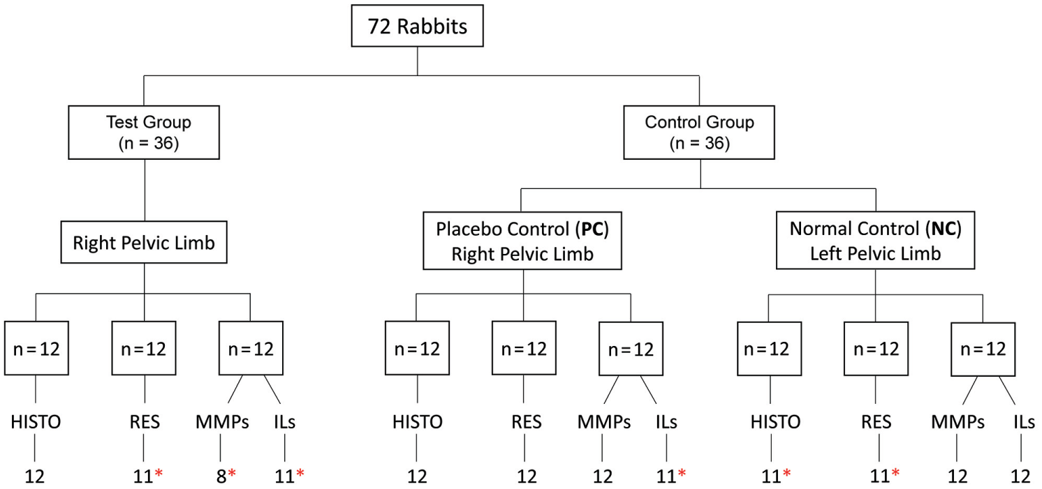
Flowchart of procedures. Histo, histology; ILs, interleukins; MMPs, metalloproteinases; RES, mechanical resistance. *Sample size <12 corresponds to specimen losses (see text).
After posterior pelvic limb trichotomy and antisepsis of the operative field with an alcohol chlorhexidine solution, exactly 2 cm proximal to the insertion of the calcaneal tendon, an angulated 26G × 0.5 in needle (Becton Dickinson) was used for intratendinous injections. The injection technique was tested on only 3 pilot animals with similar body weight to make standardized intratendinous injections without any image guidance (Figure 2). The technician prepared all syringes without the knowledge of the orthopaedist.
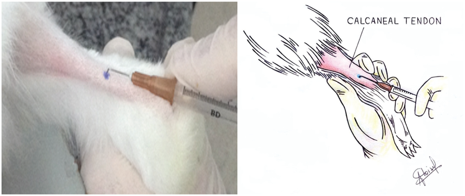
Intratendinous injection technique into the calcaneal tendon with a curved 26G × 0.5 in needle.
Dosages (1.4 mg / 0.2 mL) were based on the lowest used by Kennedy and Willis, 12 who injected 0.25 mL and 0.5 mL of betamethasone into tendons (2 groups). According to the authors, these dosages represent one-quarter to one-half of the usual adult injection, and the lowest dosages employed were associated with severe histological alterations in that study.
All animals were maintained in individual cages without mobility restrictions and with a light/dark cycle, as well as with water and standard rabbit chow (Purina) ad libitum.
Specimen Preparation
After 48 hours, all animals were euthanized with an overdose of anesthetic agents (150 mg/kg of ketamine hydrochloride and 2 mg/kg of xylazine hydrochloride); then, their calcaneal tendons were removed from the myotendinous junction to its insertion point on calcaneal bone (about 4 cm of total length). For histology, 36 tendons were fixed in 10% neutral buffered formalin for a minimum of 48 hours before processing. The specimens were then embedded in paraffin blocks, cut into 5-μm-thick sections, and stained with hematoxylin and eosin and picrosirius red. Another 36 tendons were stored in a –20°C freezer for future tensile strength tests. Finally, the other 36 tendons were transected in 2 parts and immersed in liquid nitrogen and, immediately after, in –80°C freezers for later biochemical analysis; after 4 hours of thawing at room temperature, zymography analysis for MMPs and enzyme-linked immunosorbent assay (ELISA) for interleukins were performed. Two tendons slipped off the grips of the universal testing machine. Of 144 specimens, 9 were excluded because of technical problems (energy blackout).
Biomechanical Traction Test
The mechanical resistance of the calcaneal tendon was evaluated by a previously validated method.21,36 Briefly, the tendons (all stored for <1 months) were removed from the freezer wrapped in saline-soaked gauze and thawed at room temperature for 4 hours. 21 Freezing has been shown to have little or no effect on the biomechanical properties of the ligaments. 34 Then, their circumferences were measured and the cross-sectional area calculated by the formula A = π.R2, where R is the radius and A is the cross-sectional area. The ends of the tendons were then fixed in the grips of a universal testing machine (EMIC DL 10.000), with a cell load of 1000 N and an application rate of 30 mm/min. M-Test software (v 1.01; Magtrol) was used to provide the force × deformation chart of each specimen and related mechanical properties: maximum force (N), deformation (mm), energy at maximum force (N·mm), energy per area at maximum force (N·mm/mm2), tension at maximum force (MPa), and elasticity modulus (MPa) (Figure 3).
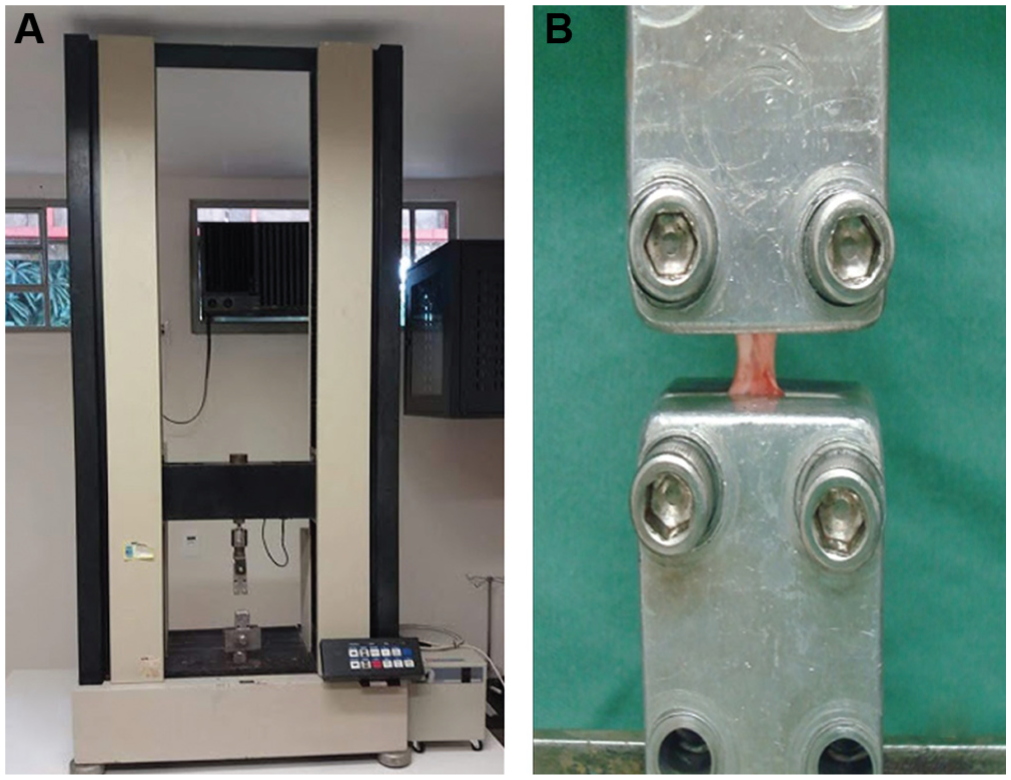
(A) Universal testing machine. (B) Tendon fixed in 2 grips during testing.
Histomorphometry
To quantify the tenocytes and inflammatory cells by hematoxylin and eosin staining, the dissector method with a Weibel reticle was used. 18 Histomorphometry by the Weibel reticulum was performed with a system of 50 traces in a known area (62,500 mm2 at 400×) in which each cell or image of collagen fiber was counted when crossing traces of the reticulum. Quantitative results were obtained by dividing total counts by 50 (Figure 4). In the present study, at least 10 fields of each tendon slide were analyzed with a quantitative histomorphometric grading system with the Weibel reticule in a randomized blinded manner. An experienced pathologist (A.T.F.) blinded to the groups performed all histologic analysis.
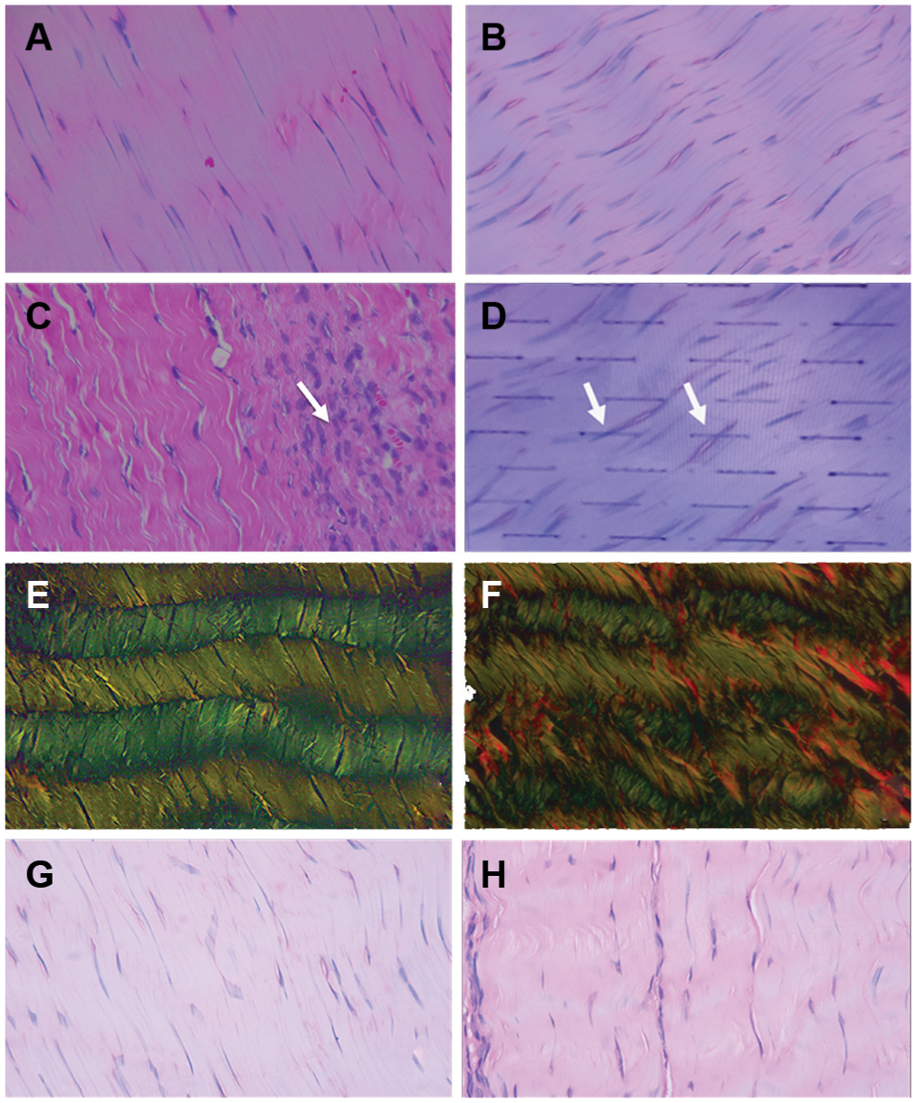
Histology of tendons (magnification, 400×). Normal control group: (A) hematoxylin and eosin stain shows preserved collagen fibers. Test group: hematoxylin and eosin shows (B) increased cellularity and (C) inflammatory cells (arrow); (D) Weibel reticle shows tenocytes crossing the traces (arrows). Normal control group: picrosirius red stain shows (E) thin collagen fibers in green and (F) thick fibers in red; (G) hematoxylin and eosin, normal aspect. Placebo control group: (H) hematoxylin and eosin.
Zymography Assay
The zymography assay was performed as previously described. 35 Briefly, after sample preparation, electrophoresis with an agarose gel was used to separate the different molecular weights. The gels were photographed with an ImageQuant LAS 4000 (GE Healthcare Life Sciences). Quantification of expression was performed by densitometry in an image analyzer (White Darkhon; GE Healthcare Life Sciences) with ImageQuant TL (v 4000) software. MMP2 was observed as 2 bands of 72 and 62 kDa (inactive and active forms). MMP1 was identified in the collagenous gels as inactive (60 kDa) and active (54 kDa) bands.
ELISA
ELISA was performed according to a protocol established by our laboratory. The antibodies were recombinant rabbit IL1 alpha (RP0 346U-005; Kingfischer Biotech, Inc) and IL6 for recombinant rabbit IL6 (SEA a79Rb; Cloud-Clone Corp), and the data were quantified in pg/mL. 16
Statistical Analysis
Sample size was calculated as 12 animals per group. We used an 80% power test and a 5% significance level based on previous studies with the same rabbit species and same equipment, 23 considering tensile strength test with an SD of 70 N and a mean difference of 70 N in maximum force, adding a dropout of 20%. Therefore, for this parameter, 11 animals were still within the calculated dropout. For the other outcomes, we considered the final sample size to be within that of similar studies. Indeed, similar experimental animal studies used 3 to 10 animals per group for histological outcomes2,5,13,29 and 8 or 9 animals for biochemical analysis,1,14 and as such, we think that our sample size for these parameters was appropriate for our conclusions.
The comparisons between groups regarding the histological and MMP and interleukin expression variables were performed with the nonparametric Kruskal-Wallis test and Dunn test for multiple comparisons. Analysis of the mechanical resistance variables of the biomechanical properties was performed with an analysis of variance parametric test after verification of the symmetry assumptions of the variables and the equal variance.
The differences were considered statistically significant if P < .05, with statistical analysis performed with SPSS software (v 21.0; IBM).
Results
All biomechanical properties were similar between the groups, such as maximum deformation (mm), energy per area at maximum force (N·mm/mm2), maximum force (N), energy at maximum force (N·mm), elasticity modulus (MPa), and tension at maximum force (MPa) (Table 1), suggesting that the calcaneal tendons had preserved functional structure 48 hours after a single CS injection.
Comparison Among Groups in Relation to the Variables of Mechanical Resistance
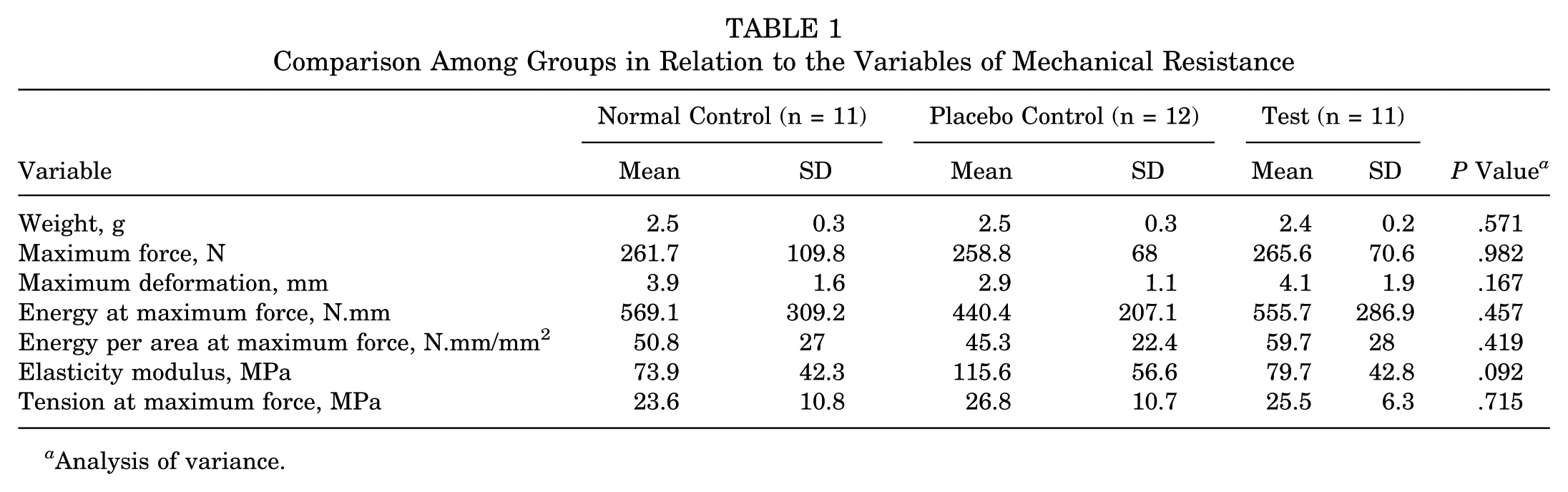
Analysis of variance.
The histomorphometric variables are shown in Figure 4 and Table 2. There were no statistically significant differences between the test and control groups. There was a statistically significant difference for thick collagen fiber counts between the normal control group (without injection) and the placebo group (after saline injection; P = .041) (Figure 5).
Comparison Among Groups in Relation to the Histological Variables (Counts/50 Traces) a
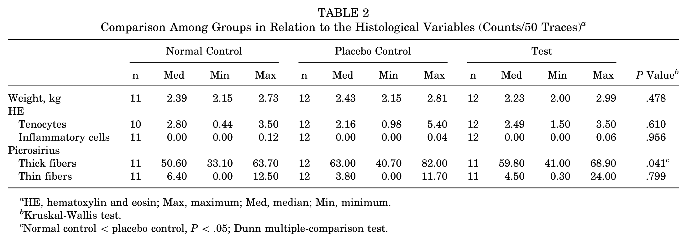
HE, hematoxylin and eosin; Max, maximum; Med, median; Min, minimum.
Kruskal-Wallis test.
Normal control < placebo control, P < .05; Dunn multiple-comparison test.
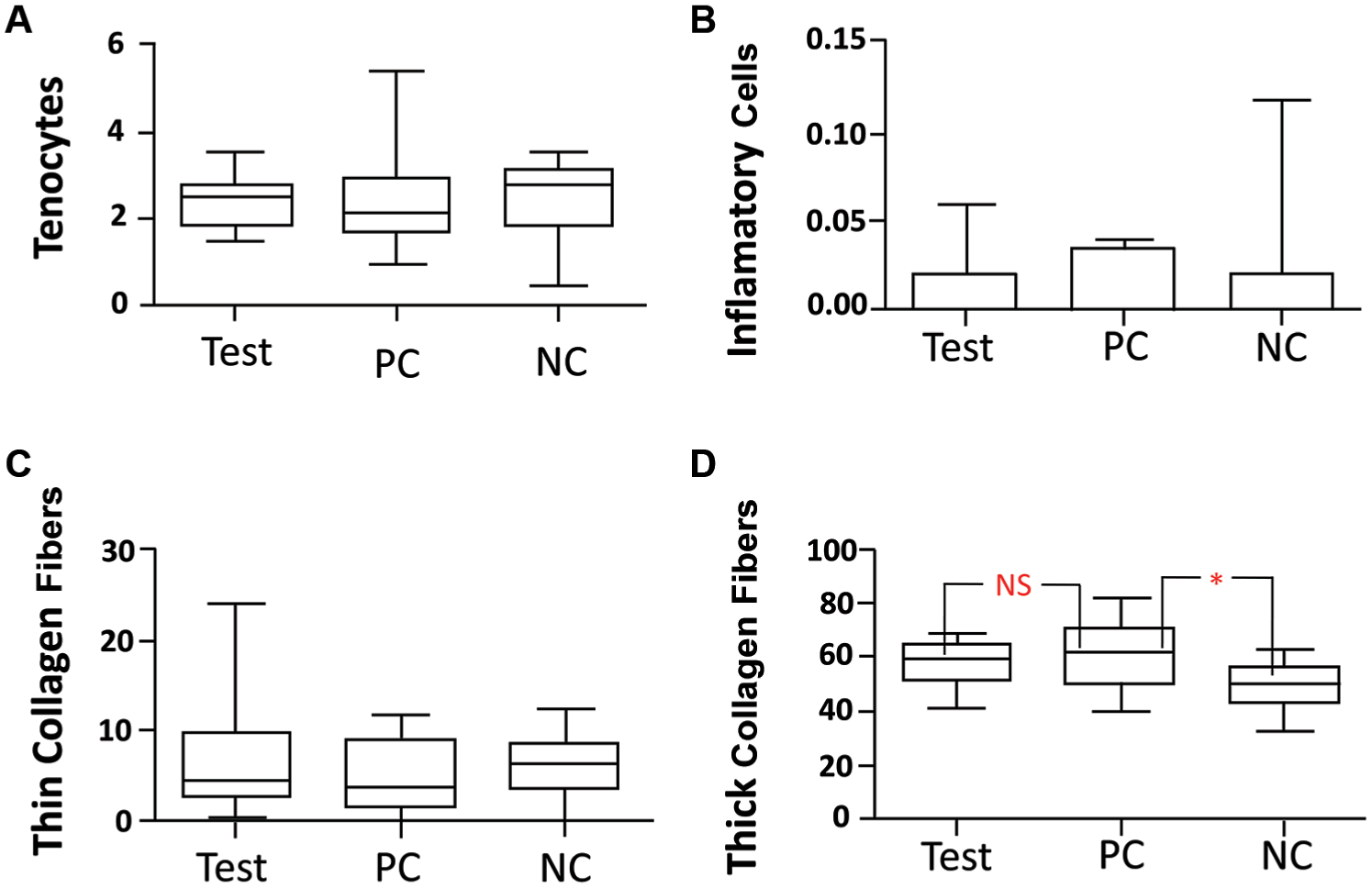
Box plots of medians of counts per 50 traces (Weibel reticle) for each histological finding. NC, normal control; NS, not significant; PC, placebo control. *P < .05.
A significant decrease in MMP2 expression (P = .027) was observed after CS injection in the test group as compared with the control groups (Table 3).
Comparison Among the Groups in Relation to MMPs and ILs a
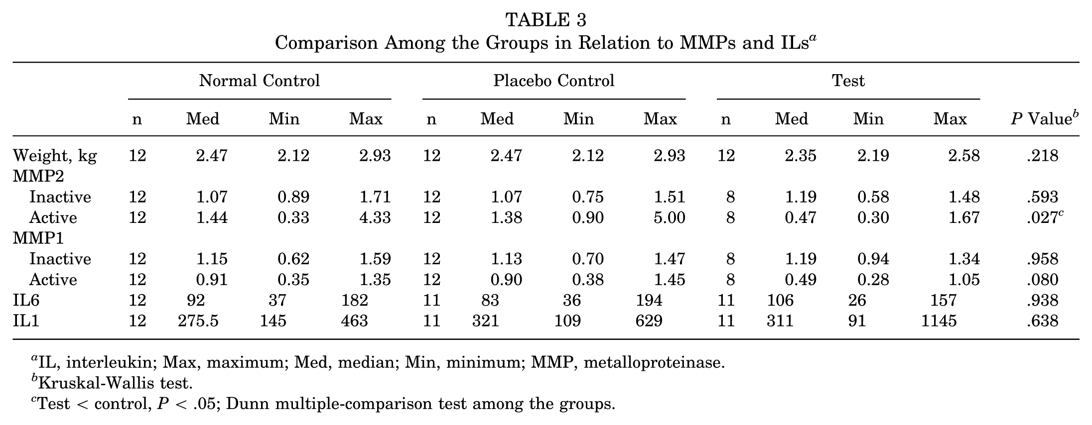
IL, interleukin; Max, maximum; Med, median; Min, minimum; MMP, metalloproteinase.
Kruskal-Wallis test.
Test < control, P < .05; Dunn multiple-comparison test among the groups.
There were no significant differences in IL1 and IL6 levels in the tendon tissues between the groups (Table 3).
Discussion
A single injection of CS into healthy calcaneal tendons did not trigger local morphological structural or biomechanical injuries at 48 hours, but it did promote a decrease in MMP2 levels, which is usually associated with a reduction in collagen degradation.
The short time point used in the present study (48 hours) was chosen to compare with the important alterations reported in pioneering studies,5,12 which raised some concerns about the use of CS injections for tendinopathies in clinical practice. These pioneering studies of single intratendinous injections of CS in the normal calcaneal tendons of rabbits showed “collagen necrosis” and a lower mechanical resistance after 48 hours, which were not confirmed in the present study, despite the use of similar drug, dosage, and volume. It is important to consider that, at the time of these pioneering studies, the needles were not disposable and did not have a sharp cutting tip and that tendon puncture might be associated with tissue injury and ruptures. 22 However, a systematic review demonstrated consistent findings in high-quality clinical controlled trials that CS injections reduced pain in the short term (0-5 days) as compared with other inteventions. 6
In another systematic review, 18 animal studies provided adequate data on mechanical properties of tendons after CS injections. 9 Of these, 6 showed a decrease in mechanical properties; 3, an increase; and the remaining 9, no significant change. Our study corroborates these last 9 studies. However, the heterogeneity in CS, dosages, injection techniques, and animals precludes any safe conclusion.
Previous histological studies of tissues in or around CS injections in rabbits did not include standard histomorphometry.2,5,12,29,31,33 Additionally, findings were variable in the calcaneal tendon, as follows: collagen necrosis,5,12,33 inflammatory processes,2,19,31 traumatic injuries owing to repetitive injections caused by needles,19,27,31 or no relevant change. 29 The present study demonstrated no significant differences in inflammation, biomechanical properties, or histological characteristics (ie, collagen necrosis).
MMPs, which are enzymes produced mainly by tenoblasts, fibroblasts, and leukocytes, play a critical role in the remodeling and repair of tendinopathies by degrading and repairing components of the extracellular matrix of tendons. In vitro studies from patient samples with tendinopathies showed both an increase 28 and a decrease 32 in MMP2 expression or demonstrated no change. 14 The expression of MMP is induced by growth factors and interleukins and the activity of phagocytosis by macrophages, among others, but it is inhibited by steroids. 13 In the present study, MMP1 and MMP2 expression analysis was chosen because of their collagen cleavage, such as collagen types I and III (most abundant in tendons). 13
Tendon remodeling is mediated by a complex inflammatory system in tendinopathies. The early response to overload is induced by macrophages, interleukins, and cyclooxygenases, as demonstrated by in vitro studies.15,20 An increase in macrophages 20 and IL6 expression 15 has been observed. In addition, an IL6 increase was found in the interstitial microdialysis liquid after calcaneal tendon rupture repair. 1 However, we did not find in the present study differences in IL1 and IL6 levels 48 hours after CS injection.
There are some limitations of this study. One is the early follow-up period (48 hours), which was chosen to compare with the results of pioneering studies that found important tendon alterations after a single CS injection into the tendon in the same period.5,12 Besides, there is evidence for the clinical effectiveness of CS injection in the short term, and the half-life of betamethasone is within this period (36-72 hours). 4 Long-term analyses would be interesting but would need the same experimental design and another 72 animals as the sample size. Another drawback is the single injection because, in clinical practice, many orthopaedic surgeons prescribe multiple injections, although tissue injury may increase with this technique. 22 Also, we lost 9 specimens during the experiment, and bias concerns would be raised. However, we considered the final sample size of each group as being appropriate based on calculations including dropouts and based on the sample sizes of previous studies. Finally, we did not use an experimental tendinopathic model instead of a normal tendon, because there is not a validated animal model 10 and the application of the findings to clinical practice would need careful judgment. Furthermore, it would be interesting to test multiple injections, increasing dosages, and long-term effects, but for the same double-blind design, it would be necessary to employ a larger number of animals.
Nevertheless, the strength of the present study was the double-blind design and the biochemical, biomechanical, and histological outcomes all together in the same experimental set. This is, to our knowledge, the first double-blind randomized controlled study with CS injection into the normal tendons of animals.
In conclusion, single injections of CS into the healthy calcaneal tendon did not trigger acute local morphological structural or biomechanical injuries at 48 hours but did promote a decrease in MMP2 levels, which is usually associated with a reduction in collagen degradation. Additional studies are needed with increased duration of follow-up, with various doses and multiple injections, and in tendinopathic models.
Footnotes
Acknowledgements
We thank Helio Rubens Nunes, PhD, for the statistical analysis, the librarians Marlucci Betini and Diva Luvizotto Gasperini Rodrigues for the bibliographic search, and Luis Edvalter Bardella for the technical assistance.
The authors declared that they have no conflicts of interest in the authorship and publication of this contribution. AOSSM checks author disclosures against the Open Payments Database (OPD). AOSSM has not conducted an independent investigation on the OPD and disclaims any liability or responsibility relating thereto.
References
Supplementary Material
Please find the following supplemental material available below.
For Open Access articles published under a Creative Commons License, all supplemental material carries the same license as the article it is associated with.
For non-Open Access articles published, all supplemental material carries a non-exclusive license, and permission requests for re-use of supplemental material or any part of supplemental material shall be sent directly to the copyright owner as specified in the copyright notice associated with the article.