
Abstract
Background:
Arthroscopic repair of delaminated rotator cuff tears (RCTs) has shown poor prognoses. Despite the importance of delaminated tears, only a few studies have compared delaminated and nondelaminated tears.
Purpose:
This study aimed to compare the clinical outcomes and structural integrity after en masse arthroscopic rotator cuff repair between delaminated and nondelaminated RCTs and to evaluate whether infraspinatus tendon involvement affects the prognosis for delaminated tears after arthroscopic cuff repair, through use of propensity score matching for precise comparison.
Study Design:
Cohort study; Level of evidence, 3.
Methods:
This study included 180 consecutive patients with medium- or large-sized RCTs who had an arthroscopic rotator cuff repair with a minimum 2-year follow-up, of whom 57 and 123 had delaminated tears (group 1) and nondelaminated tears (group 2), respectively. The en masse repair technique using a single-row or transosseous-equivalent double-row suture-bridge technique was used for of all the delaminated cases. Preoperative and postoperative visual analog scale pain scores, shoulder active range of motion, American Shoulder and Elbow Surgeons (ASES) scores, and Constant scores were assessed. Magnetic resonance imaging was performed at least 24 months postoperatively to identify retear of the repaired rotator cuffs. After propensity score matching, 32 cases in both groups were successfully matched, and the clinical and radiological results were analyzed.
Results:
Before propensity score matching, postoperative clinical outcomes were improved, showing no significant differences between the groups, excluding forward elevation (P = .011). Groups 1 and 2 had 17 (29.8%) and 11 retear cases (8.9%), respectively (P < .001). After propensity score matching, only the ASES score (72.5 vs 77.1) showed a significant superiority in group 2 (P = .038). Propensity-matched groups 1 and 2 had 8 (25.0%) and 2 (6.3%) retear cases, respectively (P = .034). No significant difference was found in structural integrity depending on whether the RCT included the infraspinatus tendon (IST). The odds ratio for retear of the delaminated tears, including IST, was 5.5 (95% confidence interval, 1.0-30.0, P = .038).
Conclusion:
Delaminated RCT was a negative prognostic factor of structural integrity after repair and could affect the functional outcome. However, whether IST tear was involved had no effect on the prognosis after repair.
Delamination of rotator cuff tears (RCTs) has been considered a negative prognostic factor after tear repair in several studies. Delamination, defined as the retraction of intratendinous fiber and horizontal intrasubstance tears, is mainly found in advanced degenerative tears.14,43 Boileau et al 4 showed that delamination of the subscapularis or infraspinatus tendon was negatively associated with tendon healing. Flurin et al 12 also reported that delamination of the supraspinatus and infraspinatus correlated with the extent of leakage of contrast medium on computed tomography or magnetic resonance imaging (MRI) arthrography. 12 However, comparative studies between delaminated and nondelaminated RCTs are rare. In addition, a recent study by Kim et al 22 comparing delaminated and nondelaminated RCTs showed that nondelaminated tears had a higher retear rate and that delamination was not a negative prognostic factor. Thus, whether delamination of the tear is a negative prognostic factor is not known.
In addition, the retear rate is higher in large and massive RCTs than in small and medium tears. 41 Because the articular layer tends to be retracted more than the bursal layer,32,43-45 repair of delaminated tears is more difficult than that of nondelaminated tears.4,12 Tanaka et al 46 reported that the larger the size of the RCT, the more frequently the delamination was limited to the posterior part. Those authors suggested the importance of the posterior and deep layer of the rotator cuff by showing that careful repair of the deep layer could prevent retear of the deep layer and improve prognosis. It is important to determine whether the retear rate is high in large and massive RCTs, including the infraspinatus, because of the effect of delamination involved in these tears. Therefore, the importance of delamination as a prognostic factor that affects retear can be confirmed by distinguishing the differences between the RCTs, including the infraspinatus tendon with and without delamination.
Thus, the purpose of this study was to compare clinical outcomes and structural integrity after arthroscopic rotator cuff repair between delaminated and nondelaminated RCTs and to evaluate whether involvement of the infraspinatus tendon affects the prognosis for delaminated tears after arthroscopic cuff repair; propensity score (PS) matching was used for precise comparison.
Methods
Study Design and Setting
This study was approved by the Institutional Review Board of Gil Medical Center, Gachon University, Incheon, Republic of Korea (GCIRB2017-232). In this retrospective cohort study, patients were selected from 1526 patients with medium or large RCT among 3572 patients who underwent arthroscopic rotator cuff repair by one senior surgeon (Y.K.K.) between February 2007 and August 2014. Among them, patients available for a minimum 2-year postoperative follow-up MRI were selected. Patients are always recommended follow-up MRI at least 2 years postoperatively. However, due to the high cost, the number of patients undergoing follow-up MRI was limited. Thus, 180 patients who underwent follow-up MRI were confirmed. The 180 confirmed patients had a completely torn supraspinatus tendon and underwent arthroscopic complete rotator cuff repair with either the transosseous-equivalent double-row suture-bridge (double-row) or single-row technique. Cases of partial-thickness RCTs, concomitant complete tears of the subscapularis tendon, and irreparable RCTs were excluded because a concomitant complete subscapularis tear was often a result of trauma rather than degeneration.
Preoperative Patient Characteristics
The patient-related factors used in this study were sex, age, dominant hand, duration of symptoms, type of work, smoking status, body mass index (BMI), diabetes mellitus (DM), and dyslipidemia, most of which have been reported to affect rotator cuff tendon healing (Table 1).30,36,37,39,47 The type of work was classified as high, medium, or low as follows: heavy manual labor requiring notable shoulder activity was considered high demand; manual labor with less activity was considered medium demand; and sedentary work was considered low demand. 6 BMI was calculated with the equation of weight in kilograms divided by height in meters squared. As a measure of relative weight, BMI is an acceptable proxy. 3 We assumed that the BMI of each patient was consistent during the study period. Patients were assigned to the DM cohort if they had been diagnosed with diabetes by their physician in accordance with the American Diabetes Association criteria as follows: fasting blood glucose level higher than 25 mg/dL or a random glucose level higher than 200 mg/dL and current use of oral or injectable antihyperglycemic medications. Patients were assigned to the non-DM cohort if they did not have a diagnosis of diabetes. 9 The National Cholesterol Education Program defines hypercholesterolemia (also known as dyslipidemia) as a blood cholesterol concentration of 240 mg/dL or higher, with concentrations between 200 and 239 mg/dL considered borderline high.1,19 To evaluate the cholesterol level, we defined the population with dyslipidemia as having a fasting total cholesterol concentration of more than 240 mg/dL.
Preoperative Data of the Patients (n = 180) a
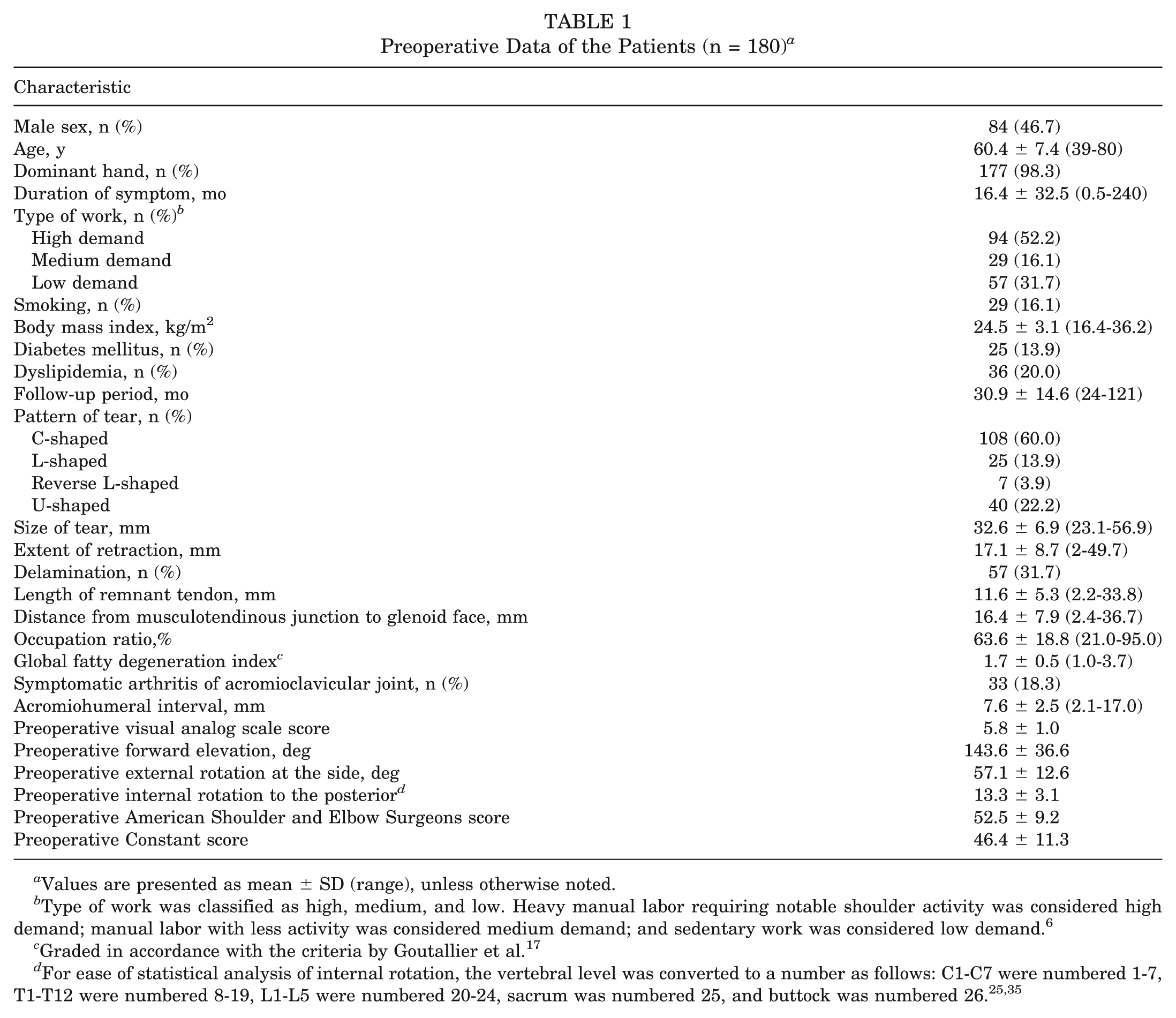
Values are presented as mean ± SD (range), unless otherwise noted.
Type of work was classified as high, medium, and low. Heavy manual labor requiring notable shoulder activity was considered high demand; manual labor with less activity was considered medium demand; and sedentary work was considered low demand. 6
Graded in accordance with the criteria by Goutallier et al. 17
Clinical Assessment
This study used 3 outcome measures: the visual analog scale (VAS) score for pain, the shoulder index of the American Shoulder and Elbow Surgeons (ASES), and the Constant score. ASES and Constant scores range from 0 to 100. Active range of motion of the shoulder was measured, including forward elevation (FE), external rotation (ER) with the arm at the side, and internal rotation (IR) at the back. We converted the vertebral level to a number for ease of statistical analysis of IR as follows: C1 through C7 were numbered 1 through 7, T1 through T12 were numbered 8 through 19, L1 through L5 were numbered 20 through 24, sacrum was numbered 25, and buttock was numbered 26.25,35
Radiological Assessment
All patients underwent preoperative and postoperative follow-up imaging with MRI with a 3.0-T scanner (Skyra; Siemens Medical). Oblique coronal, oblique sagittal, and axial T2-weighted images were acquired for the assessment of the rotator cuff tendons. The slice thicknesses on the coronal, sagittal, and axial views were 3, 4, and 3 mm, respectively. 25
All radiological assessments were performed twice by 2 board-certified orthopaedic surgeons with more than 5 years of experience. Intraobserver and interobserver measurement reliabilities were assessed by use of the Cohen κ value for the categorical variables and the intraclass correlation coefficient for the continuous variables. 24
The factors related to the tendon itself were the type of tendon that was torn, pattern of the tear, size of the tear, extent of retraction, delamination, length of the remnant tendon, and distance from the musculotendinous junction (MTJ) to the face of the glenoid. Tears were classified as C-shaped, L-shaped, reverse L-shaped, or U-shaped on the basis of the preoperative MRI and arthroscopic findings, according to Ellman 11 and Davidson and Burkhart. 8 The size of the tear was calculated as the linear distance between the anterior and posterior margins of the tear on oblique sagittal T2-weighted images. The extent of retraction was measured by use of the straight line distance between the medial margin of the footprint of the supraspinatus and the medial margin of the retracted cuff on oblique coronal T2-weighted images. 25 The length of the remnant tendon was measured as the distance from the lateral edge of the supraspinatus tendon to the MTJ, and the position of the MTJ in relation to the glenoid face line (the distance from the glenoid face and the binary variable of the medial-lateral position) was evaluated on the oblique coronal T2-weighted images. 48
Parameters for muscle atrophy and fatty change included the occupation ratio and global fatty degeneration index (GFDI). To evaluate the atrophy of the supraspinatus muscle, we calculated the occupation ratios of the muscle belly in the supraspinatus fossa by using the method of Tashjian et al. 48 The GFDI was calculated to evaluate the fatty degeneration in the supraspinatus, infraspinatus, and subscapularis muscles as the mean value of the grade for each muscle, in accordance with the method described by Goutallier et al. 18
Bony factors were symptomatic arthritis of the acromioclavicular joint (ACJ) and acromiohumeral interval (AHI). The AHI was measured as the distance between the lower margin of the acromion and the upper margin of the humeral head by use of anteroposterior shoulder radiographs, in accordance with Golding’s 16 method. ACJ arthritis was evaluated on the basis of joint space narrowing, subchondral cysts, and presence of osteophytes. Zanca view and MRI were used to aid in the evaluation. The authors confirmed symptomatic ACJ disease on the basis of ACJ tenderness, pain, and positive cross-body adduction test result. 15
RCT sizes, including the dimensions of the delamination, were evaluated on preoperative MRI. The dimension of the delamination was calculated by use of the coronal size of the delaminated portion. 20 Delaminated tear was defined as the presence of interstitial splitting of one tendon by 5 mm or more on fat-suppressed T2-weighted coronal MRI. We confirmed cleavage tearing on arthroscopy. Tendon integrity was assessed on postoperative MRI in accordance with the criteria of Sugaya et al. 44 Types 4 and 5 of Sugaya’s categories were classified as retears.
Surgical Procedures
All the surgeries were performed by a single senior author. All operative procedures were performed with the patient in the beach-chair position, sitting at an angle of 70° under general anesthesia. The standard posterior and anterior portals were used for diagnostic examinations. Appropriate treatment such as a repair or debridement was performed in a case of coexisting subscapularis leading edge tear or labral lesion. For biceps tendon lesions, either a biceps tenotomy or tenodesis was performed, depending on the patient’s age and daily activities. The size of the tear and extent of retraction, as assessed on preoperative MRI, were confirmed by measurement using a length-marked probe. If a delaminated cuff tear was present, the intratendinous cleavage was debrided with a shaver to expose the fresh tissue between the bursal and articular layers of the delaminated tendon. The repair technique, either single- or double-row, was determined based on the excursion of the torn tendon and tear pattern. If the length of the remnant tendon was less than 10 mm and the tendon mobility was not sufficient to cover the anatomic footprint despite adequate soft tissue release, single-row repair was performed. 26 Sutures were tied with the SMC (Samsung Medical Center) knot technique. Anchors (CrossFT; ConMed Linvatec) were routinely placed in the midportion of the rotator cuff footprint, halfway between the articular margin and the lateral edge of the tuberosity, in an effort to obtain good footprint coverage without excessive tension in a single-row repair (see the online Video Supplement for this technique).
In a double-row repair, anchors (CrossFT Suture Anchors) for the medial row were inserted at the sulcus just lateral to the margin of the articular surface. All suture limbs were passed 2 to 3 mm lateral to the musculotendinous junction by use of a retrograde shuttle relay technique with a suture-passing device. After the medial-row sutures were tied, knotless anchors (PopLok Knotless Suture Anchors; ConMed Linvatec) for the lateral row were placed distal to the greater tuberosity with sutures under tension to compress across the footprint.25,48
For delaminated torn tendons, sutures were passed through both the articular and bursal layers while the torn tendon was pulled with a grasper to the prepared footprint and maintained in place. Once the suture was passed through both layers, the torn tendon was attached through use of the single- or double-row technique (Figure 1).

For a delaminated torn rotator cuff, the en masse repair technique was used. (A) Single-row repair technique. (B) Transosseous-equivalent double-row suture-bridge repair technique.
Rehabilitation
An immobilizing abduction brace was applied immediately after surgery. On the first day after surgery, pendulum and deltoid isometric exercises were gently performed. At 3 weeks after surgery, passive range of motion stretching exercises were started. Active-assisted range of motion exercises were started 6 weeks after surgery. Strengthening exercises were allowed 3 months after surgery. 26
Statistical Analysis
Because the patients in this study were not randomly assigned to the group of delaminated tears, we used PS matching to reduce the selection bias and the effect of potential confounding factors. 7 PS matching included all preoperative and intraoperative parameters examined in this study that may have affected the outcome of the surgery, and the matching was performed based on the calculated propensity score. Therefore, we examined the differences in all of the pre- and intraoperative parameters between the delaminated and nondelaminated tear groups before PS matching. Before PS matching, the preoperative characteristics in the group with delaminated RCT were compared with those of the group with nondelaminated tears by use of the Student t test for continuous variables (age, symptom duration, BMI, cuff tear size, degree of retraction, remnant tendon length, MTJ-glenoid distance, occupation ratio, GFDI, AHI, and all clinical outcomes) and the chi-square or Fisher exact test for categorical variables (sex, dominant hand, labor intensity, smoking, DM, hyperlipidemia, tear pattern, symptomatic ACJ arthritis, single-row, procedure for long head of biceps tendon, subscapularis repair, arthroscopic subacromial decompression, and retear). 22 However, in several variables, more than 20% of the expected counts of each cell on the contingency table were less than 5. For those cases, the Fisher exact test was performed. The data table was larger than 2 × 2. Thus, the Freeman-Halton extension of the Fisher exact test was used, instead of the traditional Fisher exact test.13,34 After PS matching, the matched data between the delaminated and nondelaminated tear groups were analyzed through use of a paired t test for continuous variables and the McNemar test for categorical variables. The retear difference was evaluated according to whether the infraspinatus tear was included, and the Cochran-Mantel-Haenszel test was performed with a striated analysis, including the Breslow-Day test. PS matching was performed with a 1:1 greedy matching algorithm (8- to 1-digit match). After matching, the standardized mean difference was obtained for validation and was found to be less than 0.1 for all the variables. Intraobserver and interobserver reliabilities were excellent for all the parameters (intraclass correlation coefficient, >0.75; range, 0.81-0.87; Cohen’s kappa value, >0.8; range, 0.83-0.92).28,29 All statistical tests were 2-tailed, and a P value of less than .05 was considered statistically significant. Statistical analyses were performed with SAS version 9.4 (SAS Institute Inc).
Results
Before the PS matching, several preoperative characteristics between the delaminated and nondelaminated tear groups showed significant differences. Of the patients, 23 men and 34 women had a delaminated RCT, and 61 men and 62 women had a nondelaminated RCT (P = .248). The mean ± SD ages were 62.4 ± 7.0 and 59.4 ± 7.5 years, respectively (P = .012). Significant differences were found between the 2 groups in terms of tear patterns (P < .001), tear size (P < .001), extent of retraction (P < .001), distance from MTJ to the glenoid face (P < .001), and GFDI (P = .002) (Table 2).
Preoperative Comparison Between the Delaminated and Nondelaminated Groups Before and After Propensity Score Matching a
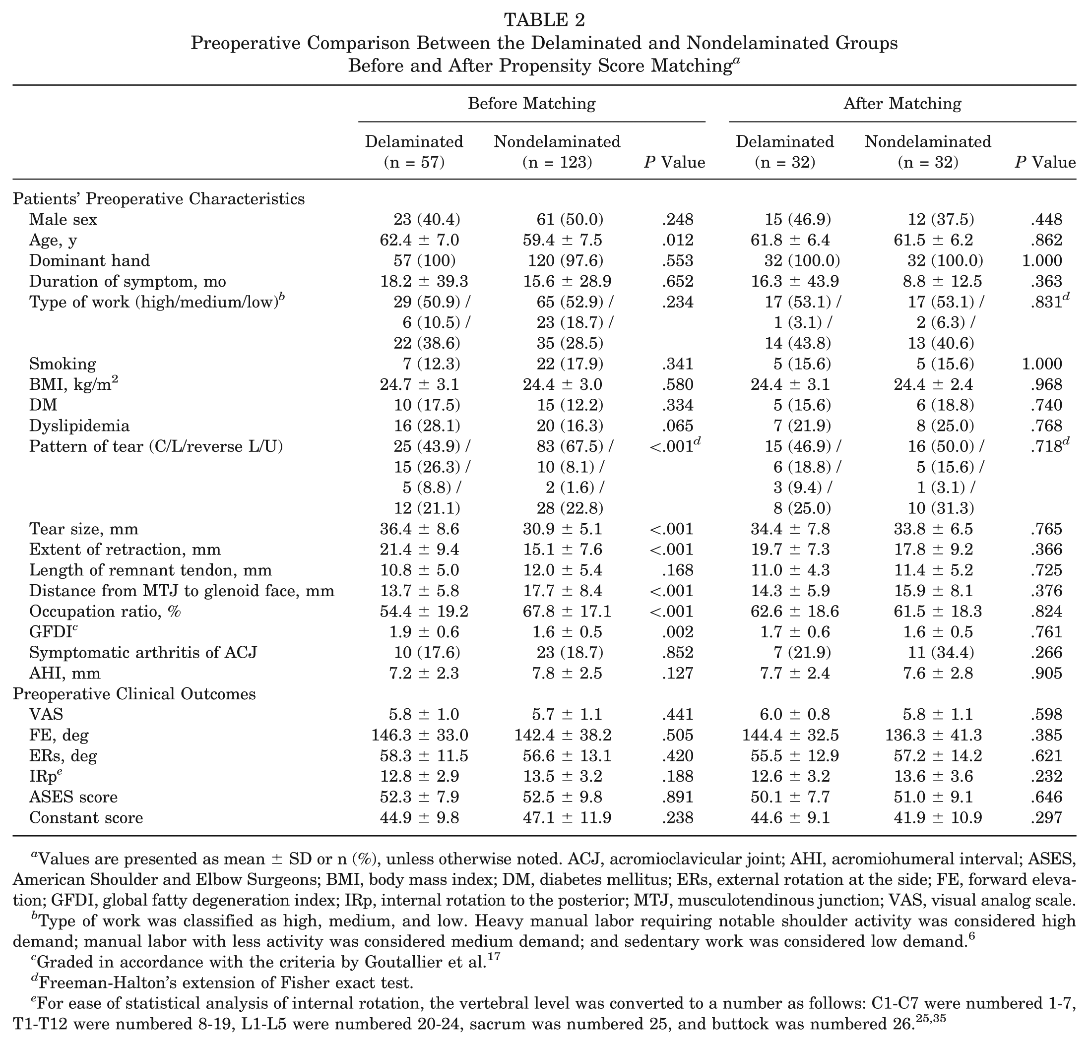
Values are presented as mean ± SD or n (%), unless otherwise noted. ACJ, acromioclavicular joint; AHI, acromiohumeral interval; ASES, American Shoulder and Elbow Surgeons; BMI, body mass index; DM, diabetes mellitus; ERs, external rotation at the side; FE, forward elevation; GFDI, global fatty degeneration index; IRp, internal rotation to the posterior; MTJ, musculotendinous junction; VAS, visual analog scale.
Type of work was classified as high, medium, and low. Heavy manual labor requiring notable shoulder activity was considered high demand; manual labor with less activity was considered medium demand; and sedentary work was considered low demand. 6
Graded in accordance with the criteria by Goutallier et al. 17
Freeman-Halton’s extension of Fisher exact test.
After PS matching, in each group, 32 patients were matched for the analysis. The preoperative baseline VAS score, ASES score, Constant scores, and range of motion did not differ significantly between the nondelaminated and delaminated tear groups (Table 2). The analysis of the intraoperative characteristics between the 2 groups showed a significant difference only in the biceps procedures (P < .001). After PS matching, no statistically significant difference was found between the 2 groups (Table 3).
Intraoperative Comparison Between the Delaminated and Nondelaminated Group Before and After Propensity Score Matching a
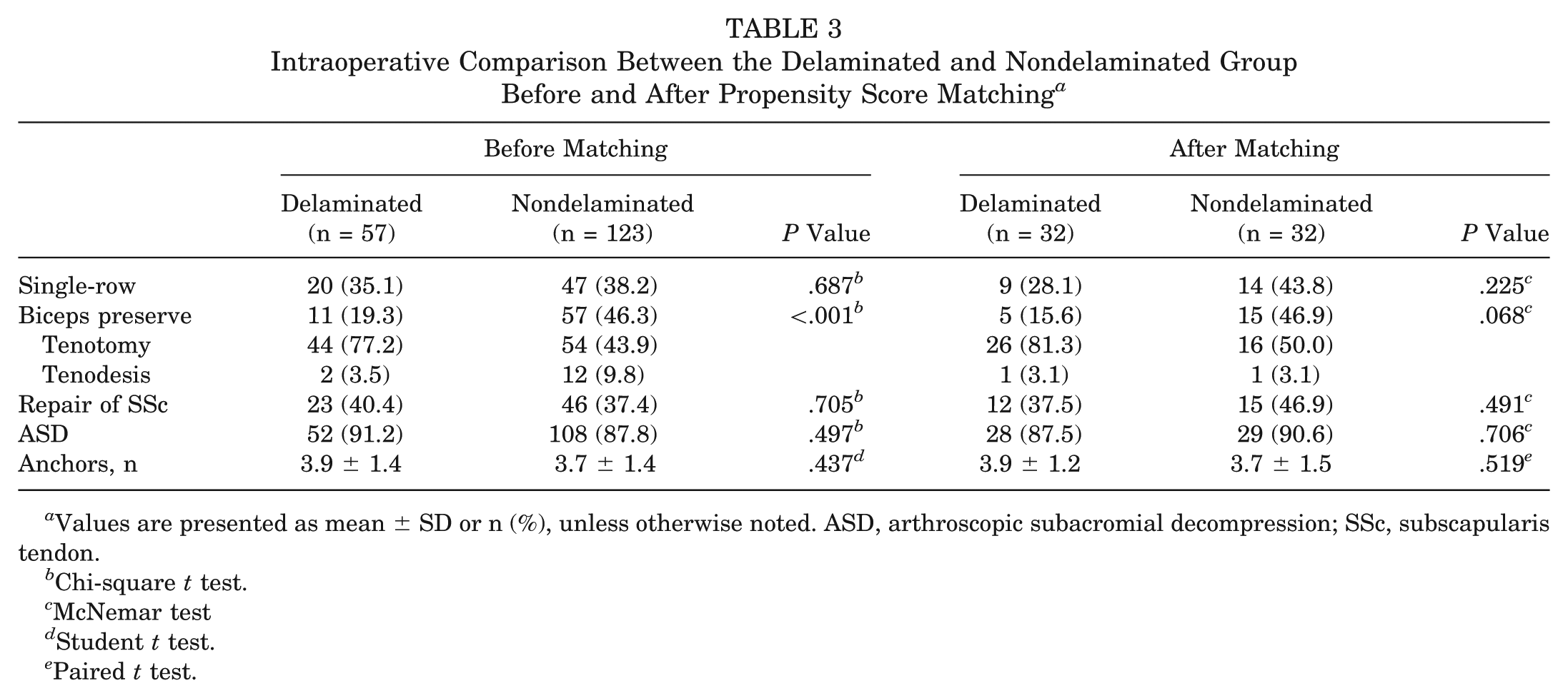
Values are presented as mean ± SD or n (%), unless otherwise noted. ASD, arthroscopic subacromial decompression; SSc, subscapularis tendon.
Chi-square t test.
McNemar test
Student t test.
Paired t test.
Single- and double-row repair groups after PS matching were also analyzed according to the delamination. After the PS matching, no statistically significant differences were found in the values of each variable depending on whether delamination was present in each group in which single- and double-row repairs were performed. Especially, the identified patterns of tear (C-shape, L-shape, reverse L-shape, and U-shape) in the patients receiving single-row repair accounted for 3 (33.3%), 5 (55.6%), 1 (11.1%), and 0 (0%) cases in the delaminated tear group and 8 (57.1%), 3 (21.4%), 0 (0%), and 3 (21.4%) cases in the nondelaminated tear group, respectively (P = .113). In the patients receiving double-row repair, the above patterns of tear were 12 (52.2%), 1 (4.4%), 2 (8.7%), and 8 (34.8%) in the delaminated group and 8 (44.4%), 2 (11.1%), 1 (5.6%), and 7 (44.5%) in the nondelaminated group, respectively (P = .816).
The postoperative characteristics of the 2 groups before PS matching showed a significant difference only in forward elevation (P = .011). After PS matching, a significant difference was found only in the ASES score (P = .038) (Table 4). Especially forward elevation was improved in both groups after operation. In addition, all functional clinical scores were improved at the final follow-up compared with the preoperative values.
Postoperative Comparison Between the Delaminated and Nondelaminated Groups Before and After Propensity Score Matching a
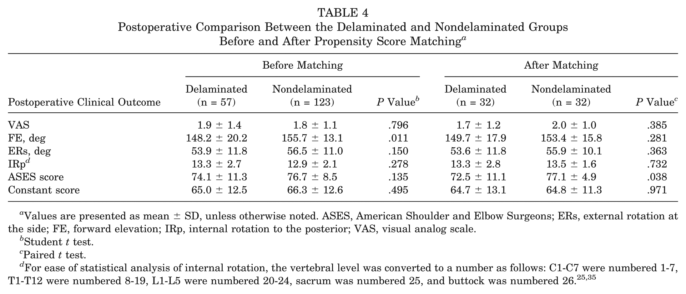
Values are presented as mean ± SD, unless otherwise noted. ASES, American Shoulder and Elbow Surgeons; ERs, external rotation at the side; FE, forward elevation; IRp, internal rotation to the posterior; VAS, visual analog scale.
Student t test.
Paired t test.
Before PS matching, the delaminated tear group had 17 retear cases (29.8%) and the nondelaminated tear group had 11 retear cases (8.9%; P < .001). After PS matching, the groups respectively had 8 (25%) and 2 retear cases (6.3%), which confirmed the significantly higher retear rate in the delaminated tear group (P = .034). We analyzed the effect of delamination on the retear according to whether the single- or double-row repair was used and whether delamination was present. The delaminated tear group showed an odds ratio of 5.4 (95% confidence interval, 1.011-28.683) for retear (P = .035), regardless of whether the single- or double-row repair was used. This means that the repair technique did not have an effect on the retear rate in delaminated tears. The odds ratio of the delaminated tear group was 5.5 (95% confidence interval, 1.013-29.976) for retear (P = .038), irrespective of infraspinatus tendon involvement. This shows that whether the infraspinatus tear was included did not affect the retear rate (Appendix Tables A1 and A2, available in the online version of this article).
Discussion
The delamination rate of RCTs has been reported with a wide range, with Sonnabend et al 42 reporting a rate of 38% after open repair, compared with Boileau et al 4 and MacDougal and Todhunter 31 reporting rates of 51% and 71%, respectively, after arthroscopic repair. Flurin et al 12 reported a delamination rate of 57% for full-thickness rotator cuff tears, whereas Matsuki et al 32 showed the rate to be as high as 82%. In the present study, the delamination rate of full-thickness tears of the rotator cuff was 32%.
In the present study, the delaminated tear group showed a higher retear rate, but the presence of infraspinatus tears did not affect the retear rate. Several studies have shown that delaminated tears contribute to a relatively high retear rate, but clinical outcomes after rotator cuff repair were tolerable. Park et al 38 reported a retear rate of 25% after the arthroscopic en masse suture bridge was used for massive RCTs, and preoperative fatty degeneration of the supraspinatus was associated with a high retear rate. Although larger retears had poor functional outcomes, smaller retears showed good outcomes. 38 Gwak et al 20 showed that the incidence rates of retear and partial defect healing were 27.4% and 16.1%, respectively, after en masse repair of the delaminated rotator cuffs. In a study that compared en masse repair versus double-layer repair of delaminated tears, Kim YS et al 27 suggested that the group who received arthroscopic conventional en masse repair of delaminated tears showed a 17% retear rate. All clinical scores were significantly improved, and no significant differences were found between the en masse group and the double-layer double-row repair group, except for VAS pain score. 27 Cha and colleagues 5 evaluated the repair integrity after dual-layer repair of delaminated cuff tears. Even though retear was found in 7.6% and 27.2% of patients in the dual-layer and single-layer repair groups, respectively, no clinically significant difference was found between the dual- and single-layer repair groups. 5 The present study showed that the retear rate in the delaminated group was 25%, which was higher than that in the nondelaminated tear group (6.3%). No significant differences were found between the delaminated and nondelaminated tear groups, except for ASES score.
Some studies have suggested that RCT including the infraspinatus tendon has worse prognosis. Agout et al 2 reported that a group with isolated supraspinatus tears showed a significantly lower retear rate than a group with RCT with posterior extension. The reason was estimated to be the significantly greater fatty degeneration in the infraspinatus than in the isolated supraspinatus, which could affect the occurrence of retear after rotator cuff repair. Shimokobe et al 41 showed that posterosuperior RCTs had a higher retear rate than anterosuperior RCTs. Especially, lower preoperative external rotation was a unique risk factor for retear, and a higher retear rate was related to tears of the infraspinatus and teres minor and the extent of their fatty degeneration. Cha et al 5 suggested that the superficial and deep layers were mainly retracted posteromedially and that the retraction of the deep and superficial layers may be affected by the infraspinatus. In the present study, however, no significant difference was found according to whether the infraspinatus was involved in the delaminated tear. In general, before PS matching, the range of RCTs included massive cuff tears; however, after matching, medium to large tears accounted for a large proportion of RCTs, but not large to massive tears. In PS matching, the 2 groups to be compared in the process of selecting a similar PS tend to include cases where the numerical values of various factors are within a similar range. Thus, the difference in the results of this study and those of other studies is considered to be due to the difference in statistical methodological characteristics. If matching was performed with a high proportion of infraspinatus and teres minor tears, the present results could have been similar to those of other studies.
Several studies have compared results among the repair techniques in delaminated cuff tears. Cha et al 5 reported that a group of patients receiving dual-layer repair had a lower retear rate than a group receiving single-layer repair in delaminated cuff tears. Most of their study subjects had medium-size tears, similar to the tear size after PS matching in this present study. Kim YS et al 27 showed no significant difference in retear rate between conventional en masse repair and separate double-layer double-row repair in delaminated tears. Kim SJ et al 23 reported no significant difference in retear rate between a group receiving all-layer suture repair and another group receiving bursal layer repair for delaminated tears. The above studies have shown varying results in retear rate according to the repair technique. However, as far as we are aware, no comparative study of single- and double-row repair has been conducted using the en masse suture technique of the current study. In several systematic reviews comparing single- and double-row repair for RCTs, most studies showed that the retear rate was higher in single-row repair.10,21,33,40,49 However, these studies showed not the results for the delaminated tears only, but also reported the results for the various spectrums of rotator cuff tear. So it is difficult to directly compare them with the present study. In the present study, whether the single- or double-row repair was used did not affect the retear rate in delaminated tears.
The strength of this study is that the clinical and radiological results between the 2 patient groups after repair of delaminated and nondelaminated tears were compared by adjusting the patients’ baseline characteristics through PS matching. This study seems to be meaningful because few clinical papers have directly compared delaminated and nondelaminated tears.
However, even though it used PS matching, the present study has a fundamental limitation due to its retrospective design. Another disadvantage is that the present study was conducted using only the en masse suture method, even though various methods are available for repairing delaminated tears. The en masse suture technique entails repairing the 2 layers of the tendon simultaneously by passing the suture through the full thickness of the cuff. 38 It is possible to supply anatomic stability without placing suture materials between articular and bursal layers. However, this could cause tendon mismatch, and several studies have shown better results by using different suture techniques than en masse suture techniques alone.5,27 Additionally, all of the procedures in the present study were performed by a single surgeon; this consistency can be considered a strength of the study, or it may pose a limitation in that the results may not be generalizable to other surgeons.
Conclusion
Delaminated RCT was a negative prognostic factor for structural integrity after repair and could affect the functional outcome. However, whether the infraspinatus tendon in the tear was involved did not affect the prognosis after repair.
Supplemental Material
DS_10.1177_0363546519838257 – Supplemental material for Comparison of Structural Integrity and Functional Outcome Between Delaminated and Nondelaminated Rotator Cuff Tears After En Masse Arthroscopic Repair: A Retrospective Cohort Study With Propensity Score Matching
Supplemental material, DS_10.1177_0363546519838257 for Comparison of Structural Integrity and Functional Outcome Between Delaminated and Nondelaminated Rotator Cuff Tears After En Masse Arthroscopic Repair: A Retrospective Cohort Study With Propensity Score Matching by Young-Kyu Kim, Kyu-Hak Jung and Hyuk-Min Kwon in The American Journal of Sports Medicine
Footnotes
The authors declared that they have no conflicts of interest in the authorship and publication of this contribution. AOSSM checks author disclosures against the Open Payments Database (OPD). AOSSM has not conducted an independent investigation on the OPD and disclaims any liability or responsibility relating thereto.
A Video Supplement for this article is available online.
References
Supplementary Material
Please find the following supplemental material available below.
For Open Access articles published under a Creative Commons License, all supplemental material carries the same license as the article it is associated with.
For non-Open Access articles published, all supplemental material carries a non-exclusive license, and permission requests for re-use of supplemental material or any part of supplemental material shall be sent directly to the copyright owner as specified in the copyright notice associated with the article.