
Abstract
Background:
Autologous chondrocyte implantation (ACI) is often performed for large cartilage defects. Because this technique has several disadvantages, including the need for second-stage surgery, cartilage repair using minced cartilage has been suggested. However, this technique could be improved using 3-dimensional scaffolds.
Purpose:
To examine the ability of chondrocyte migration and proliferation from minced cartilage in atelocollagen gel in vitro and evaluate the repairable potential of minced cartilage embedded in atelocollagen gel covered with a periosteal flap in a rabbit model.
Study Design:
Controlled laboratory study.
Methods:
Minced cartilage or isolated chondrocytes from rabbits were embedded in atelocollagen gel and cultured for 3 weeks. Chondrocyte proliferation and matrix production were evaluated in vitro. An osteochondral defect at the trochlear groove was created in 56 rabbits, which were divided into 4 groups. The defect was left empty (defect group), filled with allogenic minced cartilage (minced cartilage group), filled with isolated allogenic chondrocytes embedded in atelocollagen gel (ACI group), or filled with atelocollagen gel (atelocollagen with periosteal flap group). At 4, 12, and 24 weeks after surgery, repair of the defect was evaluated in these 4 groups.
Results:
In vitro, the number of chondrocytes and abundant matrix on the surface of the gel significantly increased in the minced cartilage group compared with the ACI group (P < .05). In vivo, the minced cartilage and ACI groups showed good cartilage repair compared with the empty defect and atelocollagen/periosteal flap groups (P < .05); there was no significant difference in the Pineda score between the minced cartilage and ACI groups.
Conclusion:
Minced cartilage in atelocollagen gel had good chondrocyte migration and proliferation abilities in vitro, and osteochondral defects were well repaired by implanting minced cartilage embedded in the atelocollagen gel in vivo. Implantation of minced cartilage embedded in atelocollagen gel showed good cartilage repair equivalent to ACI.
Clinical Relevance:
Implantation of minced cartilage embedded in atelocollagen gel as a 1-step procedure has outcomes similar to those of ACI but is cheaper and more convenient than ACI.
Among surgical procedures to restore articular cartilage, autologous chondrocyte implantation (ACI) has been conducted for large cartilage defects since Brittberg et al 6 introduced it to repair cartilage defects with hyaline cartilage. However, this technique has some problems, such as leakage of the cultured chondrocytes through a crack of the periosteum, uneven distribution of the cells by gravity, and dedifferentiation of the chondrocytes during monolayer culture. 29 To overcome these concerns, Ochi et al 29 developed a method to implant cartilage-like tissue, in which isolated chondrocytes are 3-dimensionally cultured in atelocollagen gel as scaffolds. Although these changes improved the procedure, a 2-stage surgery (harvesting of chondrocytes followed by implantation) is still needed. Moreover, isolation of chondrocytes requires enzymatic digestion, leading to the destruction of the normal cartilage matrix. A previous report showed that type 2 collagen, the main component of cartilage matrix and specific collagen, has an extremely limited turnover in an individual’s lifetime. 19
The concept by which cartilage fragments are implanted into the cartilage defect was first described in 1982, and an animal study demonstrated the significance of minced cartilage implantation as a cartilage repair procedure in 2006. 22 This procedure showed that minced cartilage could be successfully loaded onto a biodegradable scaffold and fixed by fibrin glue and then implanted into the cartilage defect. This easy procedure has been implemented in clinical settings, and good clinical results were reported.9,13 However, this procedure has only been used for cartilage defects with an area of approximately 2.75 cm2 or those with a defect size <4 cm2. 13 If minced cartilage is applied to a large cartilage defect, chondrocyte migration and proliferation from the minced cartilage within the scaffold are needed. We hypothesized that chondrocytes would migrate and proliferate in the atelocollagen gel scaffold and successfully repair the cartilage defect, which would represent a simple, less invasive, and cost-effective approach for the repair of cartilage defects. The purpose of this study was to examine the ability of chondrocyte migration and proliferation from minced cartilage in atelocollagen gel in vitro and to evaluate the repair potential of the minced cartilage embedded in atelocollagen gel covered with a periosteal flap in a rabbit model.
Methods
Animals
All procedures were performed according to the Guidelines for Animal Experimentation at Hiroshima University and with the approval of the university’s Committee of Research Facilities for Laboratory Animal Sciences, Graduate School of Biomedical Sciences. A total of 56 Japanese white rabbits weighing 2.5 to 3.2 kg were used. Under general anesthesia using ketamine hydrochloride and xylazine, surgery was performed. To evaluate the effect of the minced cartilage implantation on the osteochondral repair, the osteochondral defect model was created in both knees of 38 rabbits. Through a medial parapatellar approach, the patella was dislocated laterally, and an osteochondral defect 6 × 4 mm in width and 3 mm in depth was created using a 1-mm drill bit with low speed to avoid heat damage to the tissue according to a previous report. 21 Another 18 rabbits were used to harvest articular cartilage from the shoulder, hip, and knee joints. Harvested articular cartilage was washed 3 times using phosphate-buffered saline. Then, articular cartilage was manually minced in a petri dish using a scalpel to create cartilage measuring <1 mm3 according to a previous report. 4 Minced cartilage was used for the in vitro and vivo studies.
Culture
For the isolation of chondrocytes, cartilage specimens were treated with 0.25% trypsin (Gibco) in sterile saline for 30 minutes, followed by 0.25% collagenase type 2 (Gibco) in Dulbecco’s modified Eagle medium (Gibco), supplemented with 10% fetal bovine serum (FBS; Sigma) and antibiotics (penicillin [10,000 units] and streptomycin [10,000 units] μg/mL; Nacalai Tesque) for 4 hours at 37°C in a culture tube. The chondrocytes were washed 3 times with culture medium and then filtered through a 70-mm sterile nylon mesh (Cell Strainer; BD Biosciences Discovery Labware). Isolated chondrocytes (2.0 × 10⁵ cells) were dispersed and mixed in 100 μL atelocollagen gel (Koken). All cell mixtures were placed in a 6-well culture plate and incubated in a mixture of 5% CO2 and 95% air at a temperature of 37°C for 30 minutes to form atelocollagen composites. A culture medium containing 5% FBS and antibiotics was subsequently added into each well. Chondrocyte/atelocollagen composite was cultured for 3 weeks. The culture medium was changed every 3 days, and L-ascorbic acid (50 μg/mL) was added every 2 days. After 3 weeks, each composite was either implanted into the osteochondral defect (in vivo study) or fixed in 4% paraformaldehyde (PFA) for histological evaluation.
Then, 12.5 mg (M1 group) or 25 mg (M2 group) of minced cartilage was mixed in 100 μL atelocollagen gel and cultured for 3 weeks in the same manner described for isolated chondrocytes. After 3 weeks, minced cartilage/atelocollagen composite was fixed with PFA, and histological evaluation among 3 groups (isolated chondrocyte [IC], M1, and M2 groups) was performed.
Histological Evaluation In Vitro Study
Samples were fixed in 4% PFA at 4°C overnight and subsequently embedded in paraffin; 4 μM–thick sections were prepared and stained using hematoxylin and eosin (H&E) and Safranin O/Fast Green. Each sample was evaluated using the Bern score, as previously described. 18 The Bern score (minimum score, 0; maximum score, 9) is based on 3 items (uniformity and intensity of Safranin O staining; distance between cells/amount of matrix produced; and cell morphologic characteristics), with each item scored from 0 (minimum) to 3 (maximum).
To evaluate chondrocyte migration and proliferation in atelocollagen gel, 6 areas (500 × 500–μm grid) were randomly selected in each section, and chondrocytes were counted under a magnification of ×400.
Transplantation of Atelocollagen Composites In Vivo
A total of 38 Japanese white rabbits weighing 2.5 to 3.2 kg were used. Eight rabbits were excluded from this study because of patellar dislocation or infection. Thus, a total of 30 rabbits were finally included (Table 1). For the defect limbs, after the creation of the osteochondral defect, the patella was repositioned and the joint capsule and skin were closed. For treated limbs, the periosteal flap (7 × 5 mm) was harvested from the ipsilateral medial proximal tibia and was sutured to the peripheral cartilage rim of the defect using 5-0 nonabsorbable nylon sutures (Bear Medic Corporation) with the cambium layer facing the patella according to a previous report. 20 In the atelocollagen group, before periosteal coverage, 100 μL atelocollagen gel was placed into the defect. For the ACI group, isolated chondrocyte/atelocollagen composite cultured for 3 weeks was implanted into the defect. For the minced cartilage group, 100 μL atelocollagen gel containing about 10 mg minced cartilage without culture was implanted into the defect. Thereafter, the periosteal flap was sutured and the joint capsule and skin were closed.
Distribution of Animals by Treatment of Limbs and Time of Histologic Evaluation a
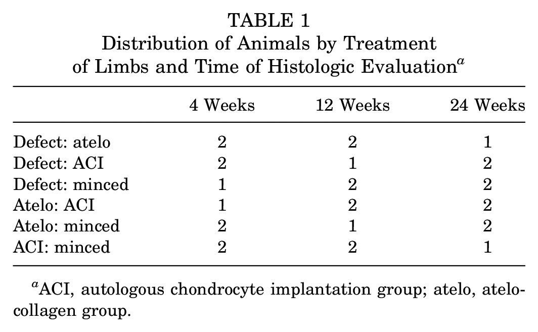
ACI, autologous chondrocyte implantation group; atelo, atelocollagen group.
Histological Evaluation In Vivo Study
At 4, 12, and 24 weeks after surgery, rabbits were sacrificed using sodium pentobarbital. At each time point, 5 knees in each group were evaluated. Knee joints were harvested and fixed with 4% PFA at 4°C overnight. Then, they were embedded in paraffin, and 4 μM–thick sections were prepared. H&E and Safranin O/Fast Green staining were performed. The Pineda score, ranging from 0 (best) to 14 (worst), was used for the semiquantitative analysis. 31 Histological grading was performed by 2 observers (T.N., M.I.) who were not aware of the source of the samples.
Immunohistochemistry
The sections were pretreated with antigen retrieval reagent (Immunoactive; Matsunami Glass Ind) for 1 hour followed by 0.3% H2O2 for 30 minutes, normal blocking serum for 30 minutes, and primary antibody against type 2 collagen (dilution, 1:100; anti-hCL (II); Daiichi Fine Chemical), type 1 collagen (dilution, 1:250; Novus Biologicals), and Ki67 (dilution, 1:400; Cell Signaling Technology) overnight at 4°C. On the next day, the sections were visualized using the avidin-biotin system (Vectastatin Elite ABC Mouse IgG kit; Vector Laboratories Inc) and 3,3′-diaminobenzidine (Peroxidase Substrate Kit; Vector Laboratories Inc), according to the manufacturer’s instructions.
For the evaluation of chondrocyte migration and proliferation in the atelocollagen gel after the implantation, 3 areas (300 × 300 μm grid) in the reparative tissue of each section were randomly selected and cells were counted under ×400 magnification. We calculated the mean rate of the Ki67-positive cells per all cells for each group.
Statistical Analysis
Values are given as the SEM. Differences in the Pineda scale in the 4 treatment groups were analyzed using the Kruskal-Wallis test and Steel-Dwass test. Differences in the Ki67-positive cell rate were analyzed using analysis of variance and the Tukey-Kramer test. P values <.05 were considered significant.
Results
In Vitro Experiments
To examine chondrocyte migration and proliferation in vitro, isolated chondrocytes or minced cartilage was embedded in atelocollagen gel and cultured for 3 weeks. In the M1 and M2 groups, minced cartilage maintained the intensity of the Safranin O staining without degeneration. Although abundant cartilage matrix on the surface of the gel was shown in all groups, the uniformity and intensity of the Safranin O of the M2 group was greater than that of other groups (Figure 1A). The Bern score was the highest in the M2 group, and there was no significant difference between the IC and M1 groups. In the IC group, chondrocytes were sparse in the gel, although there were abundant chondrocytes in the M1 and M2 groups (Figure 1B). The cell number was the highest in the M2 group (Figure 1C).
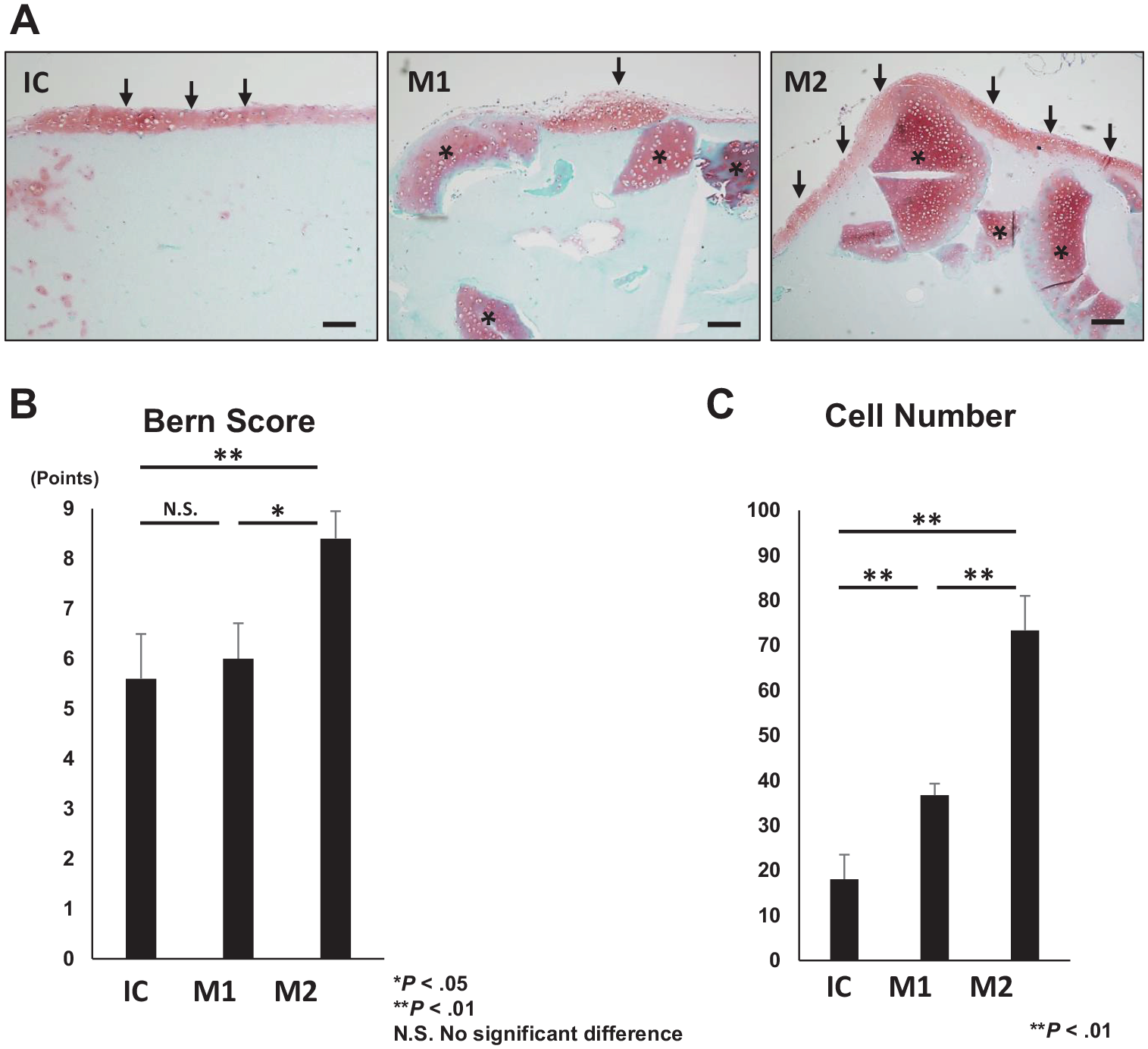
Culture of isolated chondrocytes and minced cartilage in the atelocollagen gel after 3 weeks. (A) Safranin O/Fast Green staining. Arrows indicate new cartilage layer. Asterisks indicate minced cartilage. Bar indicates 100 μm. (B) Bern score in each group. (C) Cell number in atelocollagen gel in each group. In panels B and C, *P < .05 and **P < .01. IC, isolated chondrocyte group; M1, 12.5 mg minced cartilage; M2, 25 mg minced cartilage.
In Vivo Experiments
Gross Appearance
At 4 weeks after the operation, the defect group had sparse reparative tissue in the defect, whereas the other 3 groups were filled with white fibrous tissue. However, the surface in the atelocollagen group was irregular compared with the other 2 groups. At 12 weeks, the defect remained in the defect group. In the other 3 groups, the grafted area remained covered with a white membrane and the border between the membrane and articular cartilage appeared unclear. In the minced cartilage group, the grafted area acquired an elastic consistency compared with the other groups. At 24 weeks, the defect group showed an irregular surface and concavities. The atelocollagen group exhibited a rough surface in the defect, but the ACI and minced cartilage groups showed the glossy smooth surface and the fibrous tissue with smooth connection to the adjacent cartilage. Any hypertrophy of the repaired tissues in the ACI and minced cartilage groups was not observed during the investigation period (Figure 2). There was no reaction on the surface of the opposite patellar cartilage and no sign of synovitis.
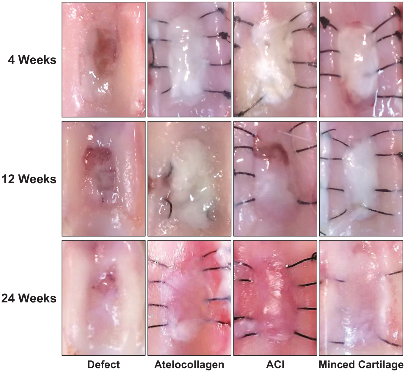
Gross appearance in each group at 4, 12, and 24 weeks after the operation. ACI, autologous chondrocyte implantation.
Histological Evaluation
At 4 weeks after the operation, the osteochondral defect was not filled substantially in the defect group. In contrast, the other 3 groups had reparative tissue with a Safranin O–positive area in the defects. In the minced cartilage group, the Safranin O–positive area was wider, although minced cartilage fragments were not detected (Figure 3). In the ACI group, bone marrow was formed in the composite, and the cartilage layer just under the periosteum was thin, although proliferation of chondrocytes was seen. Fibrous tissue was found between the composite and the bottom of the osteochondral defect. The minced cartilage group had a cartilage layer between the composite and the bottom of the osteochondral defect, and abundant chondrocyte proliferation under the periosteum was observed; bone marrow formed in the composite. Minced cartilage was not observed (Figure 4).
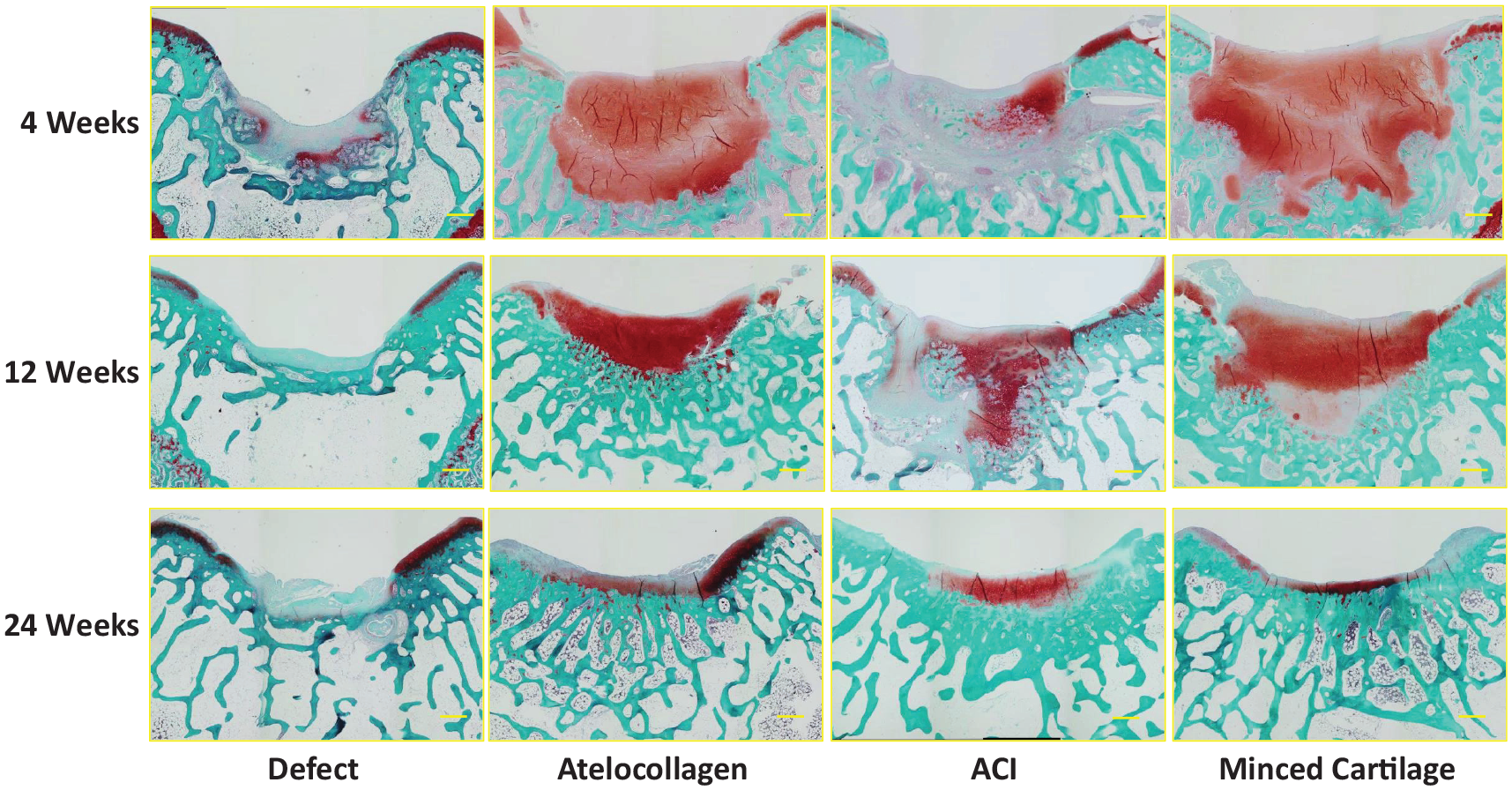
Safranin O/Fast Green staining in each group at 4, 12, and 24 weeks after the operation. The bar indicates 100 μm. ACI, autologous chondrocyte implantation.
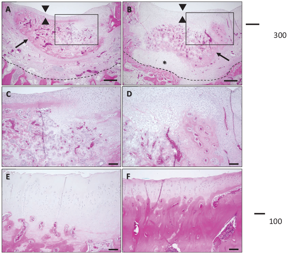
Hematoxylin and eosin staining in ACI and minced cartilage groups. (A, C, E) ACI group. (B, D, F) Minced cartilage group. Panels A-D show 4 weeks after the operation, and panels E and F are 24 weeks after the operation. Arrows indicate the composites in each group. Arrowheads show the periosteum. Panels C and D are high-magnification images of the squares in panels A and B. The asterisk indicates the chondrocyte layer. The bars in panels A and B indicate 300 μm, and those in panels C-F indicate 100 μm.
At 12 weeks after the operation in the defect group, there was no cartilaginous tissue in the defect. In the atelocollagen and ACI groups, the Safranin O–positive area had sparse chondrocytes. In the minced cartilage group, there were more chondrocytes than in the other groups (Figure 3). At 24 weeks after the operation in the defect group, the defect was filled with Safranin O–negative fibrous tissue. The osteochondral junction was not reconstructed well. In the atelocollagen and ACI groups, the defect area became thinner and chondrocytes were detected in the Safranin O–positive area. However, the subchondral bone was not repaired to the level of the surrounding subchondral bone. In the minced cartilage group, articular cartilage presented almost normal thickness, and there were abundant chondrocytes in the Safranin O–positive area. The osteochondral junction was reconstructed better than it was in the other groups (Figure 3). At 24 weeks, there was a columnar structure of chondrocytes in the ACI and minced cartilage groups. Both groups showed good repair of the cartilage layer, but the ACI group showed insufficient subchondral bone repair (Figure 4).
Pineda scores at 4 weeks after the operation were 9.2 ± 1.7, 7.0 ± 1.4, 7.2 ± 0.8, and 5.6 ± 0.5 points for the defect, atelocollagen, ACI, and minced cartilage groups, respectively. The score for the minced cartilage group was significantly lower than that for the defect group (P < .05). At 12 weeks after the operation, the mean Pineda scores of the 4 groups were 9.4 ± 1.9, 6.4 ± 1.7, 6.6 ± 2.4, and 4.8 ± 1.9 points, respectively. The score for the minced cartilage group was significantly lower than that for the defect group (P < .05). At 24 weeks after the operation, the mean Pineda scores of the 4 groups were 9.6 ± 1.4, 6.6 ± 2.9, 4.6 ± 2.6, and 1.8 ± 1.2 points, respectively. The ACI and minced cartilage groups had significantly lower scores than did the defect group (P < .05). The minced cartilage group had significantly lower scores than did the atelocollagen group (P < .05). The minced cartilage group scores tended to be lower than those of the ACI group, but there was no significant difference (Figure 5).
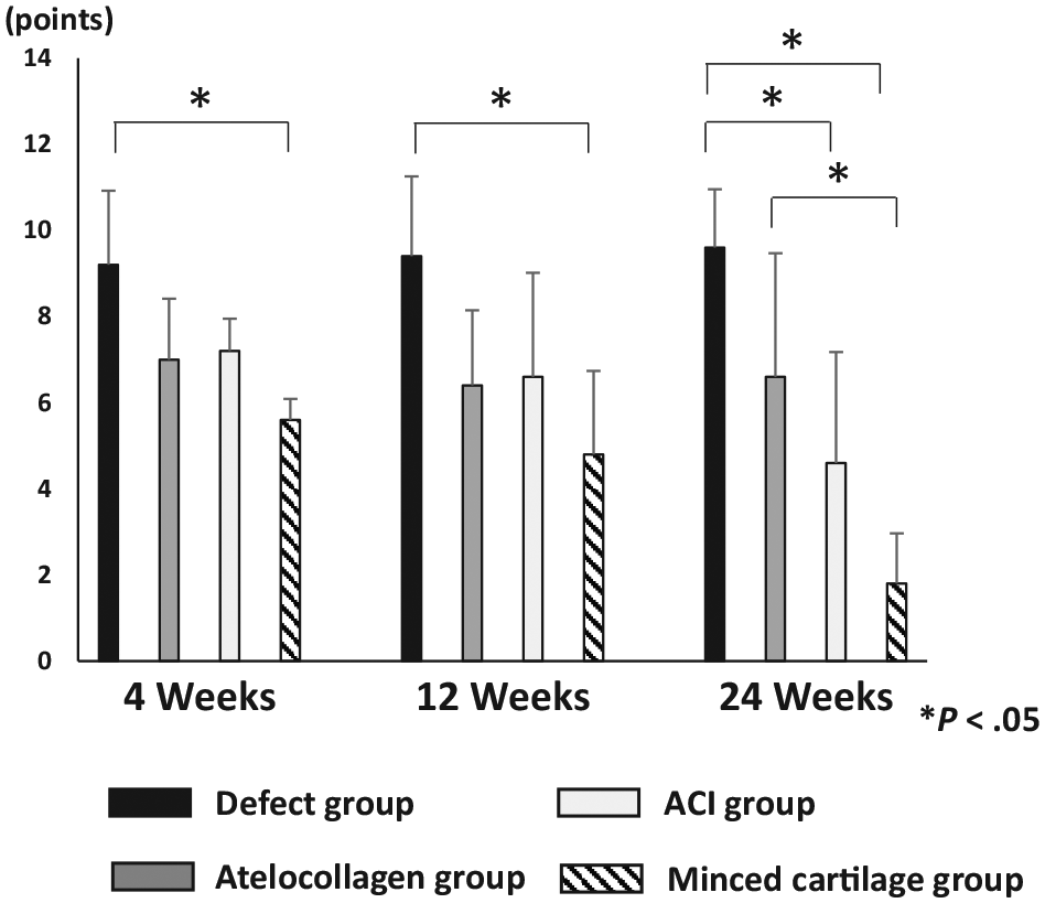
Pineda scale in each group at 4, 12, and 24 weeks after the operation. *P < .05. ACI, autologous chondrocyte implantation.
Immunohistochemistry
At 24 weeks after the operation, the reparative tissue of the defect group was well stained for type 1 collagen. However, the reparative tissue of the other groups was not stained, which means the atelocollagen gel composed of type 1 collagen was completely replaced. In contrast, in the immunohistochemistry analysis of type 2 collagen at 24 weeks after operation, the reparative tissue of the defect group was not stained, although that of the other groups was well stained (Figure 6).
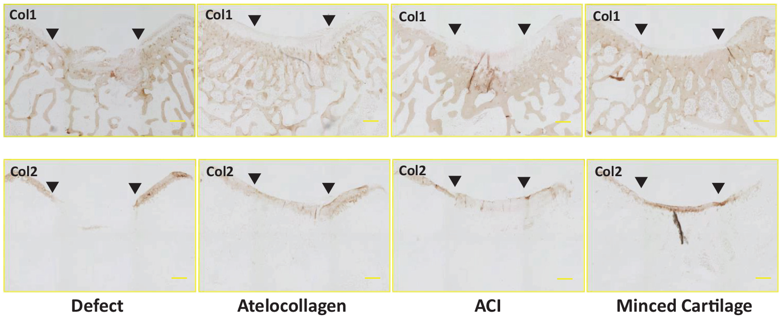
Immunohistochemistry of type 1 and 2 collagens (Col1, Col2) in each group at 24 weeks after the operation. Arrowheads indicate the border of the osteochondral defect. The bar indicates 100 μm. ACI, autologous chondrocyte implantation.
To evaluate the proliferation ability of chondrocytes, immunohistochemistry analysis of Ki67 at 4 weeks after the operation was performed. Ki67-positive cells were observed in all groups. However, the ACI and minced cartilage groups showed many Ki67-positive chondrocytes in the reparative tissues, whereas the atelocollagen and defect groups exhibited a lower number (Figure 7A). In particular, fibroblastic cells in the reparative tissue of the defect group had Ki67-positive cells. The average percentage of Ki67-positive cells in the defect, atelocollagen, ACI, and minced cartilage groups was 26.7% ± 11.8%, 47.9% ± 20.3%, 62.2% ± 24.4%, and 72.2% ± 19.3%, respectively. The percentage of Ki67-positive cells in the ACI group and the minced cartilage group was significantly higher than that in the defect group (P < .05). There was no significant difference between the ACI group and the minced cartilage group; however, the minced cartilage group tended to have higher values (Figure 7B).
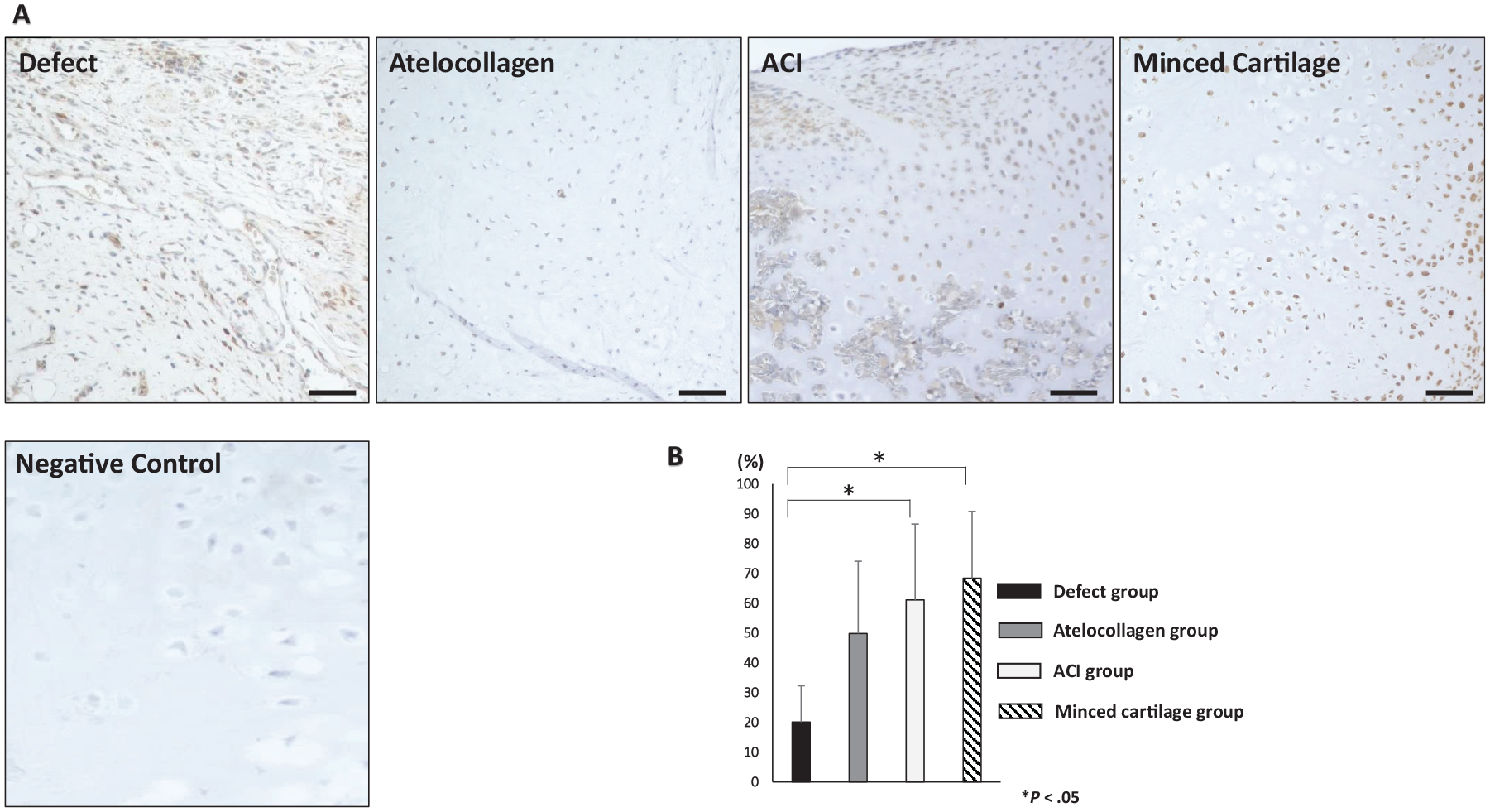
(A) Immunohistochemistry of Ki67 in each group at 24 weeks after the operation. The bar indicates 100 μm. (B) The rate of Ki67-positive cells in the reparative tissue in the osteochondral graft. *P < .05. ACI, autologous chondrocyte implantation.
Discussion
This study demonstrated that minced cartilage in atelocollagen gel had good chondrocyte migration and proliferation abilities in vitro and the osteochondral defect was well repaired by the implantation of the minced cartilage embedded in the atelocollagen gel in vivo.
In the in vitro study, the cell number and Bern score were higher in the M1 and M2 groups than in the IC group. In the in vivo study, the Pineda score in the minced cartilage group at 24 weeks after the operation was significantly better than those of the defect and atelocollagen groups. Although there was no significant difference between the minced cartilage group and the ACI group, implantation of minced cartilage is a successful procedure because of the 1-step surgery.
Cartilage repair using minced cartilage has been reported in both animal experiment and clinical settings. 5 Recent studies using large animals demonstrated that the implantation of minced cartilage exhibited good quality of cartilage repair tissue, including hyaline cartilage, and recovery of the subchondral bone for the chondral/osteochondral defect compared with other cartilage repair techniques such as the bone marrow stimulation technique.10,12 However, many studies adopted the technique in which minced cartilage was loaded onto the scaffold and fixed by fibrin glue.2,16,22,25,26 Chondrocyte outgrowth in a 3-dimensional scaffold, such as atelocollagen gel, may be superior to just loading cells on the scaffold. Moreover, fibrin glue has negative effects on cell migration and tissue repair.7,35 The 1-step procedure of the implantation of minced cartilage embedded in atelocollagen gel has the advantages of cell migration and proliferation in 3-dimensional scaffold without using fibrin glue.
Adachi et al 1 demonstrated good clinical results of ACI using atelocollagen gel for the cartilage defect of the knee at an average of 8.0 years of follow-up. In their report, the surface area of the cartilage injury was 3.6 cm2 (2.0-16.0 cm2) and the number of harvested autologous chondrocytes was 2.2 × 10⁶ (0.4-42.0 × 10⁶). These chondrocytes are embedded in atelocollagen gel and cultured for 3 to 4 weeks, and the formation of a firm gel-like material with proliferated chondrocytes is obtained before the implantation into the cartilage defect. Atelocollagen is composed of type 1 collagen with the removal of telopeptides, which are antigenic determinants on the peptide chains of type 1 collagen.17,32 Its 3-dimensional structure is superior for cell culture as a scaffold, and previous studies have shown that the chondrocyte phenotype was retained in the cell culture.20,34 In our results, the number of chondrocytes in the gel using minced cartilage was significantly higher than that using the isolated chondrocytes in vitro, which revealed that minced cartilage has greater potential for chondrocyte migration and proliferation than do isolated chondrocytes. The Bern score and the number of chondrocytes were the highest in the group using 25 mg minced cartilage. In our data using human articular cartilage, a 100-mg cartilage fragment had 2 × 10⁵ cells, which means that if the same amount of isolated chondrocytes is obtained, one-fourth of the amount of minced cartilage will be needed. 33 Conceivably, harvesting donor chondrocytes at the same amount of the normal cartilage for conventional ACI will be available to cover cartilage defects that are 4 times larger using minced cartilage-embedded atelocollagen gel. 34 In addition, a high cell number in the atelocollagen gel could produce a cartilage matrix compared with a low cell number in vitro. 20 In clinical settings, a high dose of chondrocyte in ACI showed good clinical results in an early phase trial. 27 Minced cartilage in atelocollagen gel will produce abundant cartilage matrix compared with ACI; our results revealed good cartilage repair by minced cartilage from the early phase. Even without cell culture, 1-step cartilage repair using minced cartilage embedded in atelocollagen gel could obtain results equivalent to those of ACI.
In this study, even though there was no significant difference between the minced cartilage and ACI groups, the results were positive for the minced cartilage group because of the 1-step surgery. Using minced cartilage has various advantages because it does not require cell processing and culture. Enzymic digestion to isolate chondrocyte provides cytotoxic effects to some extent. 36 Chondrocytes easily lose their phenotype as hyaline chondrocytes by culture, leading to the dedifferentiation, which is recognized as unstable for ACI. 23 Moreover, cartilage tissues, including chondrocytes, have various anabolic growth factors, such as transforming growth factor-β, and microvesicles, such as exosome containing microRNA.3,14 These factors in the minced cartilage may play an important role in the repair process of cartilage. At 4 weeks after the operation in this study, the ACI and minced cartilage groups showed bone marrow formation in the composites at the level of subchondral bone in the osteochondral defect. However, the layer with chondrocyte proliferation around the level of the original cartilage layer and the bottom of the osteochondral defect was well observed in the minced cartilage group, which may suggest that minced cartilage secretes factors that induce bone and cartilage formation. A previous report revealed that the implanted minced cartilage was present in the defect and was viable after 1 year, implying that the autocrine/paracrine effect may continue for at least 1 year. 11
This study has several limitations. First, allogenic articular cartilage fragments harvested from the knee, shoulder, and hip were used to obtain abundant cartilage fragment and reduce animal sacrifice. If autologous minced cartilage implantation is required in the clinical setting, an autologous implantation model should be used. However, it is difficult to match the condition of the osteochondral defects in the ACI group and other groups because ACI requires 3 weeks of culture using chondrocytes when creating osteochondral defect models. The concern using allogenic chondrocyte implantation is immune rejection. The immune response in isolated allogenic chondrocyte implantation induces the gradual destruction of cartilage tissue. 24 Articular cartilage has been recognized to have low immunogenicity because of the coverage of chondrocytes by the extracellular matrices. 28 Successful results in animal studies using the combination of allogenic chondrocytes and 3-dimensional scaffolds have been reported.15,30 These scaffolds could form a protective barrier around chondrocytes to block the infiltration of host immune cells into the grafts and the escape of immunogenic chondrocytes out of the graft. 8 A previous report revealed that implantation of the isolated allogenic chondrocytes embedded in atelocollagen gel into the osteochondral defect in a rabbit model did not exhibit any immune rejection. 21 In the minced cartilage embedded in atelocollagen, allogenic chondrocytes might not be sufficiently immunogenic to induce an immune rejection owing to the strong barrier of extracellular matrices and 3-dimensional scaffold. In clinical settings, the use of autologous articular cartilage fragments from the knee joint is proposed, and it will get better results than the use of allogenic articular cartilage of the knees, hips, and shoulder joints because of the nonimmune response. Second, the volume of articular cartilage fragments in the ACI and minced cartilage groups was not matched. However, 8.0 mg of minced cartilage was implanted into the osteochondral defect, and chondrocytes in the ACI group were isolated from 21.2-mg cartilage fragments. The ACI group required a larger amount of cartilage fragment than did the minced cartilage group. Third, this study used the periosteum to cover the defect, although several materials, such as collagen membrane, are available in the clinical setting. In this study, the efficacy of the minced cartilage embedded in the atelocollagen gel on cartilage repair was evaluated to improve the conventional methods of ACI using atelocollagen gel. For this aim, we used the rabbit model by Katsube et al. 21 They assessed the efficacy of the isolated allogenic chondrocytes embedded in atelocollagen gel with the periosteum patch. Our study showed good repair using the minced cartilage embedded in atelocollagen gel with the periosteum patch, and it is comparable enough with the conventional ACI. Further study is needed to evaluate the efficacy of the minced cartilage embedded in atelocollagen gel without periosteum patch on pure chondral defect. Finally, we evaluated the reparative ability of the osteochondral defect rather than the cartilage defect because the articular cartilage layer in rabbits was too thin. Evaluation of the effect of the transplantation of the minced cartilage on the cartilage injury using large animal models is necessary.
Conclusion
In conclusion, minced cartilage in atelocollagen gel demonstrated good chondrocyte migration and proliferation abilities in vitro, and the osteochondral defect was well repaired with the implantation of the minced cartilage embedded in the atelocollagen gel in vivo. Implantation of minced cartilage embedded in atelocollagen gel showed good cartilage repair equivalent to that of ACI.
Footnotes
One or more of the authors has declared the following potential conflict of interest or source of funding: This research was supported by MEXT KAKENHI Grant-in-Aid for Scientific Research (B), Grant No. 17H04314 (N.A.). AOSSM checks author disclosures against the Open Payments Database (OPD). AOSSM has not conducted an independent investigation on the OPD and disclaims any liability or responsibility relating thereto.