
Abstract
Background:
There are limited data comparing the outcomes of similarly matched patients with a medial meniscus posterior root tear (MMPRT) treated with nonoperative management, partial meniscectomy, or repair.
Purpose/Hypothesis:
The purpose was to compare treatment failure, clinical outcome scores, and radiographic findings for a matched cohort of patients who underwent either nonoperative management, partial meniscectomy, or transtibial pull-through repair for an MMPRT. We hypothesized that patients who underwent meniscus root repair will have lower rates of progression to arthroplasty than patients who were treated with nonoperative management or partial meniscectomy.
Study Design:
Cohort study; Level of evidence, 3.
Methods:
Patients who underwent transtibial medial meniscus posterior horn root repair were matched by meniscal laterality, age, sex, and Kellgren-Lawrence (K-L) grades to patients treated nonoperatively or with a partial meniscectomy. Progression to arthroplasty rates, International Knee Documentation Committee and Tegner scores, and radiographic outcomes were analyzed between groups.
Results:
Forty-five patients were included in this study (15 nonoperative, 15 partial meniscectomy, 15 root repair). Progression to arthroplasty demonstrated significant differences among treatment groups at a mean of 74 months (nonoperative, 4/15; partial meniscectomy, 9/15; meniscal repair, 0/15; P = .0003). The meniscus root repair group had significantly less arthritic progression, as measured by change in K-L grade from pre- to postoperatively (nonoperative, 1.0; partial meniscectomy, 1.1; meniscal repair, 0.1; P = .001).
Conclusion:
Meniscus root repair leads to significantly less arthritis progression and subsequent knee arthroplasty compared with nonoperative management and partial meniscectomy in a matched cohort based on patient characteristics.
Meniscus roots are a vital component of the meniscus as they act to anchor the meniscus to the tibial plateau and disperse axial loads into hoop stresses during loading. 2 Root disruption results in a loss of hoop strain resistance, increased articular cartilage contact pressure, and acceleration of degenerative changes.5,19,20 These consequences have been demonstrated in prior biomechanical studies proving that a medial meniscus posterior root tear (MMPRT) is functionally and biomechanically comparable with a total meniscectomy.2,22
Medial meniscus posterior horn root tears compose 10% to 21% of all meniscal tears.6,15 These injuries occur in both acute and chronic settings, but are most commonly found in a degenerative state with concomitant chondral defects. 23 Risk factors include increased age, increased body mass index (BMI), and female sex.5,15 Additionally, meniscal extrusion, which has been demonstrated to be an independent predictor of tibiofemoral cartilage loss and the development of subchondral bone lesions, is highly associated with meniscus root tears.4,9,11,12,28
Treatment options for an MMPRT include nonoperative management, partial meniscectomy, and root repair. Historically, patients with root tears were treated without surgery or with a partial meniscectomy; however, there is a recent shift toward meniscal preservation with the utilization of meniscus root repair. 5 The overall repair outcomes have been good1,10,20; however, limited data are available to compare these treatment methods. Therefore, the purpose of this study is to compare (1) treatment failure, (2) clinical outcome scores of the remaining patients, and (3) radiographic outcomes of a matched cohort of patients who underwent nonoperative management, partial meniscectomy, or transtibial pull-through root repair for an MMPRT. Our hypothesis is that patients who underwent meniscus root repair will have lower rates of progression to arthroplasty than patients who were treated with nonoperative management or partial meniscectomy.
Methods
After institutional review board approval (IRB ID No. 15-000601), an institutional database was retrospectively reviewed to identify patients who had a known meniscus root tear between 2005 and 2016. Patients were considered to have a root tear if advanced imaging demonstrated either a complete medial meniscus posterior root avulsion or a complete radial tear within 9 mm of the root attachment and the patient had acute onset of clinical symptoms that correlated with the magnetic resonance imaging (MRI) findings. These patients were included if they were treated with nonoperative management, partial meniscectomy, or transtibial pull-through root repair of their root tear. Root repair was initially performed in 2010, so patients treated before that received partial meniscectomy or nonoperative management. An algorithm based on currently published data and this institution’s experience is provided to assist in treatment selection (Figure 1). The algorithm was used to assist in the treatment decision process; however, each patient treated was considered on a case-by-case basis.
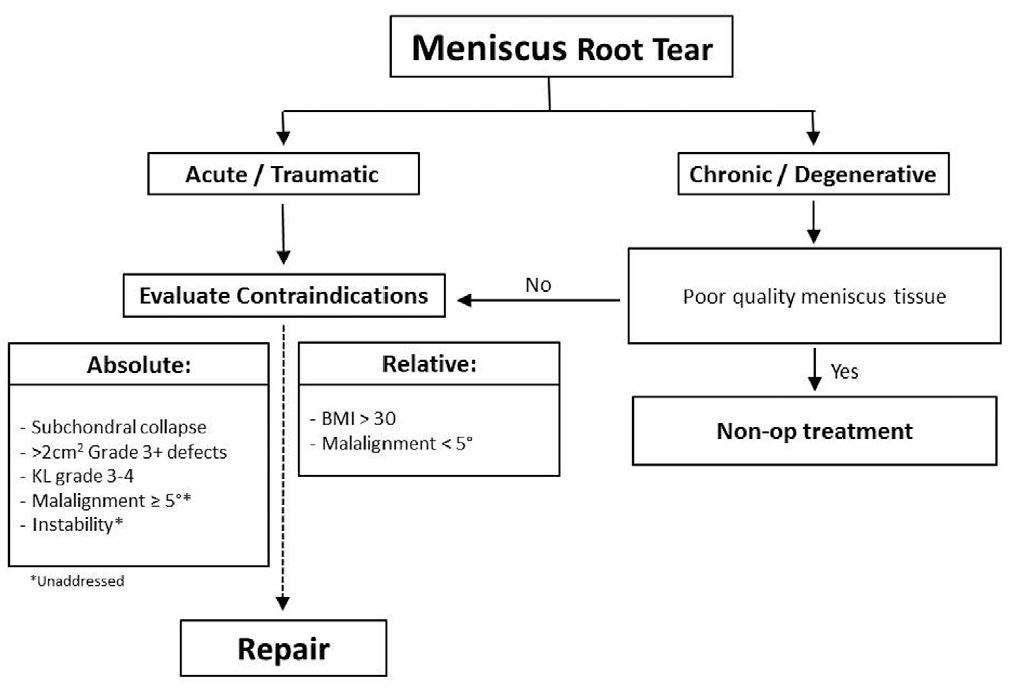
Treatment algorithm for meniscus root tear. BMI, body mass index; K-L, Kellgren-Lawrence.
The root repair cohort was matched to patients treated either nonoperatively or with partial meniscectomy. There were 15 patients who underwent a meniscus root repair that had the same laterality (medial vs lateral meniscus) as a cohort of patients with meniscus root tears that were treated either nonoperatively or with a partial meniscectomy. There were 26 potential matches each in the partial meniscectomy group and in the nonoperatively treated group. Patients with root repair were then blindly matched to both a patient treated with partial meniscectomy and a patient treated nonoperatively using the criteria of age (±8 years), sex (same), and Kellgren-Lawrence (K-L) grade (±1). Forty-five patients were identified for imaging and clinical history analysis, with 15 patients included from each treatment modality.
Individual medical records were reviewed to obtain patient information including age, sex, BMI, and laterality. Additional information collected included patient history and clinical progression, and subsequent knee arthroplasty.
Radiographic assessment of the knee was performed to determine the severity of osteoarthritis (OA) utilizing the K-L grading system. 16 Weightbearing anteroposterior radiographs at baseline and the most recent date were evaluated with arthritis graded according to the following scale: none (grade 0), doubtful (grade 1), minimal (grade 2), moderate (grade 3), or severe (grade 4). Additionally, MRI scans were reviewed for the presence or absence of meniscal extrusion. Meniscal extrusion was defined as subluxation of the peripheral margin of the medial meniscus beyond the medial tibial plateau, excluding osteophytes. 17 Meniscal extrusion exceeding 3 mm was considered to be major. 11
Failure was defined as progression to knee arthroplasty. These patients were excluded from subjective outcome score analysis but were included in radiographic and risk failure analysis. Patients were contacted to complete International Knee Documentation Committee (IKDC) subjective evaluations and Tegner scores.
All surgical procedures were performed at the same academic institution using a previously described transtibial suture technique.14,30 All repairs were performed with the patient in the supine position. Standard anterolateral and anteromedial portals were established adjacent to the patellar tendon and proximal to the meniscus to allow for instrument passage to the posteromedial and posterolateral compartments. Diagnostic arthroscopy was completed and the root tear identified. A transtibial drill guide (Arthrex) was inserted through the anterior portal. The guide tip was positioned at the center of the posterior horn root footprint. A 3.5-mm wire was inserted through the tibia and then exchanged with a 6 mm–diameter Flip Cutter (Arthrex). The Flip Cutter was used to create a 5-mm deep socket with exposed bleeding bone.
Three 0-Fiberlink (Arthrex), simple cinch (luggage tag) sutures were placed in the meniscus root using a self-retrieving device (Knee Scorpion; Arthrex). These sutures were then pulled through the tibia using a shuttling technique. The sutures were tensioned and slack was removed by cycling the knee. The sutures were then fixed to the tibia with a knotless suture anchor (4.75-mm Swivelock anchor; Arthrex).
Minimal partial weightbearing or nonweightbearing with 2 crutches and with a knee brace locked at 0° to 90° of flexion occurred for the first 6-week period. After 6 weeks, use of the brace was discontinued and the patient could begin full weightbearing and full knee range of motion. Knee loading at flexion angles greater than 90° was not allowed until 4 months postoperatively, at which point patients were typically allowed to return to activity as tolerated.
Statistical Analysis
Statistics (mean, standard deviation, median, range, and frequencies) were analyzed for patient characteristics, including BMI, age, and sex. Imaging data, clinical outcomes, and progression to arthroplasty rates were also analyzed. Patients were matched to those who underwent transtibial pull-through root repair by meniscal laterality (same side), age (±8 years), sex (same), and K-L grade (±1). Sample size was taken into account for all calculations. Wilcoxon signed rank analysis was used to compare means of continuous variables, and the Fisher exact test and chi-square test were used to compare nominal variables, when appropriate. Kaplan-Meier survival analysis was used to evaluate the time-dependent rate of conversion to arthroplasty. All analyses were performed with Microsoft Excel (2010) and JMP Pro (v 14.0.0; SAS Institute Inc). P values <.05 were considered statistically significant.
Results
Patient characteristic analysis was conducted on the 45 patients included in the study. Mean age and BMI as well as proportion of male and female patients were found to not be significantly different between groups (Table 1).
Patient Characteristics by Different Treatment Modalities a
BMI, body mass index; F/M, female/male.
Progression to total knee arthroplasty was significantly different among the treatment groups, including 9 patients treated with partial meniscectomy, 4 patients treated nonoperatively, and 0 patients treated with meniscal repair (P = .0003) (Figure 2). The average time for progression to arthroplasty was not significantly different between nonoperative (75.2 months) and partial meniscectomy (66.2 months) groups (P = .64).
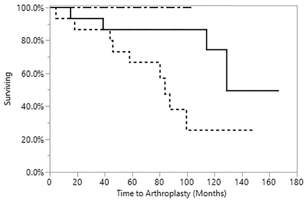
Kaplan-Meier survival analysis demonstrating conversion to knee arthroplasty by root repair (dashed and dotted line), partial meniscectomy (dotted line), and nonoperative (solid line) treatment.
Outcome scores were measured via IKDC and Tegner activity level scores for patients who did not progress to arthroplasty (11 nonoperative, 6 partial meniscectomy, 15 root repair) at a mean of 49 months (nonoperative, 53 months; partial meniscectomy, 58 months; meniscal repair, 40 months). No statistically significant differences were found between treatment groups for IKDC scores (nonoperative, 57.7; partial meniscectomy, 74; meniscal repair, 72.3; P = .38). Tegner activity scores (nonoperative, 3.8; partial meniscectomy, 4.3; meniscal repair, 4.1; P = .82) also were not significantly different between groups.
Radiographic data were analyzed in the form of preinjury or preoperative, most recent, and change in K-L grades. The mean preoperative or preinjury K-L grades were not significantly different between treatment groups (Table 2). Additionally, baseline MRI scans were reviewed to evaluate for the presence of major meniscal extrusion. There were no significant differences in the proportion of patients with major meniscal extrusion (nonoperative, 13/15; partial meniscectomy, 10/15; meniscal repair, 13/15; P = .30).
Radiographic Characteristics by Different Treatment Modalities a
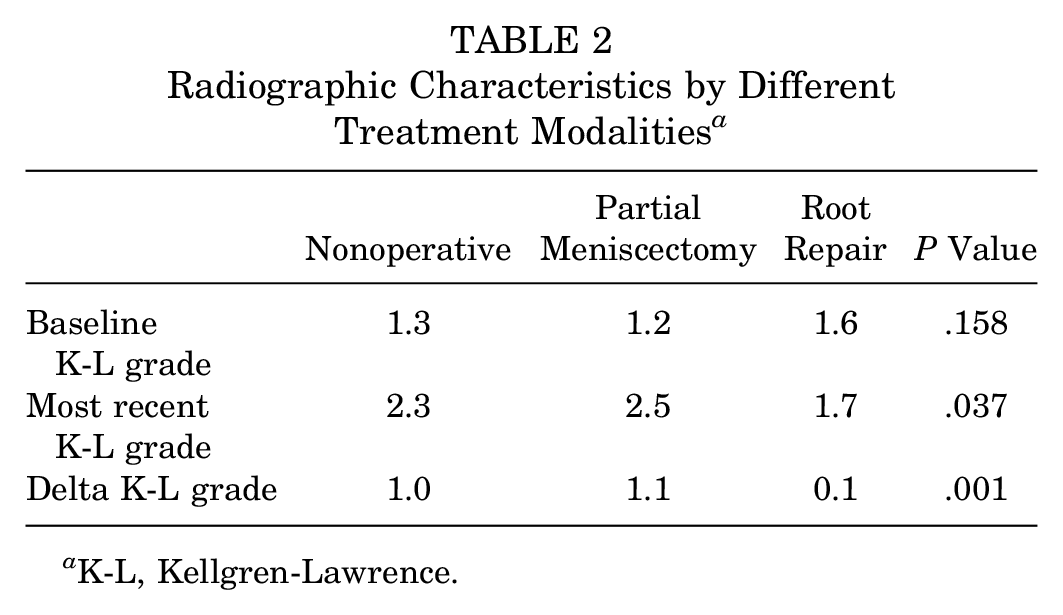
K-L, Kellgren-Lawrence.
Discussion
The major finding of our study was that patients undergoing meniscus root repair had significantly less arthritis progression and need for knee arthroplasty compared with those undergoing nonoperative management or partial meniscectomy at a mean 74 months of follow-up. This finding confirmed our hypothesis that patients who underwent meniscus root repair will have lower rates of progression to arthroplasty than patients who were treated with nonoperative management or partial meniscectomy.
Meniscectomy for a root tear is associated with a very high rate of progression to knee arthroplasty. Krych et al 18 compared the rate of failure, or progression to arthroplasty, between patients with medial meniscus root tears treated nonoperatively or with partial meniscectomy and found that 54% of partial meniscectomy and 34.6% of nonoperative patients progressed to total knee arthroplasty at a mean of 54.3 and 30.2 months, respectively. Chung et al 10 similarly found that for patients who underwent partial meniscectomy, the treatment failed in 35% of the cases at the 6-year follow-up. Conversely, they found at a similar follow-up time point, no patients treated with meniscal repair had progressed to total knee arthroplasty. Our study reflected these findings, as we similarly found 40%, 27%, and 0% progression to knee arthroplasty in partial meniscectomy, nonoperative, and meniscus root repair matched cohorts, respectively. The matched cohorts based on patient characteristics demonstrated superior outcomes with meniscal repair. However, without these contributing factors, Lee et al 21 demonstrated that in patients with well-aligned knees and minimal to no radiographic arthritis at baseline, partial meniscectomy may be a reasonable treatment option for MMPRT.
Subjective outcome scores after partial meniscectomy have traditionally been somewhat variable. Krych et al 18 reported mean IKDC scores of 67.8 for those undergoing partial meniscectomy. Lee et al 21 similarly reported mean Lysholm scores of 81.3, but Chung et al 10 reported mean IKDC and Lysholm scores of 49.3 and 62.8, respectively. Previous studies have demonstrated improvements in objective outcome scores after partial meniscectomy; however, the interpretation of “improvement” has been questioned because of the outcomes of Sihvonen et al 27 and others demonstrating the failure of partial meniscectomy to outperform “sham surgery.” These objective measures are still part of a “gold standard” to assess the efficacy of meniscal procedures. Chung et al 10 used IKDC and Lysholm scores to compare partial meniscectomy and meniscal repair groups and found significant differences in both scores, with meniscal repair being superior. Ahn et al 1 compared patients who underwent root repair with patients treated nonoperatively for a medial meniscus root tear and demonstrated that patients treated with root repair had better outcomes. In our study, we were unable to demonstrate significant differences among any of the 3 treatment groups in subjective knee scores. However, this may be a result of the analysis being underpowered. These differences in outcome scores among treatment groups would be clinically significant.
Meniscectomy is a known risk factor for progression of radiographic OA. 26 Englund et al 13 found that patients undergoing partial meniscectomy had a 43% rate of OA at an average follow-up of 16 years, while in comparison a control group was found to have only 9%. Furthermore, numerous studies have demonstrated that the amount of meniscus resected has a statistically significant effect on the progression of radiographic OA.3,7,10,25 Prior biomechanical studies have demonstrated that repair of a torn medial meniscus posterior root restores the ability of the meniscus to absorb hoop stresses and decreases tibiofemoral contact pressures comparable to those of the native knee.22,24 Unfortunately, meniscal repair has not been shown to prevent progression of OA clinically. A meta-analysis by Chung et al 8 found 6 studies that evaluated radiographic follow-up for meniscal repair, all of which demonstrated progression of OA from preoperative values. However, what is also known from the systematic reviews involving both partial meniscectomy and meniscus repair is that the factors affecting progression of OA are multifactorial. There are no studies to our knowledge that attempt to compare between partial meniscectomy and meniscal repair by controlling some of these additional factors by comparing matched cohorts. This is exactly what we were able to do in this study, and we found that there was a trend toward reduced radiographic progression of OA when comparing meniscal repair with partial meniscectomy and nonoperative treatment. We found that both the most recent and delta K-L grades were significantly better for patients who underwent meniscal repair compared with partial meniscectomy or nonoperative treatment. Chung et al 10 similarly demonstrated that patients treated with root repair had significantly less K-L grade progression and medial joint space narrowing compared with patients treated with partial meniscectomy.
Limitations of this study include the following: (1) The relatively small number of patients included. Given that treatment algorithms have more recently been developed that tend to “self-select” treatment for patients based on their clinical and patient characteristics (age, presence of OA, BMI, etc), it was difficult to find a large number of patients matched on characteristics who had undergone different definitive treatments. (2) Lack of matching based on limb alignment, which has been shown in previous studies to be protective for improved outcome after surgical outcome for medial meniscus root tears. 21 A recent biomechanical study has demonstrated that varus alignment results in increased medial compartment peak pressures, but a medial meniscal tear did not affect peak pressure even in varus alignment. 29 (3) We did not perform second-look arthroscopy or follow-up MRI in any patient group. It is unclear how many root repairs healed, or if the preoperative extrusion is improved with root repair. (4) Determining and analyzing specific tear types at the meniscus root was not investigated in this study, so the effect that certain tear types may have on patient outcomes remains unclear.
Conclusion
Meniscus root repair leads to significantly less arthritis progression and subsequent knee arthroplasty compared with nonoperative management and partial meniscectomy in a cohort matched based on patient characteristics. Further studies are needed to help define the ideal treatment for patients with a medial meniscus posterior horn root tear based on their clinical, patient, and radiographic characteristics.
Footnotes
One or more of the authors has declared the following potential conflict of interest or source of funding: C.L.C. has received general payments from Arthrex Inc (travel and lodging, education) and Zimmer Biomet Holdings Inc (travel and lodging). M.J.S. has received research support from Arthrex and Stryker; IP royalties from Arthrex; consulting fees from Arthrex; and hospitality payments from Gemini Medical LLC. B.A.L. has received IP royalties from Arthrex and VOT Solutions; consulting fees from Arthrex; and speaking fees from Linvatec and Smith & Nephew. D.B.F.S. has received consulting fees from Smith & Nephew, Genzyme, and Tigenix; and speaker fees from Smith & Nephew. A.J.K. has received research support from Aesculap/B. Braun, Arthritis Foundation, Ceterix, and Histogenics; consulting fees from Arthrex Inc, Vericel, and DePuy; IP royalties from Arthrex Inc; a grant from Exactech; and honoraria from Vericel and Musculoskeletal Transplant Foundation. AOSSM checks author disclosures against the Open Payments Database (OPD). AOSSM has not conducted an independent investigation on the OPD and disclaims any liability or responsibility relating thereto.
Submitted April 1, 2019; accepted September 26, 2019.