
Abstract
Background:
Particulated juvenile allograft cartilage (PJAC) has demonstrated good clinical efficacy in repairing articular cartilage defects, but the related repair mechanism after transplant and the biological characteristics of the transplanted cells are still unclear.
Purpose:
To study the efficacy of PJAC in repairing full-thickness cartilage defects and the specific fate of donor cells to provide experimental evidence for its clinical application.
Study Design:
Controlled laboratory study.
Methods:
Twenty female Guizhou minipigs were randomly divided into an experimental group and a control group. An 8-mm cylindrical full-thickness cartilage defect was created in the femoral trochlea of 1 knee in all minipigs. The experimental group received transplant of PJAC from 5 male juvenile Guizhou minipigs (PJAC group; n = 10) and the control group received autologous cartilage chips (ACC group; n = 10). Follow-up assessments were conducted at 1 month and 3 months to track the transplanted cells by the male-specific sex-determining region Y-linked (SRY) gene; tissue sections were hybridized in situ, and O’Driscoll histological scoring was performed according to hematoxylin and eosin staining, safranin O and fast green staining, and toluidine blue O staining, as well as immunohistochemical evaluation of aggrecan and Sry-type HMG-box 9 (SOX9).
Results:
All 20 Guizhou minipigs were followed; no infection or incision healing disorder occurred after the operation. By SRY in situ hybridization, the SRY signal of the transplanted cells was positive in the repaired tissue of the defect, and the SRY positive signal could still be detected in repaired tissue at 3 months postoperatively. The average number of positive cells was 68.6 ± 11.91 at 1 month and 32.6 ± 3.03 at 3 months (confocal microscope: ×400), and the difference was statistically significant. The O’Driscoll histological scores were 14 ± 0.71 in the ACC group and 9.8 ± 0.84 in the PJAC group at 1 month, and 18 ± 1.20 in the ACC group and 17.4 ± 1.14 in the PJAC group at 3 months. The scores were statistically significant between the ACC group and PJAC group at 1 month. The positive rates of SOX9 in the PJAC and ACC groups at 1 month were 67.6% ± 3.78% and 63.4% ± 5.30%, respectively, and the difference was not statistically significant (P > .05). The positive rates of SOX9 in the PJAC and ACC groups at 3 months were 68.8% ± 2.69% and 17.1% ± 1.26%, respectively, and the difference was statistically significant (P < .05). The positive rates of aggrecan in the PJAC and ACC groups at 1 month were 40.5% ± 2.78% and 42.4% ± 0.54% respectively, and the difference was not statistically significant (P > .05). The positive rates of aggrecan in the PJAC and ACC groups at 3 months were 40.8% ± 1.50% and 30.1% ± 2.44%, respectively, and the difference was not statistically significant (P > .05).
Conclusion:
An animal model was established with Guizhou minipigs, and the cartilage defect was repaired with PJAC from male minipigs. The SRY gene positive signal could be detected from the repaired tissue by in situ hybridization, indicating that the transplanted cells survived at least 3 months. The key genes of cartilage formation, SOX9 and aggrecan, were expressed at 1 month and 3 months, and SOX9 expression was stronger in the PJAC group than the ACC group at 3 months.
Clinical Relevance:
This study suggests that it is feasible to study the biological characteristics of transplanted cells in the cartilage region by the sex-determining gene.
Keywords
Articular cartilage is often damaged due to traumatic and degenerative factors, causing cartilage defects. Given the lack of blood supply to articular cartilage, its self-healing ability is extremely low. In recent years, techniques such as microfracture, cell transplant, and autologous cartilage chip (ACC) transplant have been applied to cartilage repair and have achieved certain therapeutic effects; however, these techniques also have drawbacks. Researchers are continuously exploring the field of cartilage repair, and a new treatment strategy using the particulated juvenile allograft cartilage (PJAC) has emerged. This technique entails preparing PJAC cubes from donors younger than 13 years and storing the cubes at low temperature. The cubes are then applied with fibrin glue to the cartilage defects.
The commercial PJAC product, DeNovo NT, has been applied to some clinical cases and has achieved good therapeutic effects.3,33 Farr et al 13 used DeNovo NT for the clinical treatment of knee cartilage defects; the 2-year follow-up results showed that knee function was significantly improved compared with that before surgery. Other cartilage defects in the ankle, elbow, and hip joints treated with PJAC also showed good repair effects.16,17,29 Relevant animal experiments have been used to study the efficacy of the PJAC technique. Bonasia et al 4 established adult New Zealand rabbit models to compare the effect of ACC transplant and PJAC technique on adult rabbit cartilage defects; the results showed no statistical difference between the 2 techniques. Our previous study used Guizhou minipigs to establish a large animal model and compared the repair effects of the 2 techniques; the results showed no significant difference in the repair effects between the 2 groups at 6 months after transplant. 2
Although PJAC has a good repair effect on cartilage defects, several factors remain unclear: the long-term survival of chondrocytes after transplant, the specific number of survived chondrocytes, and the related repair mechanism. The purpose of this study was to use conventional molecular biological techniques such as in situ hybridization to investigate whether male-derived donor cells exist in the repaired tissue of cartilage defects after PJAC transplant by detecting the male-specific sex-determining region Y-linked (SRY) gene. We hypothesized that positive male-specific SRY signals would be found in repaired tissues of cartilage defects in female hosts by in situ hybridization.
Methods
Animal Preparation
The PJAC donors were 5 male juvenile Guizhou minipigs younger than 2 months, weighing 5.4 to 6.3 kg and aged 1.3 to 1.7 months. A further 20 mature and healthy adult female Guizhou minipigs, weighing 27.4 to 32.1 kg and aged 11 to 12 months, were used to create a cartilage defect model. Among them, 10 received PJAC transplant (PJAC group) and 10 underwent ACC transplant (ACC group). All animals in this study were provided by the Large Animal Experimental Center of the Army Medical University and were raised according to the Experimental Animal Care Specifications. The study was approved by Laboratory Animal Welfare and Ethics Committee of the Army Medical University (Third Military Medical University) (No. AMUWEC2017147).
Surgical Procedures
PJAC Preparation
The experiments were performed in 5 batches. In the PJAC group, 1 juvenile male minipig and 2 adult female minipigs were used in each batch. The male juvenile minipig was anesthetized and euthanized with sodium pentobarbital (3%, 1 mL/kg; Huamu Pharmaceutical), and the knee was cut under sterile conditions; the articular cartilage was obtained from the articular surface of the distal femur. Particulated articular cartilage pieces of about 1 mm3 were cut with a scalpel and stored aseptically in a sterile test tube, mixed with a small amount of saline to keep them moist. They were stored in a refrigerator at 4°C. Preparation of fibrin glue was as follows: fibrinogen (500 mL, 20 mg/mL; Sigma), CaCl2 (20 mL, 50 mmol/L; Solarbio), and thrombin (500 mL, 250 m/mL; Sigma) were separately put into 3 aseptic centrifuge tubes for further use. When needed, the 3 reagents, together with the PJAC, were formulated into cartilage chip–fibrin glue.
Cartilage Defect Creation
The adult minipigs received an intramuscular injection of xylazine (0.1 mL/kg; Huamu Pharmaceutical) and atropine (1.5 mL for each minipig; Huamu Pharmaceutical) to induce muscle relaxation, followed by intravenous injection of sodium pentobarbital (3%, 1 mL/kg; Huamu Pharmaceutical) and fentanyl (0.15 mL/kg/h; Janssen Pharmaceutica) for anesthesia. The surgery did not start until the anesthetic effect was stable. All surgeries were performed by the same senior surgeon (X.D.) in our Center for Joint Surgery. An incision was made in the medial epidermis of the knee joint. The joint capsule and medial patellar retinaculum were cut off to dislocate the patella outward and expose the femoral trochlea. An 8-mm trephine was used to create a cartilage defect perpendicular to the surface of the trochlea without damaging the subchondral bone. The defect was debrided by use of a curette, and sterile saline was applied to wash the defect area to ensure that no more cartilage debris remained (Figure 1).
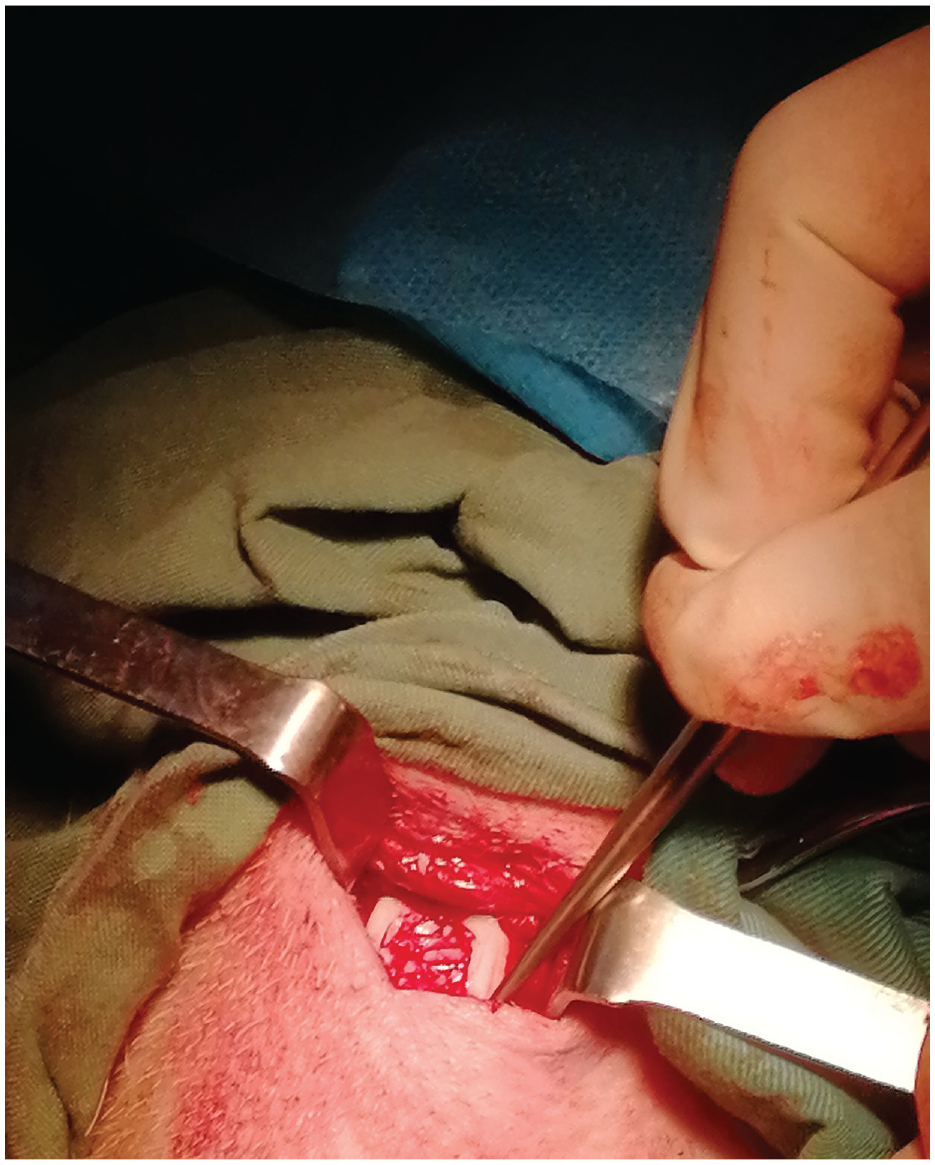
The surgical process.
PJAC and ACC Transplant
The PJAC, freshly prepared on the day of surgery, was mixed with the above-mentioned reagents that were prepared in advance to formulate cartilage chip–fibrin glue for transplant of the cartilage defect. After the glue was set for about 5 minutes, the dislocated patella was restored, and flexion and extension of 45° of the knee joint were conducted. The patellofemoral joint was examined to ensure that the particulated cartilage was fixed at the defect. Absorbable material was used for suture layer by layer. In the ACC group, the autologous articular cartilage extracted from the cartilage defect model was cut into cartilage chips of about 1 mm3 and then transplanted back into the defect. After the surgery, the minipigs were sent back to the animal center, and free movement was allowed (no movement limitation measures were taken). Each minipig received an intramuscular injection of procaine penicillin (0.03 mL/kg; Harbin Pharmaceutical) twice a day for 1 week.
Sample Collection and Processing
Follow-up assessments were performed at 1 and 3 months postoperatively. A total of 5 minipigs were euthanized in each group at each follow-up time point, and 5 samples were collected for processing. The collected samples were fixed with 4% paraformaldehyde solution for a duration of >12 hours and then decalcified with EDTA solution for 2 weeks. The decalcified sample tissue was embedded in paraffin and cut into slices of 5 µm using a microtome (Leica Rm2016). A total of 5 slices were selected for each sample for the subsequent in situ hybridization detection.
Tissues were dissected and conventionally dehydrated, embedded, prepared, and stained with hematoxylin and eosin (HE), safranin O and fast green, and toluidine blue O. The tissues were then observed under an optical microscope (Nikon Eclipse ci Imaging System; Nikon Digital Sight DS-FI2) and described. Images of different types of lesions were taken.
Design and Synthesis of mRNA Probes
The SRY gene of the Guizhou minipig was searched, and a specific probe (Qiagen) was designed based on the specific gene fragments of the minipig’s SRY gene. Probe information is as follows:
SRY probe (5′ and 3′ DIG) ACTCTGAACATAGCAGAA GCA
Negative control probe (5′ and 3′ DIG) GTGTAACACGTC TATACGCCCA
Steps of the Fluorescence in Situ Hybridization
The paraffin was sliced, dewaxed, and dehydrated. The sections were boiled in repair solution (pH 6.0) and then cooled naturally. Proteinase K (20 µg/mL) was added at 37°C for 20 minutes of digestion and then washed with phosphate-buffered saline (PBS). The prehybridization buffer was added, and samples were incubated at 37°C for 1 hour. The hybridization buffer with SRY probe (Tong Yong) was added and incubated at 37°C for the night. The hybridization buffer was washed with saline sodium citrate (SSC) after hybridization. Slices were incubated with 4′,6-diamidino-2-phenylindole dihydrochloride solution for 10 minutes and then sealed with antifluorescence quenching sealing tablets after washing. The slices were observed and photographed under the microscope.
Steps of the Digoxigenin-Labeled in Situ Hybridization
The paraffin was sliced, dewaxed, and dehydrated. The slices were boiled in repair solution (pH 6.0) and then cooled naturally. Proteinase K (20 µg/mL) was added at 37°C for 20 minutes of digestion, and then sections were washed with PBS for 3 times. Next, 3% methanol-H2O2 was added, and sections were incubated in the dark at room temperature for 15 minutes. The slides were washed with PBS 3 times, each for 5 minutes. Prehybridization buffer (Qiagen) was added, and sections were incubated at 37°C for 1 hour. The hybridization buffer with SRY probe (Qiagen) was added and incubated at 37°C for the night. The hybridization buffer was washed with SSC. A blocking serum, bovine serum albumin (BSA), was added to the section, and it was incubated at room temperature for 15 minutes. The blocking solution was removed, and anti-digoxigenin horseradish peroxidase was added. The sections were incubated at 37°C for 60 minutes and then washed with PBS. The sections were dried slightly, and freshly prepared Diaminobenzidine (DAB) chromogenic reagent was added to the marked tissue, and then the developing reaction was stopped by washing with pure water. The nucleus was dyed with Harris hematoxylin staining solution, treated with hydrochloric acid for differentiation, and dyed back to blue by ammonium hydroxide. The slices were dehydrated, sealed, and then observed and photographed under the microscope.
Steps of Immunohistochemical Tests
The paraffin was sliced, dewaxed, and dehydrated. The sections were roasted in a microwave oven with repair solution (pH 6.0) and then cooled naturally. The slides were immersed in 3% H2O2, incubated at room temperature for 25 minutes to block the endogenous peroxidase, and then washed with PBS. A circle was drawn with the immunostaining pen, and the slices were covered with 3% BSA at room temperature for 30 minutes. Slides were incubated with primary antibodies of anti-SOX9 (Sry-type HMG-box 9; ab185966, 1:200; Abcam) and anti-aggrecan (ab3773, 1:100; Abcam), placed in the refrigerator for the night at 4°C, and washed with PBS. The objective tissue was covered with secondary antibody labeled with horseradish peroxidase and incubated at room temperature for 50 minutes; the slides were washed with PBS. Freshly prepared DAB chromogenic reagent (G1212-200, 1:5000) was added to the marked tissue under a microscope until the nucleus showed brown-yellow and had a clean background, which indicated positive; then the developing reaction was stopped by washing with pure water. The tissues were counterstained in the nucleus with hematoxylin staining solution, treated with hydrochloric acid for differentiation, and stained back to blue by ammonium hydroxide. The slices were dehydrated, mounted with resin mounting medium, and observed and photographed under the microscope.
Statistical Analysis
O’Driscoll histological scoring was performed according to tissue staining; the number of positive cells in the fluorescent in situ hybridization and the positive rate of the immunohistochemical test were calculated. Statistical analysis was performed by SPSS 23.0, and measurement data were expressed as mean ± SEM to analyze whether the data conformed to the normal distribution. A paired t test was used for statistical analysis. P < .05 was considered statistically significant.
Results
Results of Fluorescence in Situ Hybridization
All minipigs were followed, and there were no abnormalities (such as wound infection or incision healing disorder). We analyzed 3 fluorescent in situ hybridization pictures of different samples. The average number of positive cells was 68.6 ± 11.91 at 1 month and 32.6 ± 3.03 at 3 months (confocal microscope: ×400). The number of positive cells at 1 month was more than that at 3 months, and the difference was statistically significant (Appendix Figures A1 and A2, available in the online version of this article).
Histological Results
At 1 month postoperatively, the cartilage defect area in the PJAC group was repaired by fibrous tissues and chondrocyte-like cells. The repaired area was thinner than the surrounding cartilage, the transplant boundary was clear, the interface between the new tissue and the host tissue coupled well, the chondrocytes around the injury were unevenly distributed, and chondrocyte aggregation was visible. At 1 month in the ACC group, the cartilage defect area was repaired by round hyaline chondrocyte-like cells and a small amount of connective tissue. The number of cells increased and the arrangement was irregular. The repaired area was thicker than that of the PJAC group at 1 month. The surface was smooth and flat, the boundary between normal cartilage and the transplanted tissue was clear, and there were more chondrocytes than in the PJAC group at 1 month.
At 3 months postoperatively, the cartilage defect area in the PJAC group was repaired by round hyaline chondrocyte-like cells and a small amount of connective tissues. The number of cells increased, the arrangement was irregular, and bone lacuna was seen locally. More areas were repaired in the same group than at 1 month. At 3 months in the ACC group, the cartilage defect area was repaired by round hyaline chondrocyte-like cells and a small amount of connective tissue. The number of cells increased, the arrangement was irregular, and bone lacuna was seen locally. The surface was smooth and flat, and there were more chondrocyte-like cells at the defect than in the PJAC group at 1 month.
At 1 month postoperatively, the safranin O and fast green staining and toluidine blue O staining in the PJAC group were weakly positive. The staining results of the ACC group were darker than those of the PJAC group at 1 month, and the staining results were further darkened in both PJAC group and ACC group at 3 months.
According to the scoring standard established by O’Driscoll et al, 27 the specimens were histologically evaluated by double-blind method. The scores were 14 ± 0.71 in the ACC group and 9.8 ± 0.84 in the PJAC group at 1 month, and 18 ± 1.20 in the ACC group and 17.4 ± 1.14 in the PJAC group at 3 months. The scores were statistically different between the ACC group and PJAC group at 1 month, and there was no statistical difference at 3 months (Figure 2).
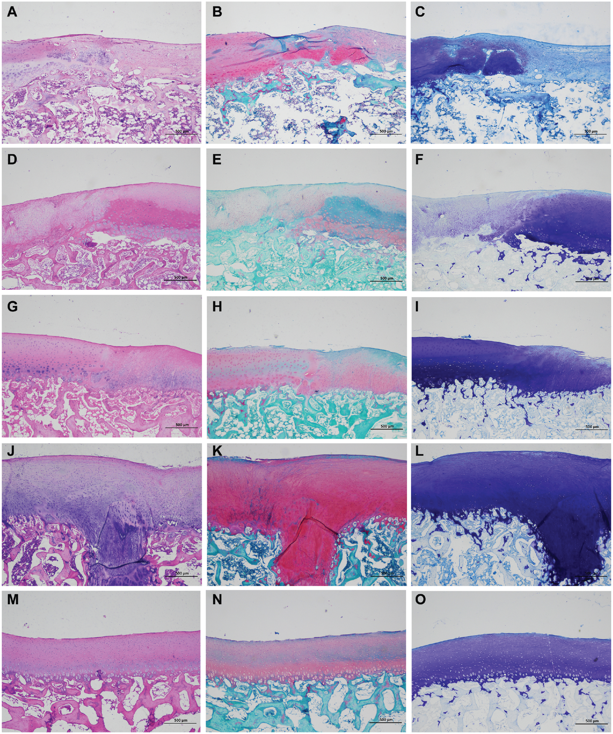
Histological results. (A) Hematoxylin and eosin (HE) staining, (B) safranin O and fast green staining, and (C) toluidine blue O staining in the particulated juvenile allograft cartilage (PJAC) group at 1 month. (D) HE staining, (E) safranin O and fast green staining, and (F) toluidine blue O staining in the autologous cartilage chip (ACC) group at 1 month. (G) HE staining, (H) safranin O and fast green staining, and (I) toluidine blue O staining in the PJAC group at 3 months. (J) HE staining, (K) safranin O and fast green staining, and (L) toluidine blue O staining in the ACC group at 3 months. (M) HE staining, (N) safranin O and fast green staining, and (O) toluidine blue O staining of normal cartilage (light microscope: ×40 for all images).
Immunohistochemical Results
We performed immunohistochemical testing of aggrecan and SOX9 at 1 and 3 months and saw positive cells at both time points. To describe the changes in immunostaining, we calculated the positive cell rates in 3 transplanted areas from different samples and conducted semi-quantitative analysis by Image-Pro Plus 6.0 (Media Cybernetics) to obtain the number of positive cells and the total number of cells in each group. The percentage of positive cells was then calculated. The positive rates of SOX9 in the PJAC and ACC groups at 1 month were 67.6% ± 3.78% and 63.4% ± 5.30%, respectively, and the difference was not statistically significant (P > .05). The positive rates of SOX9 in the PJAC and ACC groups at 3 months were 68.8% ± 2.69% and 17.1% ± 1.26%, respectively, and the difference was statistically significant (P < .05), indicating that SOX9 had a stronger expression in the PJAC group than in the ACC group at 3 months. The positive rates of aggrecan in the PJAC and ACC groups at 1 month were 40.5% ± 2.78% and 42.4% ± 0.54%, respectively, and the difference was not statistically significant (P > .05). The positive rates of aggrecan in the PJAC and ACC groups at 3 months were 40.8% ± 1.50% and 30.1% ± 2.44%, respectively, and the difference was not statistically significant (P > .05) (Figures 3 -6; Appendix Figure A3, available online).
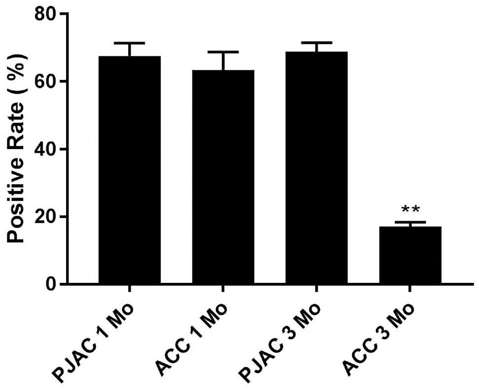
SOX9 positive rate.**P < .05 indicates a statistically significant difference. ACC, autologous cartilage chip; PJAC, particulated juvenile allograft cartilage.
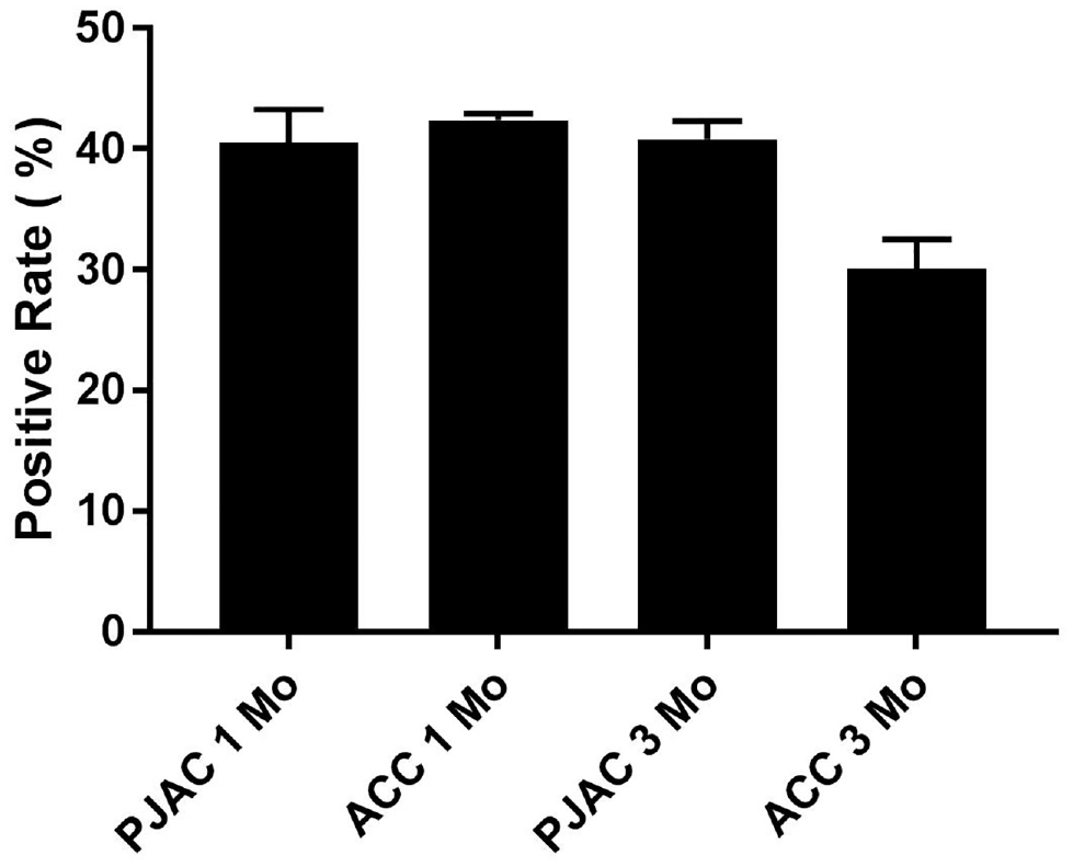
Aggrecan positive. ACC, autologous cartilage chip; PJAC, particulated juvenile allograft cartilage.
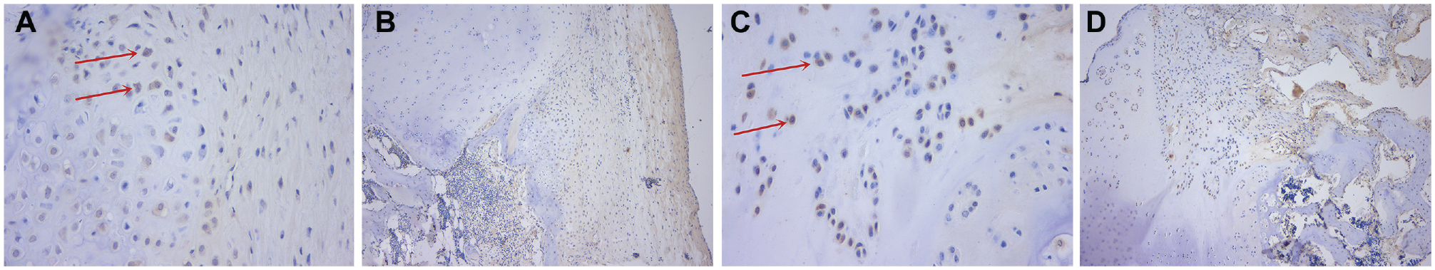
Aggrecan immunohistochemical results. (A) Particulated juvenile allograft cartilage (PJAC) group at 1 month; the red arrows show the positive cells (light microscope: ×400). (B) PJAC group at 1 month (light microscope: ×100). (C) Autologous cartilage chip (ACC) group at 1 month; the red arrows show the positive cells (light microscope: ×400). (D) ACC group at 1 month (light microscope: ×100).
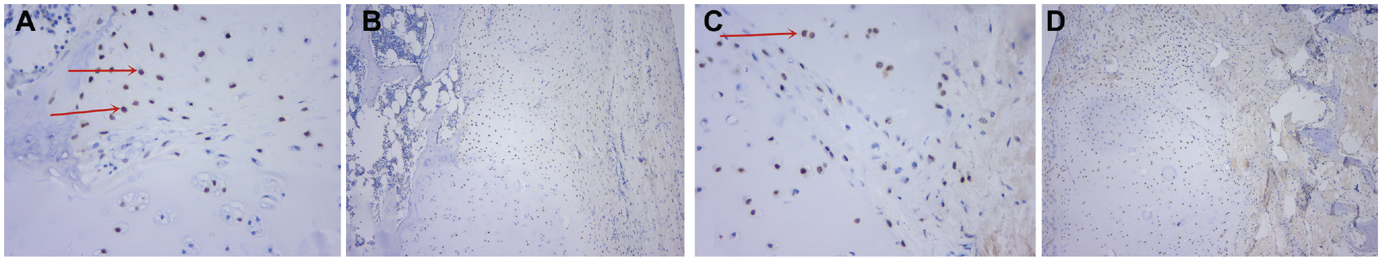
SOX9 immunohistochemical results. (A) Particulated juvenile allograft cartilage (PJAC) group at 1 month; the red arrows show the positive cells (light microscope: ×400). (B) PJAC group at 1 month (light microscope: ×100). (C) Autologous cartilage chip (ACC) group at 1 month; the red arrow shows the positive cells (light microscope: ×400). (D) ACC group at 1 month (light microscope: ×100).
Discussion
This study established a large animal model and detected a positive signal of SRY gene in transplanted cells from the repaired tissue by in situ hybridization, indicating that the transplanted cells survived at least 3 months. SOX9 and aggrecan had protein expression at 1 and 3 months, and the SOX9 expression was stronger in the PJAC group than in the ACC group. So far, about 10,000 people have received PJAC surgery worldwide, and the long-term clinical effects of PJAC surgery have been confirmed.10,13,15,16,17,29,37 In 2013, Tompkins et al 37 applied the PJAC technique to treat 16 patients with patellar cartilage disease. After 28 months, the patients’ International Knee Documentation Committee (IKDC) scores had improved significantly, and 89% and 73% of the patients’ MRI results showed normal or near-normal cartilage repair, respectively, at the repaired defect area. In 2014, Farr et al 13 followed 25 patients for 24 months after PJAC surgery; the patients’ IKDC, Knee injury and Osteoarthritis Outcome Score, and visual analog scale pain scores all improved, and the biopsy of 8 patients’ repair tissue showed that the tissue was composed of glassy degeneration and fibrocartilage. In 2017, Grawe et al 15 conducted an MRI analysis of 45 PJAC patients; the investigators found that 75% of the patients had good or moderate filling of cartilage defects at 24 months, and the repair tissue gradually matured over time. In 2018, Van Dyke et al 38 applied the PJAC technique to treat 9 patients who had first metatarsal cartilage injury; at 3 years after surgery, the patients’ pain scores had decreased and the American Orthopaedic Foot and Ankle Society scores had increased, and 8 of the 9 patients were satisfied or very satisfied with the results. Increasing clinical evidence has shown that the long-term efficacy of PJAC is quite good, especially in clinical outcomes and radiological results. However, few studies have examined the histologic and repair mechanisms of PJAC in large animal experimental studies. When one is evaluating the clinical treatment effect of PJAC, it is necessary to consider whether the chondrocytes survive after transplant, whether the key genes function, and what is the mechanism of the cartilage repair. The large animal study we describe here was a necessary complement to clinical research that analyzed the therapeutic effects of PJAC from the perspectives of histology and related mechanisms.
In this study, we investigated whether the donor cells existed in the repaired tissue cells of the PJAC transplanted cartilage defect model. To distinguish between the transplanted chondrocytes and host cells, we used PJAC derived from male Guizhou minipigs and transplanted it into the cartilage defect of female hosts; then we conducted in situ hybridization using a male-specific SRY gene. ‡
The SRY gene was discovered in 1990. 34 It is a highly conserved sequence of genes on the Y chromosome that exists only in male patients. On the basis of this biological characteristic, the SRY gene can be used as a marker. 7 Ostrander et al 28 applied the SRY gene to track transplanted chondrocytes and confirmed that male donor cells existed in the repaired tissue for at least 28 days after transplant, although the number of donor cells decreased over time. Pilichi et al 32 conducted a 4-year follow-up on sheep embryonic stemlike cells that had been engrafted into sheep femoral condyle osteochondral defects. After 4 years, the transplanted chondrocytes could still be detected. Those investigators selected SRY gene as a marker for the transplanted tissue, and the results showed that the method was effective and reliable. Cook et al 12 performed allograft cartilage transplant in adult dogs and tracked the transplanted chondrocytes for 6 months using the SRY gene.
To our knowledge, the study we report here is the first attempt to apply the SRY gene in situ hybridization technique to study PJAC in repairing cartilage defects and track the transplanted cells. Positive in situ hybridization signals were detected in the repaired tissue cells at 1 month postoperatively, and these positive signals still existed at 3 months. This indicated that PJAC contributed to the repair of host cartilage defects. Previous studies have shown that PJAC has a strong ability to regenerate, which is better than adult allograft cartilage transplant and has a better repair effect. In this study, in terms of histological scores, a statistical difference was seen between the PJAC group and ACC group at 1 month postoperatively and no statistical difference between the PJAC group and ACC group at 3 months, indicating that the repair effect of the PJAC group was similar to that of the ACC group over time. The statistical difference at 1 month might be due to immunological rejection, apoptosis, and other mechanisms that led to cell death, but the allograft cells gradually stabilized over time. It is hoped that relevant studies can provide a scientific basis for producing better therapeutic effects in the clinical application of PJAC.
We conducted immunohistochemistry tests 1 (ie, by the reaction of antigen and antibody) to analyze the protein expression of key genes in cartilage formation. By analyzing the SOX9 positive cell rate, we found an interesting phenomenon. At 3 months, the expression of SOX9 protein was stronger in the PJAC transplant than in the ACC transplant, which might be due to the continuous enhancement of the repair and proliferation ability of PJAC. In contrast, the decrease of SOX9 staining in the ACC group may be because the expression of different genes in the cartilage repair is sequential; that is, the gene expression changes dynamically. The gene expression of SOX9 is of great significance in the early repair of cartilage. The early repair progress in the ACC group was faster and then there was a downregulation, whereas the early repair process in the PJAC group was slower and longer lasting. Thus, at 3 months, a high expression of SOX9 remained in the PJAC group. The specific repair mechanism requires further study. Recent studies have shown that as an upstream gene, the transcription factor SOX9 can initiate cartilage formation and even coordinate signal transduction pathways such as TGF-β/Smad, Wnt/β-catenin, and HIF-1. 8 SOX9 protein is a powerful transcription activator both in vivo and in vitro, which can activate the transcription of target genes. SOX9 binds to specific sites on Col2al introns, directly regulating the expression of chondrocyte-specific marker Col2al 24 ; the regulation of SOX9 expression plays a central role in the cartilage repair process.6,8,23,39,40 The increasing expression of SOX9 protein may continue to cause an increase in Col2al and produce the same repair effect as the ACC transplant. 2
Adult chondrocytes have very weak proliferative capacity due to fewer blood vessels and lymphoid tissues. ACC transplant requires 2 surgeries because of limited tissue source, which would cause greater trauma and unstable phenotype. In recent years, it has been found that the long-term treatment efficacy of microfractures is not good, and the proliferative capacity of PJAC is stronger than that of adult cells, which creates a promising treatment plan. 10
Although the application of PJAC has achieved some effects on the repair of cartilage defects, whether the donor cells participate in the process of cartilage defect repair from a histological perspective still needs to be analyzed. To explore the mechanism of PJAC repair, we chose minipigs as a large animal model to simulate the cartilage transplant process of PJAC. The size of the knee joint, weightbearing requirements, and cartilage thickness of pigs are much more similar to human beings than those of smaller animal models and are physiologically and physically similar to humans. The potential for juvenile cartilage proliferation and repair in pigs is similar to that in humans; thus, it is the best large animal model to simulate human cartilage repair and transplant with high clinical reference value.9,11,24,31
This study used a specific in situ hybridization technique to demonstrate that allograft chondrocytes can survive for at least 3 months in the donor body. We also confirmed that SOX9 and aggrecan were expressed and that their expressions were stronger in the PJAC group than in the ACC group at 3 months. This may cause the increase of type II collagen, thereby continuously enhancing the repair process. Our study explained the mechanism of the improvement in imaging results and clinical symptoms and is an important supplement to the clinical research of PJAC.
This study also has limitations, such as a small number of animals and a relatively short observation period of <3 months. The juvenile cartilage we transplanted consisted of fresh juvenile cartilage particles, which are different from the clinically used particles that are stored at low temperature for a certain period, and could not completely imitate the clinical application of the PJAC technique.
Conclusion
In this study, a large animal model was established with Guizhou minipigs, and the cartilage defect was repaired with male PJAC. The SRY gene positive signal was detected from the repaired tissue by in situ hybridization, indicating that the transplanted cells survived for at least 3 months. SOX9 and aggrecan were expressed in repaired cartilage at 1 and 3 months after transplant, and the SOX9 expression in the PJAC group was stronger than that in the ACC group at 3 months. The PJAC technique has good application prospects for repairing cartilage defects, but the related repair mechanism needs further research.
Supplemental Material
DS_10.1177_0363546520958700 – Supplemental material for Donor Cell Fate in Particulated Juvenile Allograft Cartilage for the Repair of Articular Cartilage Defects
Supplemental material, DS_10.1177_0363546520958700 for Donor Cell Fate in Particulated Juvenile Allograft Cartilage for the Repair of Articular Cartilage Defects by Changgui Zhang, Yunong Ao, Jin Cao, Liu Yang and Xiaojun Duan in The American Journal of Sports Medicine
Footnotes
Acknowledgements
The authors thank Xin Chen from the Center for Joint Surgery, Southwest Hospital, Army Medical University, for providing language support for this article.
Submitted March 6, 2020; accepted June 18, 2020.
One or more of the authors has declared the following potential conflict of interest or source of funding: This work was financially supported by the National Natural Science Foundation of China (grant No. 81071484) and Key Research Project of Southwest Hospital (grant No. SWH2017JSZD-05). AOSSM checks author disclosures against the Open Payments Database (OPD). AOSSM has not conducted an independent investigation on the OPD and disclaims any liability or responsibility relating thereto.
References
Supplementary Material
Please find the following supplemental material available below.
For Open Access articles published under a Creative Commons License, all supplemental material carries the same license as the article it is associated with.
For non-Open Access articles published, all supplemental material carries a non-exclusive license, and permission requests for re-use of supplemental material or any part of supplemental material shall be sent directly to the copyright owner as specified in the copyright notice associated with the article.