
Abstract
Background:
Deficits in sporting performance after Achilles tendon repair may be due to changes in musculotendinous unit structure, including tendon elongation and muscle fascicle shortening.
Purpose/Hypothesis:
The purpose was to discern whether Achilles tendon rupture reduces triceps surae muscle force generation, alters functional ankle range of motion, or both during sports-related tasks. We hypothesized that individuals who have undergone Achilles tendon repair lack the functional ankle range of motion needed to complete sports-related tasks.
Study Design:
Descriptive laboratory study.
Methods:
The study included individuals 1 to 3 years after treatment of Achilles tendon rupture with open repair. Participants (n = 11) completed a heel-rise task and 3 jumping tasks. Lower extremity biomechanics were analyzed using motion capture. Between-limb differences were tested using paired t test.
Results:
Pelvic vertical displacement was reduced during the heel-rise (mean difference, −12.8%; P = .026) but not during the jumping task (P > .1). In the concentric phase of all tasks, peak ankle plantarflexion angle (range of mean difference, −19.2% to −48.8%; P < .05) and total plantar flexor work (defined as the area under the plantar flexor torque – ankle angle curve) (range of mean difference, −9.5% to −25.7%; P < .05) were lower on the repaired side relative to the uninjured side. No significant differences were seen in peak Achilles tendon load or impulse with any of the tasks. There were no differences in plantar flexor work or Achilles tendon load parameters during eccentric phases.
Conclusion:
Impaired task performance or increased demands on proximal joints were observed on the repaired side in tasks isolating ankle function. Tasks that did not isolate ankle function appeared to be well recovered, although functional ankle range of motion was reduced with rupture. Reduced plantar flexor muscle-tendon unit work supports previous reports that an elongated tendon and shorter muscle fascicles caused by Achilles tendon rupture constrain functional capacity. Achilles tendon peak load and impulse were not decreased, suggesting that reduced and shifted functional ankle range of motion (favoring dorsiflexion) underlies performance deficits.
Clinical Relevance:
These findings point to the need to reduce tendon elongation and restore muscle length of the triceps surae after Achilles tendon rupture in order to address musculature that is short but not necessarily weak for improved performance with sports-related activities.
Achilles tendon rupture has substantial implications for an individuals’ ability to engage in sporting activity. Up to 80% of individuals with Achilles tendon rupture are able to return to some amount of sporting activity. 41 In elite athletes, the return to sports rates tend to be lower, with between 60% and 70% of National Basketball Association and National Football League athletes returning to sports in the same division,3,29,38 although return to play rates seem to be sport-dependent.13,38,39 Sports performance is also impaired upon return to sport. Professional athletes have been found to have diminished performance ratings after injury compared with baseline3,13,29,38 and lower performance ratings in the first season after return from Achilles tendon rupture when compared with their uninjured peers.3,29,38 From a biomechanical standpoint, Achilles tendon ruptures permanently reduce performance during running, jumping, and participating in sports in recreationally active people.8,21,31,40,43,44
Achilles tendon ruptures stimulate gross structural changes in both the tendon and triceps surae musculature. After rupture, the Achilles tendon heals in a permanently elongated position22,33,36 and never regains the material properties of healthy tendon tissue.2,9,12,42 Achilles tendon rupture not only affects the tendon itself but also disrupts the whole muscle-tendon unit. At presentation with Achilles tendon rupture, the triceps surae muscles have shorter fascicle length and increased pennation angles, which persist throughout the first 14 weeks after injury 19 and have been suggested to drive tendon elongation as the musculotendinous unit tries to restore tension on a healing tendon. 18 Fascicle remodeling and increased pennation persist 3 to 6 months after injury.1,30 In addition to shorter and more pennate muscle fascicles, 24 muscle atrophy15,16,24,32,44 and fatty infiltration15,16 have been reported beyond 1 year after injury.
Whether structural disruption of the triceps surae muscles after Achilles tendon rupture reduces the ability of these muscles to generate force, alters functional ankle range of motion (defined as the available ankle excursion over which the plantar flexors are able to generate adequate force for task completion), or a combination of both during higher level, sports-related tasks is not well-described. Previous work linked muscle-tendon structural changes after Achilles tendon rupture with functional deficits during isokinetic strength testing,7,11,20 heel-rise tests,5,36 and plyometric activity. 8 The purpose of this study was to determine the effects of Achilles tendon ruptures treated with surgical repair on tendon loading parameters during a combination of clinical tasks to begin to disentangle the effects of muscle contractility and functional ankle range of motion on higher level task performance.
Given the elongation of the tendon and the shortening of muscle resting length after Achilles tendon rupture, we hypothesized that individuals who have undergone Achilles tendon repair lack the functional ankle range of motion needed to complete heel-rise and jumping tasks (Figure 1). Decreased functional ankle range of motion would result in no differences between peak force generation of the muscle (Achilles tendon impulse, peak Achilles tendon load) on the repaired compared with the uninjured side. However, functional ankle range of motion would be reduced and shifted toward more dorsiflexed positions to pre-tension the muscle. We hypothesized that alterations in functional ankle range of motion would be reflected in reduced work (defined as the area under the plantar flexor torque – ankle angle curve) done by the plantar flexor muscles (as the peak force would be similar but would occur over a shorter fascicle length). In summary, we hypothesized that the ruptured side would have decreased functional ankle range of motion relative to the uninjured side as evidenced by lower ankle excursion, lower peak ankle angle, and lower ankle work as well as no side-to-side difference in peak Achilles tendon load or impulse.
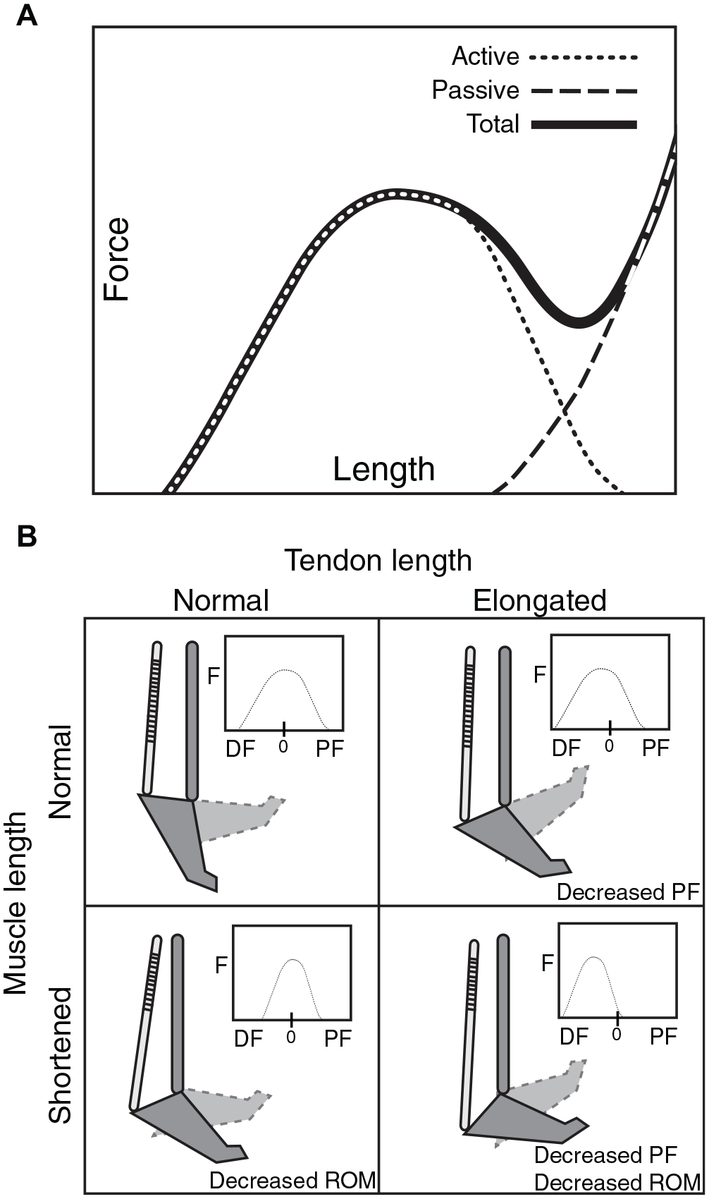
Study hypothesis. (A) Muscle and tendon length directly affect the ability of the plantar flexors (PF) to generate force (F). After Achilles tendon rupture, the tendon is elongated and the muscle fascicles are shortened, decreasing the excursion over which the muscle is able to contract. (B) To compensate for an elongated tendon and adequately preload the muscle, the ankle moves into additional dorsiflexion (DF), decreasing the functional ankle range of motion (ROM) and shifting it toward dorsiflexion. This conceptual framework is supported by literature linking kinetic ankle deficits during concentric angle tasks with structural deficits in both patients 19 and computational models. 5
Methods
Study Design and Participants
This is a cross-sectional, retrospective analysis of prospectively collected data. Participants included in this study were enrolled between April and November 2017. This study was approved by the University of Delaware Institutional Review Board. To be included, participants needed to have undergone Achilles tendon rupture treated with open surgical repair 1 to 3 years earlier. This time frame was based on literature suggesting that the majority of functional gains are made by 1 year after injury, with no additional gains between 1 and 2 years after injury 27 and limited gains 1 to 7 years after injury. 8 Exclusion criteria were history of postoperative complications including deep vein thrombosis or rerupture, and an inability to jump unilaterally because of pain or injury aside from the Achilles tendon rupture. Participants’ surgical and postoperative rehabilitative treatment was administered according to the recommendations of their healthcare providers, and participants did not receive standardized intervention as part of the study.
All data were collected in a single session via questionnaires (the Achilles Tendon Total Rupture Score 26 and Physical Activity Scale 14 ) to characterize participants’ self-reported function and physical activity. Muscle and tendon characteristics were assessed by ultrasonographic examination and have been previously reported. 43 Participants then completed functional testing in which each test was performed unilaterally, including a heel-rise task, countermovement jump (CMJ), drop countermovement jump (DJ), and hopping, as these are lower extremity functional tasks that have been found to be valid and reliable in individuals with Achilles tendon injuries.25,28,35,37 Muscle activation and lower limb biomechanics (but not Achilles tendon load) have been previously reported for the hopping task, 43 and muscle activation has been previously reported for the CMJ task. 44
The right limb was tested first for all participants to quasi-randomize testing of the repaired and uninjured sides. For the heel-rise task, participants were positioned standing unilaterally on a 10° incline box. Participants were allowed 2-finger support on an external bar for balance. Participants performed 5 heel rises at a rate of 1 heel rise per second (Figure 2). For the CMJ, participants were instructed to stand unilaterally with their arms folded across their chest and then perform a maximal, vertical jump, trying to land in the same place on the force plate. For the DJ, participants again stood unilaterally with their arms folded across their chest on top of a 10-cm box. They were instructed to try to slide off of the front of the box and, upon landing on the floor, perform a maximal vertical jump. They completed 3 repetitions on each leg for the CMJ and DJ tasks. For hopping, participants stood unilaterally with their arms to their sides as if jumping rope. They performed 25 rhythmic jumps at a self-selected pace, trying to stay within the same place. Trials were repeated if participants jumped off of the force plate. Participants completed 2 repetitions of the hopping task on each leg.
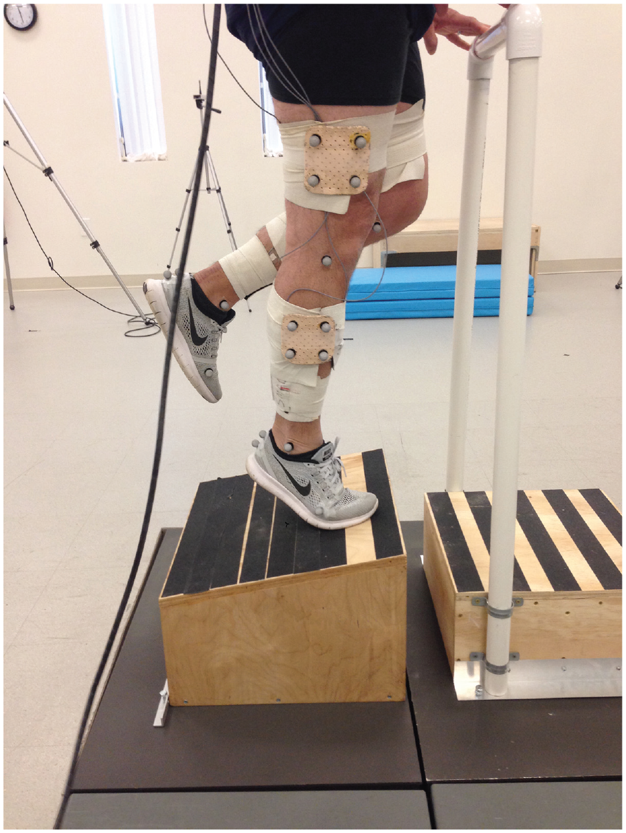
Heel-rise task.
Biomechanical Analysis
Lower limb and pelvic kinematics and kinetics were assessed using 8 cameras (Nexus; Vicon) and 2 in-ground force plates (Bertec Corporation). Kinematic results were collected at 120 Hz and kinetic results were collected at 1080 Hz. Retroreflective markers were placed on the foot (first and fifth metatarsal heads, posterior heel), ankle (medial and lateral malleoli), knee (medial and lateral femoral condyles), and hip/pelvis (greater trochanter, anterior superior iliac spine) with tracking clusters on the shank, thigh, and sacrum. 10
Lower limb kinematics and kinetics were calculated by means of a constrained kinematic model using a previously described approach. 4 Briefly, a generic musculoskeletal model (gait 2392) 34 was scaled using each participant’s bodyweight and markers placed over anatomic landmarks. After scaling body segments of each participant, we moved these models into the anatomic position by fitting the experimental data collected during a standing trial using best practices. 17 Next, inverse kinematics quantified lower extremity motion, and inverse dynamics quantified lower extremity kinetics during each of the movement trials. The change in pelvic height during each activity was calculated to quantify movement performance. Pelvic height was defined as the vertical distance of the center of the pelvis from the ground. We used OpenSim’s pelvic body coordinate system origin, which is based on the anterior superior iliac spine and posterior superior iliac spine landmarks. 34 Using this peak height, we analyzed the concentric and eccentric phases of each activity and resampled the phases to 101 data points (MATLAB; The Mathworks). Impulse was calculated as the area under the load-time curve and work as the area under the load-displacement curve (trapz function in MATLAB). To characterize joint kinetics and Achilles tendon loading across participants, kinetic variables were normalized by participant bodyweight for Achilles tendon peak load and impulse and by participant bodyweight multiplied by participant stature for joint kinetics including peak torque and joint work. After calculating the ankle joint moments generated during these activities, we calculated Achilles tendon load by dividing these moments by an Achilles tendon moment arm of 5 cm (per Matijevich et al 23 ) and normalized tendon load by participant bodyweight. Achilles tendon peak load and impulse loading are reported to characterize the loads applied to the tendon. Because we did not quantify triceps surae muscle shortening dynamics with ultrasound imaging, we decided to report joint-level work.
Statistical Analysis
Descriptive statistics (mean ± SD) are reported for each of the variables of interest. Differences in Achilles tendon load parameters between repaired and uninjured sides were examined using 1-tailed paired t tests. A 1-tailed test was used because it is understood that ankle performance is impaired after Achilles tendon rupture. Differences in hip and knee joint biomechanics between repaired and uninjured sides were examined using 2-tailed paired t tests. Significance was established a priori as an alpha level of .05. A conservative estimate indicated that 10 participants were needed to power the study at an alpha level of .05 and power of 0.95, based on previously reported effect sizes observed in between-limb biomechanical comparisons of individuals after Achilles tendon rupture. 40
Results
Participant Characteristics
The participants (10 male and 1 female) had a mean ± SD body mass index of 25.9 ± 1.0 and were 43.6 ± 2.7 years of age. Participants were 17 ± 6 months from repair at the time of study participation, with a mean time from rupture to surgery of 11 ± 10 days. The right side was ruptured in 5 participants, and the left side was ruptured in 6 participants. Participants scored a mean of 88.8 ± 4.5 out of 100 points on the Achilles Tendon Total Rupture Score and 5.0 ± 0.5 on the Physical Activity Scale, corresponding to the description “moderate exercise at least 3 hours a week, eg, tennis, swimming, jogging, etc.”
Heel-Rise Ankle Performance
Participants had significantly less pelvic displacement (the change in vertical pelvic position from the start of the movement to the peak vertical position of the movement) during the heel-rise task on the injured relative to the uninjured side (mean difference = −12.8%, P = .026). During the concentric phase, peak ankle angle and ankle excursion were significantly lower on the repaired side (peak – mean difference = −48.8%, P = .001; excursion – mean difference = −24.4%, P = .002) (Figure 3; Table 1). Total plantar flexor work was lower on the repaired side, but there were no statistically significant differences in peak Achilles tendon load (mean difference = −2.2%, P = .372) or Achilles tendon impulse (mean difference = −1.0%, P = .528) between injured and uninjured sides (Figure 4; Table 1).
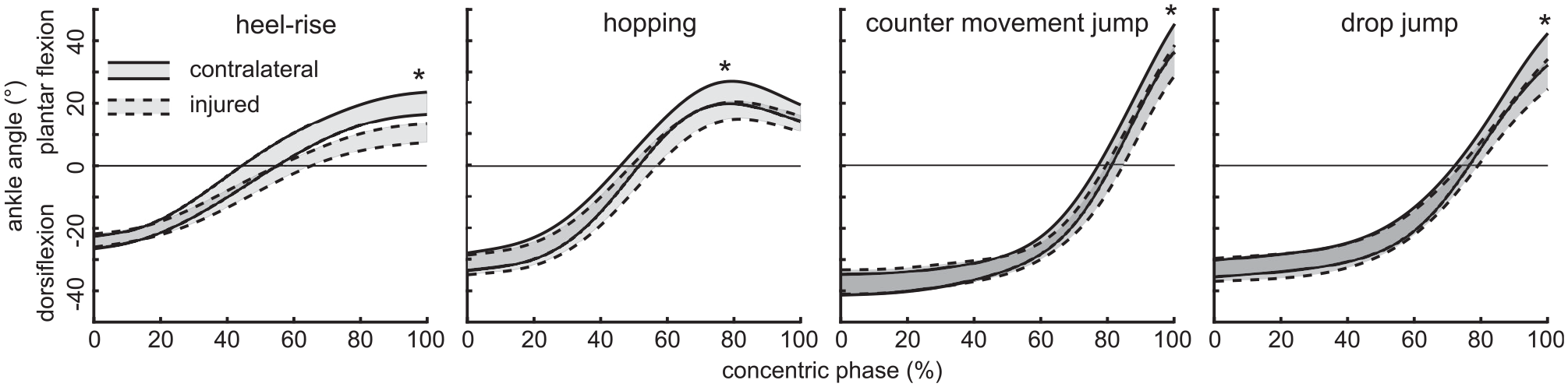
Plantar flexor angle during the concentric phase for each of the clinical tasks. Plantarflexion is positive; shaded, outlined areas represent bootstrapped 95% CIs. *P < .05 for between-limb comparison.
Ankle Mechanics During Concentric and Eccentric Phases of Functional Tasks a
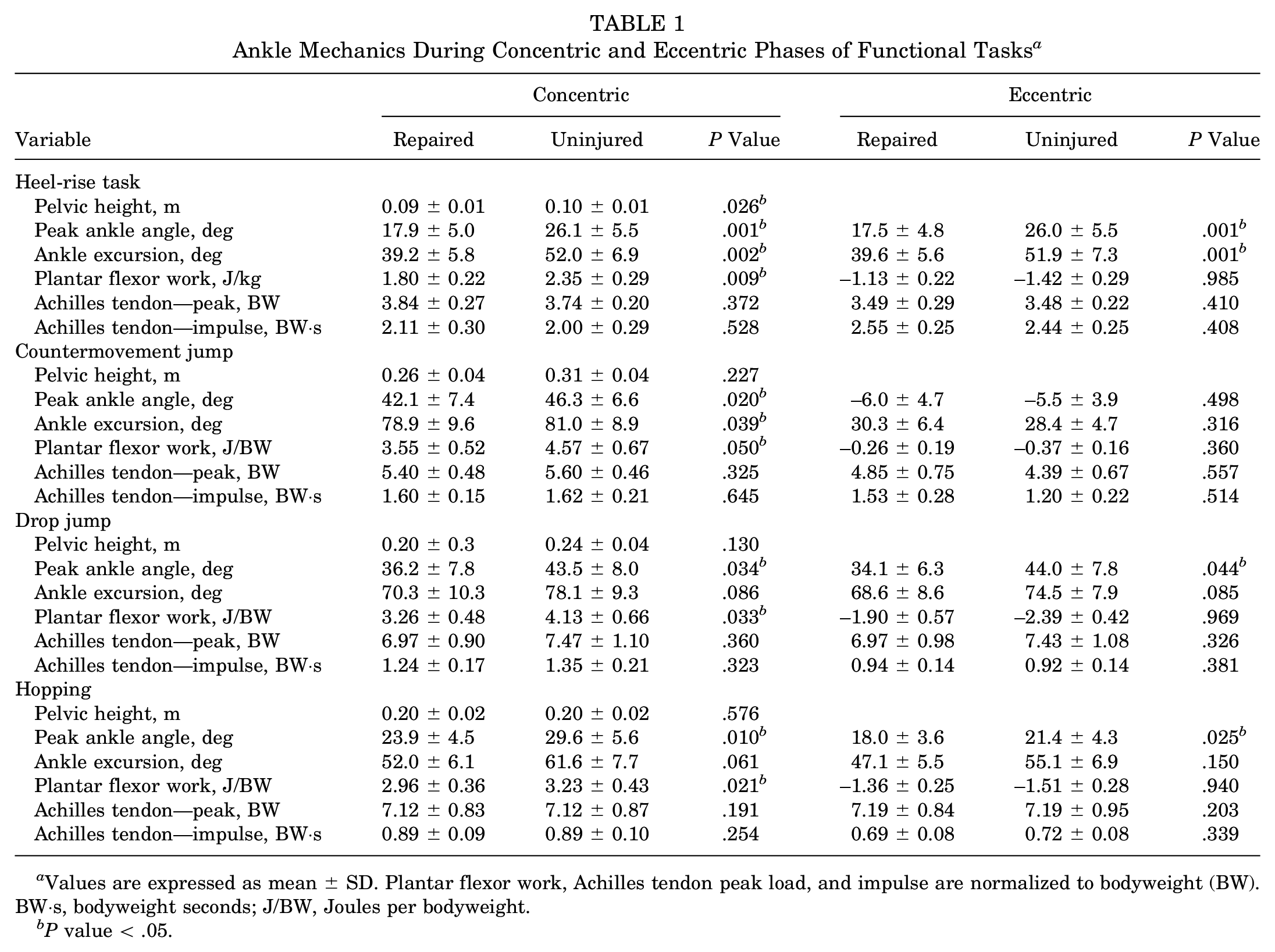
Values are expressed as mean ± SD. Plantar flexor work, Achilles tendon peak load, and impulse are normalized to bodyweight (BW). BW·s, bodyweight seconds; J/BW, Joules per bodyweight.
P value < .05.
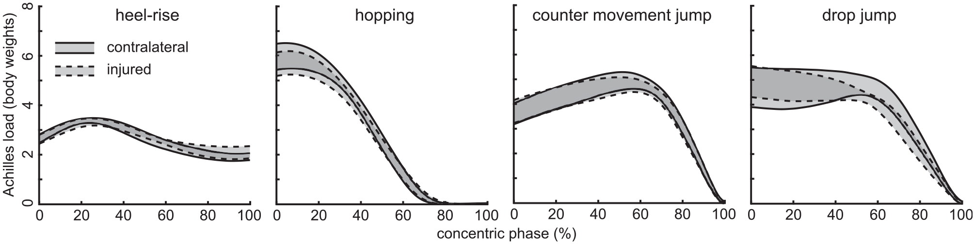
Achilles tendon load profiles for each of the clinical tasks. Plantarflexion is positive; traces represent bootstrapped 95% CIs.
Similar to the concentric phase, during the eccentric phase the injured ankle had significantly lower peak ankle angle (mean difference = −49.4%, P = .001) and ankle excursion (mean difference = −24.7%, P = .001) (Table 1). No statistically significant differences were found for total plantar flexor work (mean difference = −23.7%, P = .985), peak Achilles tendon load (mean difference = −1.3%, P = .410), or Achilles tendon impulse (mean difference = −2.0%, P = .408) between injured and uninjured sides (Table 1).
Jumping Ankle Performance
There were no differences in pelvic vertical trajectory during jumping on the injured compared with uninjured side during any of the jumping tasks (mean difference, P value – CMJ: −6.7%, P = .227; DJ: −10.5%, P = .130; hop: 0.8%, P = .576) (Table 1).
For the concentric phase of the CMJ, ankle angle peak (mean difference = −19.1%, P = .020) and excursion (mean difference = −11.5%, P = .039) were significantly lower on the repaired side (Table 1). Plantar flexor work was lower on the repaired side (mean difference = −19.5%, P = .050), but there were no differences in Achilles tendon peak load (mean difference = −4.6%, P = .325) or impulse (mean difference = −2.6%, P = .645). No between-limb differences were observed in any measure during the eccentric phase (Table 1).
For the concentric phase of the DJ, ankle angle peak (mean difference = −22.1%, P = .034) but not excursion (mean difference = −10.6%, P = .086) was significantly lower on the repaired side (Table 1). Concentric work was lower on the repaired side (mean difference = −24.3%, P = .033), but there were no between-limb differences in Achilles tendon peak load (mean difference = −5.3%, P = .360) or impulse (mean difference = −6.7%, P = .323). Ankle angle peak (mean difference = −19.0%, P = .044) was lower on the repaired side during the eccentric phase, but we found no between-limb differences in ankle peak, plantar flexor work, or Achilles tendon peak load or impulse during the eccentric phase (Table 1).
For the concentric phase of the hopping task, ankle angle peak (mean difference = −28.1%, P = .010) but not excursion (mean difference = −10.8%, P = .061) was significantly lower on the repaired side. Concentric plantar flexor work was lower on the repaired side (mean difference = −20.7%, P = .021), but no differences were seen in peak Achilles tendon load (mean difference = −7.0%, P = .191) or Achilles tendon impulse (mean difference = −6.6%, P = .254). In the eccentric phase, peak ankle angle was significantly lower on the repaired side (mean difference = −24.5%, P = .025), but no between-limb differences in ankle excursion were noted (mean difference = −7.0%, P = .150). Similar to the other jumping tasks, there were no between-limb differences in plantar flexor work or Achilles tendon peak load or impulse during the eccentric phase (Table 1).
Hip and Knee Contributions to Heel-Rise and Jumping Performance
We found no differences in knee or hip work between sides during the concentric phase of any task with the exception of combined proximal lower extremity (hip and knee together) work during the concentric phase of hopping (between-limb comparison for combined lower extremity: heel-rise task, mean difference 7.0%, P = .369; CMJ, mean difference 18.1%, P = .496; DJ, mean difference 2.5%, P = .664; hop, mean difference 41.6%, P = .006) (Table 2).
Hip and Knee Work During Concentric Phase of Functional Tasks a
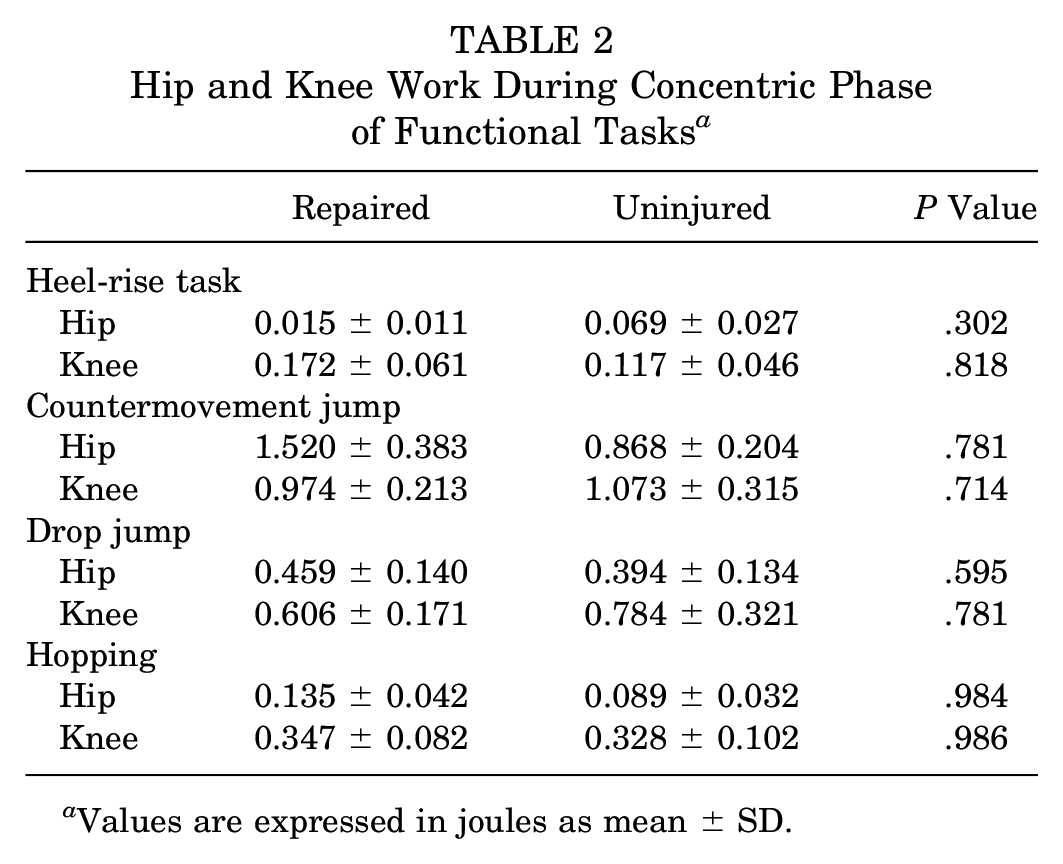
Values are expressed in joules as mean ± SD.
Discussion
This study is the first to disentangle functional ankle range from plantar flexor strength by assessing lower extremity biomechanics during higher level strength and plyometric tasks commonly used in the rehabilitation progression to return individuals to sporting activities after Achilles tendon rupture. In tasks that isolate ankle function, such as the heel-rise and hopping tasks, participants demonstrated either impaired task performance (evidenced by lower pelvic displacement during the heel-rise task) or increased demands on the more proximal joints (evidenced by comparable jump height between sides but increased proximal limb peak load and work on the repaired side). Tasks that did not isolate ankle function—such as the CMJ and DJ—appeared to be well recovered, although functional ankle range of motion was reduced by 19% to 48% on the repaired side.
The concentric phase was consistently more affected by Achilles tendon rupture across all tasks, with a 20% to 26% deficit in plantar flexor work on the repaired side. Achilles tendon peak load and impulse were not decreased, however. Taken together, these findings suggest that reduced and shifted functional ankle range of motion (favoring a more dorsiflexed range) underlie heel-rise and jumping performance deficits in a group of individuals who had recovered well based on self-reported function after injury. This points to the need to reduce tendon elongation and restore muscle length of the triceps surae after Achilles tendon rupture, in order to address musculature that is short but not necessarily weak, for improved performance with sports-related activities.
A combination of shortened muscle fascicle length and tendon elongation is a potential explanation for impaired functional ankle range of motion because the muscle has less excursion to contract, particularly affecting performance with tasks like heel rises that emphasize end-range plantarflexion.5,6,11 Functional deficits resulting from this imbalance in length-tension relationships of the myotendinous unit are supported by moderate-to-strong relationships between muscle fascicle length and isometric plantarflexion torque at 14 weeks after injury. 19 Functionally, isolated calf strength deficits that are more pronounced in greater amounts of plantarflexion have been observed with dynamometry, correlating strongly with muscle atrophy. 15 Tendon elongation has been suggested to underlie changes in walking,2,42 running,8,21,40 and jumping mechanics throughout the course of recovery.31,40,43,44 Additionally, investigators have observed moderate-to-strong relationships between tendon elongation and deficits in triceps surae muscle isometric strength, particularly at end-range plantarflexion, even in individuals >10 years after injury. 16 Combined with the results of the present study, these data indicate that the triceps surae musculature continues to have capacity to produce force, but over a limited range, pointing to the need to intervene with the goal of restoring muscle and tendon length, likely very early after rupture.
From a clinical standpoint, it seems important that the rehabilitation approach for returning to sport incorporates a variety of sport-specific maneuvers to develop task-related motor plans, given the differences in compensatory strategies observed across tasks. The results of this study support the importance of restoring functional ankle range of motion after Achilles tendon rupture treated with repair. Determining whether to focus rehabilitation on restoring functional ankle range of motion or on compensatory strategies may require consideration not only of tendon length but also muscle fascicle length as a prognostic indicator of rehabilitation potential. The extent to which muscle functional length can be remediated by rehabilitation is not known, so proximal lower extremity strengthening may be particularly important in individuals with short muscle fascicle length and elongated tendon length in order to prepare those individuals who will require proximal compensatory strategies to engage in sports activity. This is in line with previous literature supporting the need for proximal strengthening resulting from increased loads at the knee during plyometric tasks after Achilles tendon rupture.40,43 Additionally, because muscle and tendon length become abnormal very early after Achilles rupture, developing new surgical techniques to restore muscle fascicle length may help improve muscle functional length later in recovery.
A few limitations to this study are important to consider. This study was designed using the uninjured side as a within-patient control. An uninjured control group was not included so as to limit variables that could confound the analysis (eg, physical activity level, body mass index, age). It is possible that individuals in this study could have modified their heel-rise or jumping strategy bilaterally in response to their Achilles rupture, reducing the effect of presence of rupture in this analysis. Compared with previously published data in healthy participants, 4 the participants included in this study had similar or slightly increased peak Achilles tendon load (percentage difference based on means from repaired side in this group to controls: heel-rise, 28%; CMJ, 10%; DJ, 27%; hop, 7%) and similar or reduced Achilles tendon impulse (percentage difference based on means from repaired side in this group to controls: heel-rise, 2%; CMJ, −33%; DJ, −59%; hop, −32%). Additionally, although all participants underwent open surgical repair and some form of postoperative rehabilitation, surgical technique and rehabilitation were not standardized. Including individuals treated by standard of care, rather than intervention delivered, likely reflects the clinical reality of patients with Achilles tendon rupture treated with open repair. We estimated Achilles tendon load by dividing ankle torque by a constant length moment arm of 5 cm. 23 However, we do not expect this approximation to affect the findings because we used paired t tests.
In summary, the findings of this study suggest that individuals with well-recovered, self-reported function continue to demonstrate substantial deficits in concentric ankle excursion and work after Achilles tendon repair, although Achilles impulse and peak Achilles tendon force are restored to the uninjured side. Taken together, these findings suggest that even 1 year after injury, at a time when most athletes have returned to sporting activity, 41 altered length-tension relationships within the triceps surae musculotendinous unit limit the functional ankle range of motion available to an athlete. Future studies are needed that investigate restoring muscle and tendon length-tension relationships early after injury and promoting recovery of functional ankle range of motion throughout the course of recovery in order to optimize athlete performance, particularly in tasks isolating ankle plantar flexor function.
Footnotes
Submitted October 8, 2020; accepted February 19, 2021.
One or more of the authors has declared the following potential conflict of interest or source of funding: This study was funded by the National Institute of Arthritis and Musculoskeletal and Skin Diseases (award number R01AR072034, K01AR075877) and the National Institute of Diabetes and Digestive and Kidney Diseases (award number F32 DK123916) of the National Institutes of Health. This research was also supported by the Foundation for Physical Therapy and the University of Delaware Research Foundation. The funders had no role in the design of the study; in the collection, analysis, or interpretation of data; in the writing of the manuscript; or in the decision to publish the results. AOSSM checks author disclosures against the Open Payments Database (OPD). AOSSM has not conducted an independent investigation on the OPD and disclaims any liability or responsibility relating thereto.