






Abstract
Background:
Poor tendon-to-bone healing in chronic rotator cuff tears (RCTs) is related to unsatisfactory outcomes. Exosomes derived from mesenchymal stem cells reportedly enhance rotator cuff healing. However, the difficulty in producing exosomes with a stronger effect on enthesis regeneration must be resolved.
Purpose:
To study the effect of exosomes derived from kartogenin (KGN)-preconditioned human bone marrow mesenchymal stem cells (KGN-Exos) on tendon-to-bone healing in a rat model of chronic RCT.
Study Design:
Controlled laboratory study.
Methods:
Exosome-loaded sodium alginate hydrogel (SAH) was prepared. Moreover, exosomes were labeled with 1,1′-dioctadecyl-3,3,3′,3′-tetramethylindotricarbocyanine iodide (DiR) or 1,1′-dioctadecyl-3,3,3′3′-tetramethylindocarbocyanine perchlorate (Dil) for in vivo tracking. Bilateral rotator cuff repair (RCR) was conducted in an established chronic RCT rat model. A total of 66 rats were randomized to control, untreated exosome (un-Exos), and KGN-Exos groups to receive local injections of pure SAH, un-Exos, or KGN-Exos SAH at the repaired site. The presence of DiR/Dil-labeled exosomes was assessed at 1 day and 1 week, and tendon-to-bone healing was evaluated histologically, immunohistochemically, and biomechanically at 4 and 8 weeks.
Results:
Both un-Exos and KGN-Exos exhibited sustained release from SAH for up to 96 hours. In vivo study revealed that un-Exos and KGN-Exos were localized to the repaired site at 1 week. Moreover, the KGN-Exos group showed a higher histological score and increased glycosaminoglycan and collagen II expression at 4 and 8 weeks. In addition, more mature and better-organized collagen fibers with higher ratios of collagen I to collagen III were observed at 8 weeks in the tendon-to-bone interface compared with those in the control and un-Exos groups. Biomechanically, the KGN-Exos group had the highest failure load (28.12 ± 2.40 N) and stiffness (28.57 ± 2.49 N/mm) among the 3 groups at 8 weeks.
Conclusion:
Local injection of SAH with sustained KGN-Exos release could effectively promote cartilage formation as well as collagen maturation and organization for enthesis regeneration, contributing to enhanced biomechanical properties after RCR.
Clinical Relevance:
KGN-Exos injection may be used as a cell-free therapeutic option to accelerate tendon-to-bone healing in chronic RCT.
Rotator cuff tears (RCTs) around the shoulder joint are commonly associated with shoulder pain and disability. Despite advances in surgical techniques and apparatuses for rotator cuff repair (RCR) in recent years, the overall retear rate after repair is still high, which is of concern to both patients and surgeons. 12 Currently, biological augmentation methods have been developed to promote healing between the torn tendon and the bone and may be the key to prevent retear. 43 A tendon-to-bone interface, commonly referred to as the enthesis, is composed of 4 layers: the tendon, the unmineralized fibrocartilage, the mineralized fibrocartilage, and the bone. 5 However, it is challenging to regenerate a native-like enthesis after RCR. 35 Especially, the reestablishment of fibrocartilage layers is paramount but also poses great difficulty. Instead, an intervening layer of fibrovascular scar tissue gradually forms and ultimately results in fibrous integration with low biomechanical properties between the tendon and bone. 13
Mesenchymal stem cells (MSCs) have been widely applied in tissue repair owing to their robust differentiation ability. 2 Different types of MSCs have been successfully used for the enhancement of enthesis healing.13,31,44 However, the potential risks of exogenous MSCs, such as tumorigenicity and immunogenicity, have limited their clinical application.10,23,24 Recently, noncellular therapies associated with MSCs have attracted much attention. The extracellular matrix, cytokines, and extracellular vesicles produced by stem cells are the main active components for tissue regeneration.37,40 Exosomes are small extracellular vesicles with a diameter ranging from 30 to 200 nm, which are secreted by cells and contain a variety of molecules, like RNAs, proteins, and other bioactive regulators. 21 MSC-derived exosomes have been applied in basic research of regenerative sports medicine and are considered a promising cell-free therapeutic option for clinical translation.28,32,40 For instance, bone marrow has been used as a cell source to extract exosomes and enhance cartilage regeneration for cartilage defects, osteoarthritis, and tendon-to-bone enthesis injury.17,18 Nonetheless, treatment of exosomes derived from bone marrow MSCs (BMSCs) has not always been successful. 27 The performance of exosomes needs to be optimized by endowing them with stronger specific effects. Recently, Wu et al 36 reported that exosomes derived from magnetically actuated BMSCs had more significant effects on tendon-to-bone healing than BMSC-derived exosomes. Feng et al 11 found that exosomes derived from genetically modified Scleraxis-overexpressing PDGFRα(+) BMSCs could reduce osteolysis during tendon-bone healing. However, the authors did not focus on the specific effect of exosomes in chondrogenesis for enthesis regeneration.
Kartogenin (KGN) is a novel small heterocyclic compound identified from >22,000 drug-like molecules. 20 Owing to its excellent regenerative capabilities, it has drawn considerable attention. For instance, KGN has been applied to accelerate enthesis regeneration of the rotator cuff and Achilles tendon in several studies.6,34,45,46 However, due to the low water solubility of KGN, drug administration and accurate dose control are difficult to perform. Notably, high doses of KGN can induce hyperchondrogenesis. 41 More recently, KGN was used to pretreat human umbilical cord MSCs (hUCMSCs) to produce specific exosomes, and exosomes derived from these KGN-preconditioned hUCMSCs were shown to promote chondrogenesis in a full-thickness cartilage defect rabbit model because of the unique enrichment of a set of miRNAs. 19 In the present study, we aimed to investigate the effect of exosomes derived from KGN-preconditioned human bone marrow mesenchymal stem cells (KGN-Exos) on tendon-to-bone healing of the rotator cuff in a rat chronic RCT model. It was hypothesized that KGN-Exos may lead to better tendon-to-bone healing after RCR than untreated exosomes.
Methods
Stem Cell Culture and KGN Precondition
Human bone marrow mesenchymal stem cells (hBMSCs) in Passage 0 from a 38-year-old man were purchased from ScienCell Research Laboratories. 7 Cells were cultured in fresh growth medium (α-MEM with 10% fetal bovine serum as well as 1% penicillin and streptomycin solution) at 37°C in 5% (vol/vol) CO2 in a cell culture dish.
For KGN preconditioning, hBMSCs at the third passage were seeded onto culture dishes. After reaching approximately 80% confluence, the cells were incubated with fresh growth medium containing 1 µM KGN (BioGems) for 3 days. 19 After aspiration, cells were rinsed repeatedly to remove residual KGN, and medium containing exosome-depleted fetal bovine serum (Invitrogen) was added. Then the cells were incubated for another 48 hours to obtain the culture supernatants. hBMSCs cultured in medium without KGN preconditioning were used as a negative control.
Exosome Isolation and Characterization
Exosomes were extracted from culture supernatants by gradient ultracentrifugation. 40 In brief, the following differential centrifugation protocol was used to remove remaining cells and cellular debris: 300g for 5 minutes, 2000g for 30 minutes, and 10,000g for 30 minutes. The supernatants were centrifuged at 100,000g for 70 minutes to obtain the exosome pellet. Subsequently, the pellet was washed with phosphate-buffered saline (PBS) to remove contaminating proteins and then ultracentrifuged repeatedly at 100,000g for 70 minutes and resuspended in PBS for collection of the KGN-Exos or untreated hBMSCs (un-Exos).
For characterization, the morphological features of the exosomes were observed using an FEI Tecnai G2 Spirit Transmission Electron Microscope and visualized using an AMT CCD camera (Advanced Microscopy Techniques). The size distribution was determined by nanoparticle tracking analysis using ZetaView PMX 120 (Particle Metrix). The expression of the TSG101, ALIX, HSP70, and Calnexin of cells (BMSCs and KGN-BMSCs) and exosomes (un-Exos and KGN-Exos) was analyzed with Western blotting.
Preparation and In Vitro Release of Exosome-Loaded Hydrogels
Un-Exos or KGN-Exos solution was prepared for injection. To track exosomes in vivo, they were prestained with 1,1′-dioctadecyl-3,3,3′,3′-tetramethylindocarbocyanine perchlorate (Dil; Sigma-Aldrich) or 1,1′-dioctadecyl-3,3,3′,3′-tetramethylindotricarbocyanine iodide (DiR; Thermo Fisher Scientific). In brief, the exosomes were incubated with Dil or DiR at 37°C for 20 minutes and then washed with PBS. The Dil-labeled exosomes were obtained by ultracentrifugation (100,000g for 20 minutes) at 4°C. To prolong the retention and localized release of exosomes, an exosome-loaded sodium alginate hydrogel (SAH) was prepared for injection in accordance with a previous study. 15 Under gentle agitation, dry sodium alginate crystals (Sigma-Aldrich) were mixed with distilled water, and exosomes (1% wt/vol) were added. The solution was then mixed with a calcium chloride solution (5% wt/vol) and placed in an incubator for 48 hours until it became gel-like. To investigate the release profile of un-Exos or KGN-Exos, the number of exosomes was measured at 1, 3, 6, 9, 12, 24, 48, 72, and 96 hours. Exosome-loaded SAH was placed in a 24-well plate and immersed in serum-free culture medium (1 mL per well) at 37°C. The samples were then quantified using an ExoELISA CD63 Kit (System Biosciences) at each time point in accordance with the manufacturer’s instructions.
Model Establishment and Surgical Procedure
Our study was approved by the Animal Care and Experimental Committee of Shanghai Jiao Tong University. In total, 66 Sprague-Dawley rats (male; 12 weeks old; weight, 270-310 g) were randomized into the group that received SAH (control group; n = 16), the group that received SAH containing untreated exosomes (un-Exos group; n = 25), and the group that received SAH containing exosomes derived from the KGN-preconditioned hBMSCs (KGN-Exos group; n = 25) (Figure 1). First, to establish a chronic RCT model, animals were anesthetized with pentobarbital sodium (40 mg/kg) intraperitoneally, and the bilateral supraspinatus in both shoulders of each animal was sharply resected from the greater tuberosity of the humeral head. The end of the tendon was marked with a suture for relocation in the following RCR procedure. Four weeks after the RCT, RCR was conducted. In brief, the supraspinatus tendon was exposed and the previous scar was incised. A blade knife was used to debride the native enthesis of the footprint. A 22-gauge needle was used to create bone tunnels in a crossed fashion at the anterior and posterior margins of the insertion site. The supraspinatus tendon was reattached to the greater tuberosity using a modified Mason-Allen technique with a No. 4-0 Prolene suture (Ethicon). The 23-gauge needle was inserted into the repair site. After the wound was closed in a standard layered fashion, the needle was connected to a syringe containing SAH or SAH + exosomes, and injection was then performed. The needle was removed with a delay of 4 to 5 seconds after the injection.
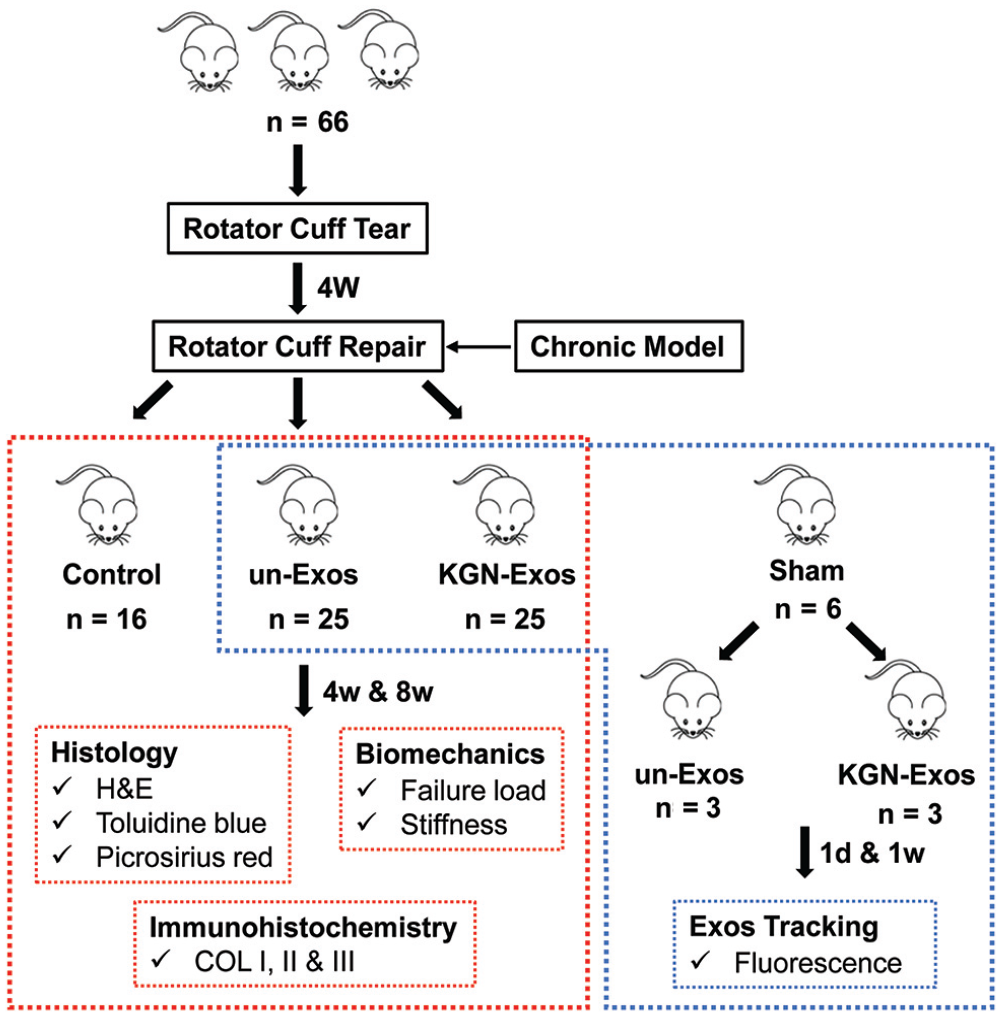
Flowchart for the study design. COL, collagen; control, group that received SAH; H&E, hematoxylin and eosin; KGN-Exos, group that received SAH containing exosomes derived from kartogenin-preconditioned human bone marrow mesenchymal stem cells; SAH, sodium alginate hydrogel; un-Exos, group that received SAH containing untreated exosomes.
To track the exosomes in vivo, SAH containing Dil-labeled un-Exos or Dil-labeled KGN-Exos was injected into the repaired site of the rotator cuff and was also injected into the normal supraspinatus tendon-to-bone junction of shoulders after sham surgery as a control. One week later, the rats were euthanized, and frozen coronal sections were prepared. The presence of Dil-labeled exosomes at the tendon-to-bone interface was observed using a fluorescence microscope. Moreover, a fluorescent imaging system Visque InVivo Smart (Vieworks) was used to observe the retention of DiR-labeled exosomes in vivo after injection at 1 day and 1 week.
Histological Evaluation
The supraspinatus tendon-humeral complexes were obtained 4 and 8 weeks after RCR surgery, 10 supraspinatus tendon-humeral complexes at each time point. Samples were fixed in 4% paraformaldehyde and then decalcified at 37°C for 2 weeks with 0.5 M ethylenediaminetetraacetic acid. After dehydration and embedding in paraffin, the samples were sectioned with a thickness of 5 µm. These samples were stained with hematoxylin and eosin (H&E), toluidine blue (TB), and picrosirius red. A light microscope (IX71SBF-2; Olympus) was used to examine the H&E- and TB-stained slides. The metachromasia area in the TB-stained slides was measured using the ImageJ software (National Institutes of Health). 14 A polarized light microscope (Eclipse E800; Nikon) was used to detect the collagenous tissue of the tendon near the greater tuberosity in the picrosirius red slides. Ten rectangular areas (50 µm × 50 µm) were randomly selected, and grayscale values were measured using the ImageJ software. Higher values indicate higher levels of collagen maturation.4,22 A histological scoring system was used to semiquantitatively assess the interface between the tendon and bone (Appendix Table A1, available in the online version of this article). 38 Higher histological scores suggest better tendon-to-bone healing. The histological evaluation was performed by 2 independent observers (J.X. and Z.Y.), who were blinded to group allocation.
Immunohistochemical Analysis
Immunohistochemical staining was performed to determine the expression of collagen I, II, and III in the tendon-to-bone area. The paraffin sections were treated with primary antibodies against collagen I, II, and III overnight at 4°C and incubated with secondary antibodies for 1 hour at room temperature. After developing with chromogen for 10 minutes at room temperature and rinsing in running water for 5 minutes, all sections were counterstained with hematoxylin. Semiquantitative analysis was performed using the ImageJ software by 2 independent observers (J.X. and Z.Y.), who were blinded to group allocation. In brief, the integrated optical density/area of each group was calculated and was then normalized to that of the control group at 4 weeks.
Biomechanical Testing
Supraspinatus tendon-humerus complexes of each group (n = 6) were harvested 4 and 8 weeks after RCR surgery. The proximal humerus was fixed in polymethylmethacrylate, whereas the supraspinatus tendon was braided with sutures and polyester cloth as previously described. 39 All the specimens were tested using an electronic universal materials testing system (Instron 5569). In brief, the specimens were preconditioned with 0.1 N and then were loaded to failure under uniaxial tension at an elongation rate of 10 mm/min. The failure loads were noted, and stiffness was calculated from the load-deformation curve.
Statistical Analysis
Sample size was calculated based on our preliminary study. To detect a statistical difference concerning failure load between un-Exos and KGN-Exos groups at 8 weeks, a minimum of 6 rats per group were needed according to a power analysis with β = .8 and α = .05. Continuous data were expressed as the mean ± SD. One-way analysis of variance was used to compare the data among the groups using GraphPad Prism 7.0 (GraphPad Software), and statistical significance was set at P < .05.
Results
Identification and Characterization of Exosomes
The presence of un-Exos and KGN-Exos for injection was identified (Figure 2). Transmission electron microscopy displayed the characteristic sphere-shaped bilayer membrane structures of un-Exos and KGN-Exos (Figure 2A). Nanoparticle tracking analysis revealed that the particles of both un-Exos and KGN-Exos mainly ranged from 100 to 150 nm in size (Figure 2B). Western blotting showed presence of positive exosome markers TSG101, ALIX, and HSP70 as well as the absence of negative exosome marker Calnexin in both un-Exos and KGN-Exos (Figure 2C). Furthermore, the release of un-Exos and KGN-Exos from the SAH was detected (Figure 2D). The results showed that both un-Exos and KGN-Exos exhibited a sustained release from SAH for up to 96 hours with an initial burst release. A release platform appeared after 9 hours.
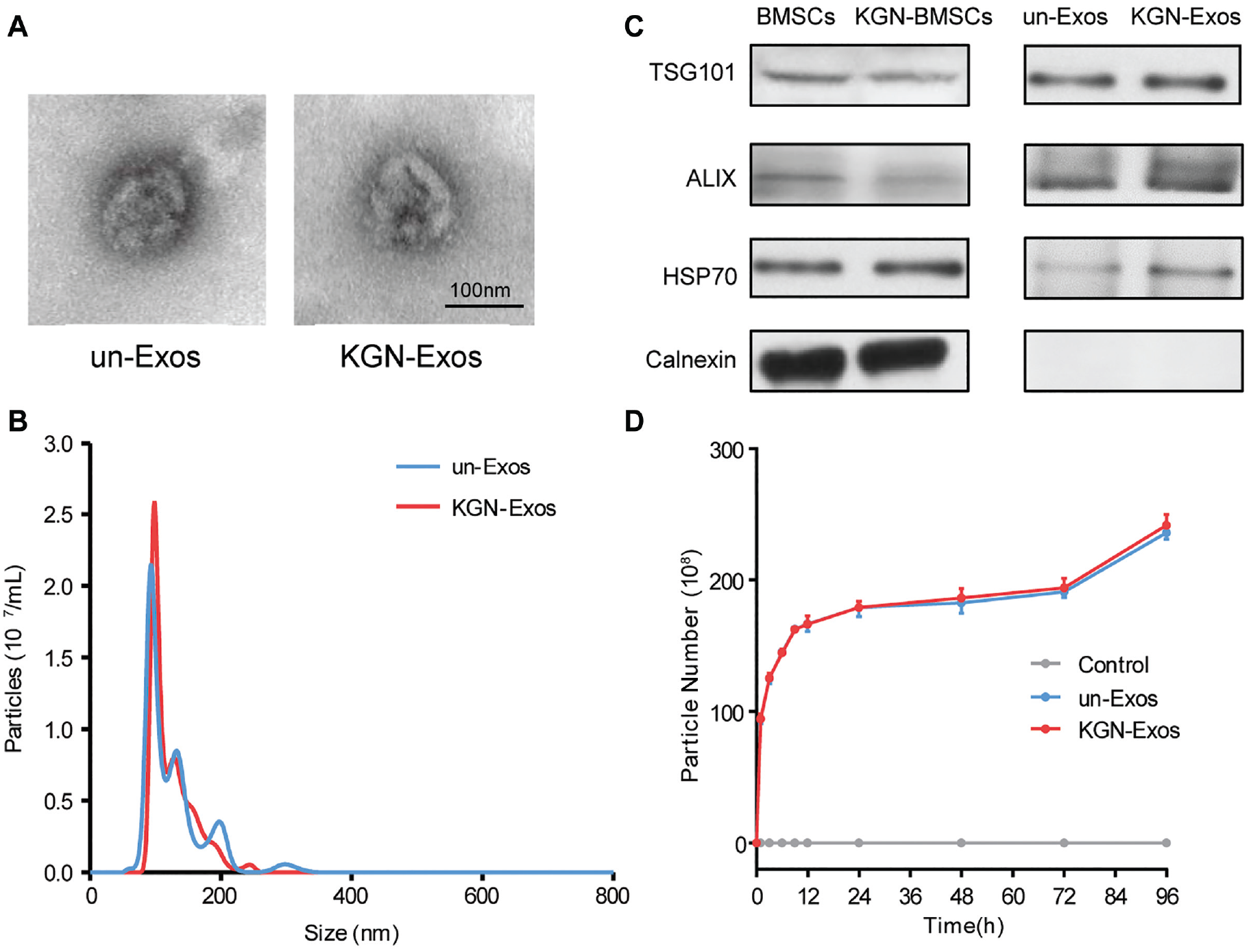
(A) Transmission electron microscopy observation and (B) size distribution of un-Exos and KGN-Exos. (C) Western blotting for TSG101, ALIX, HSP70, and Calnexin of BMSCs, KGN-BMSCs, un-Exos, and KGN-Exos. (D) Releasing curve of SAH (control), and un-Exos and KGN-Exos released from SAH at 1, 3, 6, 9, 12, 24, 48, 72, and 96 hours. BMSC, bone marrow mesenchymal stem cell; Control, group that received SAH; KGN-BMSCs, kartogenin-preconditioned bone marrow mesenchymal stem cells; KGN-Exos, group that received SAH containing exosomes derived from kartogenin-preconditioned human bone marrow mesenchymal stem cells; SAH, sodium alginate hydrogel; un-Exos, group that received SAH containing untreated exosomes.
Histological and Immunohistochemical Findings
Tracking of Exosomes
To track the exosomes in vivo, the retention of DiR-labeled un-Exos and KGN-Exos in the shoulder was assessed at 1 day and 1 week after surgery. Figure 3A shows that the DiR-labeled un-Exos and KGN-Exos were localized to the shoulder joint for at least 1 week after surgery. Moreover, the distribution of Dil-labeled un-Exos and KGN-Exos at supraspinatus tendon-to-bone sites was assessed 1 week after surgery in the sham and RCR groups (Figure 3B). Few Dil-labeled un-Exos and KGN-Exos were seen in the sham group, whereas numerous un-Exos and KGN-Exos were localized to the repaired site in the RCR group.
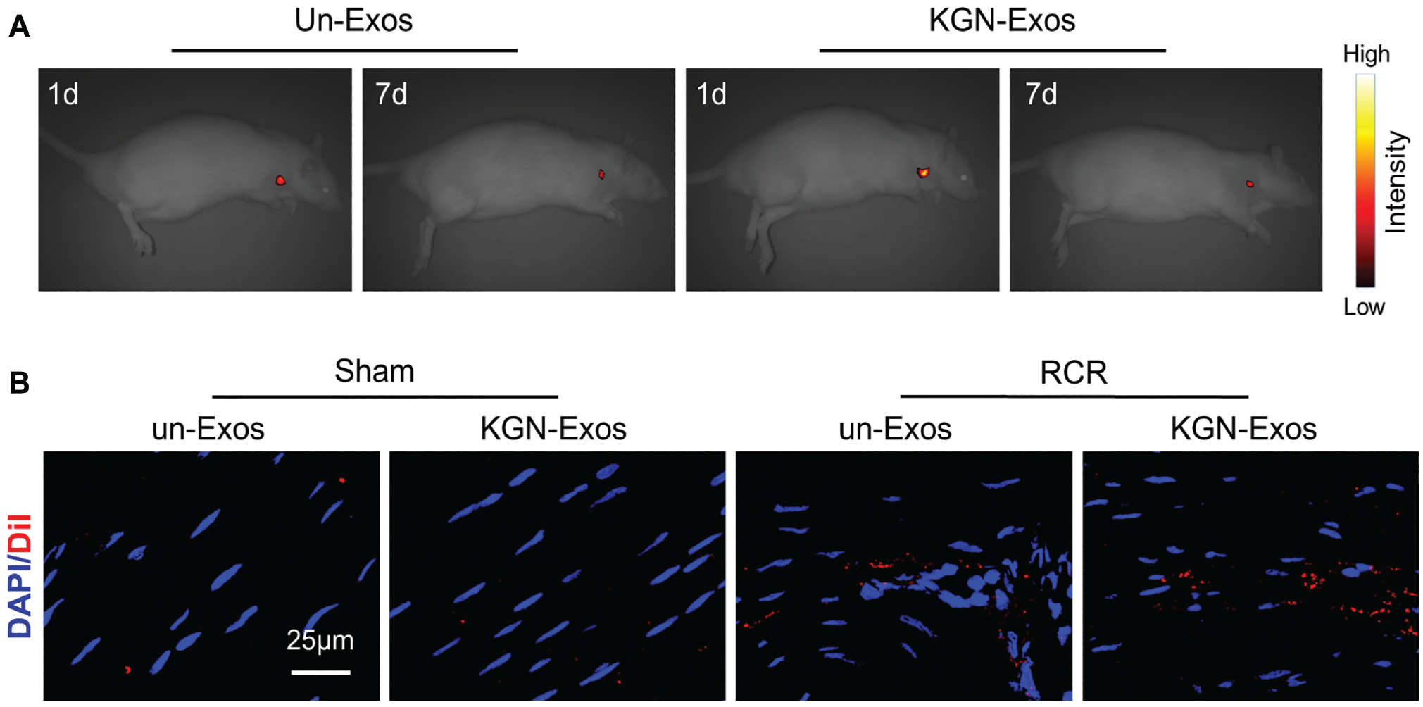
Tracking of exosomes in vivo. (A) Fluorescent imaging of rats with DiR-labeled un-Exos and KGN-Exos in the RCR group at 1 day and 1 week postoperatively. (B) Fluorescent frozen sections of Dil-labeled un-Exos and KGN-Exos at 1 week postoperatively in the sham and RCR groups. DAPI, 4′,6-diamidino-2-phenylindole; Dil, 1,1′-dioctadecyl-3,3,3′,3′-tetramethylindocarbocyanine perchlorate; DiR, 1,1′-dioctadecyl-3,3,3′,3′-tetramethylindotricarbocyanine iodide; KGN-Exos, exosomes derived from kartogenin-preconditioned human bone marrow mesenchymal stem cells; RCR, rotator cuff repair; un-Exos, untreated exosomes. The red areas indicate the labeled exosomes.
General Morphologic Characteristics
At 4 weeks, the control group showed poorly organized, highly cellular, and fibrovascular tissues at the interface between the tendon and bone, whereas the Exos and KGN-Exos groups presented several aligned collagen fibers with lower cellularity (Figure 4). The interfaces in the 3 groups were progressively remodeled over time. At 8 weeks, the KGN-Exos group presented a better organized collagen accompanied by more oriented cells along the collagen fibers compared with the other 2 groups. Accordingly, the KGN-Exos group had the highest histological score among the 3 groups (14.3 ± 1.3 [control], 18.9 ± 1.4 [un-Exos], 24.8 ± 1.5 [KGN-Exos]), indicating the most mature interface with the best collagen fiber alignment and minimal cellularity. Moreover, the histological score in the un-Exos group was significantly higher than that in the control group at 4 and 8 weeks.
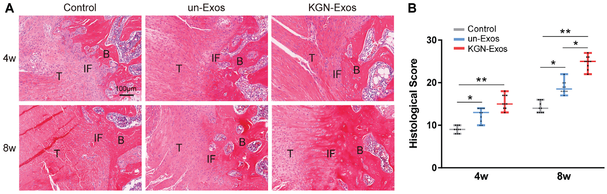
(A) Hematoxylin and eosin staining and (B) histological score for the control, un-Exos, and KGN-Exos groups at 4 and 8 weeks postoperatively. *P < .05, **P < .01. B, bone; IF, interface; KGN-Exos, exosomes derived from kartogenin-preconditioned human bone marrow mesenchymal stem cells; T, tendon; un-Exos, untreated exosomes.
Cartilage Regeneration
TB staining of glycosaminoglycan was semiquantitatively determined according to the metachromasia area. The un-Exos and KGN-Exos groups had significantly larger areas than the control group at 4 weeks (14,110 ± 2428 µm2 [un-Exos] vs 2632 ± 361 µm2 [control], P = .001; 28,940 ± 6853 µm2 [KGN-Exos] vs 2632 ± 361 µm2 [control], P < .001) and 8 weeks (65,080 ± 6320 µm2 [un-Exos] vs 6315 ± 626 µm2 [control], P < .001; 91,900 ± 5631 µm2 [KGN-Exos] vs 6315 ± 626 µm2 [control], P < .001) (Figure 5, A and B). Moreover, the KGN-Exos group showed the largest metachromasia area at the interface at 4 and 8 weeks. Similarly, the un-Exos and KGN-Exos groups showed increased collagen II expression compared with the control group at 4 and 8 weeks, and the KGN-Exos group had the highest collagen II expression at the interface at 4 and 8 weeks (Figure 5, C and D). Considered together, KGN-Exos significantly facilitated cartilage regeneration at the interface between the tendon and bone.
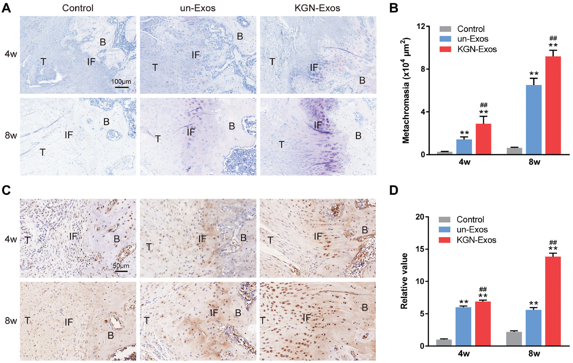
(A) Toluidine blue staining and (B) semiquantitative analysis for the control, un-Exos, and KGN-Exos groups at 4 and 8 weeks postoperatively. (C) Collagen II immunohistochemical staining and (D) semiquantitative analysis for the control, un-Exos, and KGN-Exos groups at 4 and 8 weeks postoperatively. **P < .01 vs control group, ##P < .01 vs un-Exos group. B, bone; IF, interface; KGN-Exos, exosomes derived from kartogenin-preconditioned human bone marrow mesenchymal stem cells; T, tendon; un-Exos, untreated exosomes.
Collagen Maturation and Organization
At 4 weeks, the collagen fibers in both the control and the un-Exos groups were more irregular and less organized than those in the KGN-Exos group (Figure 6A). Semiquantitative analysis based on brightness revealed that the brightness in the KGN-Exos group was higher than that in the control group (Figure 6D). At 8 weeks, the collagen fibers tended to be more organized and mature with stronger birefringence, whereas the un-Exos and KGN-Exos groups had better outcomes than the control group. Moreover, among the 3 groups, the KGN-Exos group exhibited the highest brightness. In addition, immunohistochemical staining for collagen I and collagen III demonstrated that the KGN-Exos group had the highest ratio of collagen I to collagen III ratio at 4 and 8 weeks among the 3 groups (Figure 6, B, C, and E). Collectively, KGN-Exos promoted collagen maturation and organization of the enthesis after RCR.
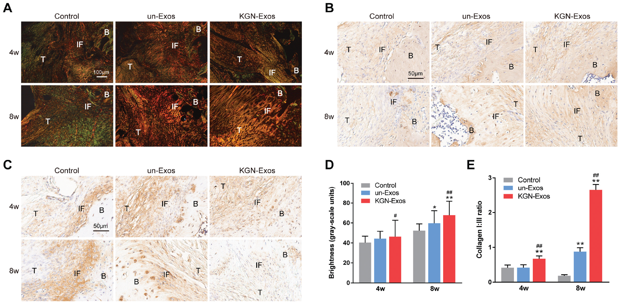
(A) Picrosirius red staining and (B) collagen I and (C) collagen III immunohistochemical staining for the control, un-Exos, and KGN-Exos groups at 4 and 8 weeks postoperatively. (D and E) Semiquantitative analysis of (D) picrosirius red staining and (E) collagen I and collagen III immunohistochemical staining for the control, un-Exos, and KGN-Exos groups at 4 and 8 weeks postoperatively. *P < .05 vs control group, **P < .01 vs control group, #P < .05 vs un-Exos group, ##P < .01 vs un-Exos group. B, bone; IF, interface; KGN-Exos, exosomes derived from kartogenin-preconditioned human bone marrow mesenchymal stem cells; T, tendon; un-Exos, untreated exosomes.
Biomechanical Testing
All the failure modes (tendon-to-bone interface tear or tendon substance tear) were recorded in the 3 groups at 4 and 8 weeks (Appendix Table A2, available online). In the control group, all specimens failed at the tendon-to-bone interface at 4 weeks, and most specimens (4/6) failed at the interface at 8 weeks. Most specimens (5/6) failed at the tendon-to-bone interface in both un-Exos and KGN-Exos groups at 4 weeks, whereas 3 of 6 specimens in the un-Exos and 5 of 6 in the KGN-Exos group failed within the tendon substance at 8 weeks, suggesting relatively robust tendon-to-bone healing.
As shown in Figure 7, although no significant difference in failure load was detected at 4 weeks among the 3 groups, the failure load of the un-Exos (23.53 ± 3.02 N) and KGN-Exos (28.12 ± 2.40 N) groups was significantly higher at 8 weeks compared with the control group (19.32 ± 1.82 N). Moreover, the failure load of the KGN-Exos group at 8 weeks was the highest. Similarly, the stiffness of the un-Exos (22.88 ± 4.08 N/mm) and KGN-Exos (28.57 ± 2.49 N/mm) groups was significantly higher than that of the control group at 8 weeks (18.17 ± 1.54 N/mm). The stiffness of the KGN-Exos group at 8 weeks was the highest.
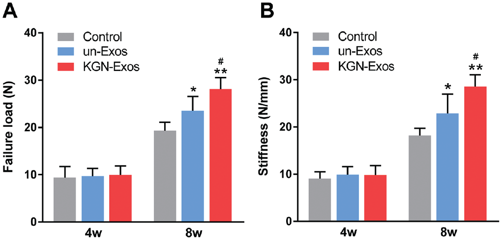
(A) Failure load and (B) stiffness of the control, un-Exos, and KGN-Exos groups at 4 and 8 weeks postoperatively. *P < .05 vs control group, **P < .01 vs control group, #P < .05 vs un-Exos group. KGN-Exos, exosomes derived from kartogenin-preconditioned human bone marrow mesenchymal stem cells; un-Exos, untreated exosomes.
Discussion
As is well known, good prognosis after RCR is associated with robust tendon-to-bone healing.1,29 So far, various strategies have been used to promote tendon-to-bone healing after RCR. 25 In the present study, exosomes were used to accelerate tendon-to-bone healing in vivo. One of the most important findings of this study was that the exosomes injected into the repaired rotator cuff persisted for at least 1 week and played a positive role in tendon-to-bone healing. In particular, we first applied KGN-Exos to RCR and investigated whether the therapeutic effect of exosomes could be improved by extrinsic KGN preconditioning for tendon-to-bone healing. It was demonstrated that KGN-Exos could better facilitate cartilage regeneration and collagen organization for enthesis regeneration in a rat chronic RCT model, compared with un-Exos.
Exosomes derived from stem cells are emerging as a powerful noncellular strategy in regenerative medicine because they can provide outcomes similar to stem cell therapy without ethical concerns. 8 Moreover, exosomes can reduce the safety limitations of cellular therapy in clinical application. 26 However, free exosomes in the shoulder joint are difficult to retain in the repaired site and may undergo rapid clearance. 42 To prolong the retention and localized release of exosomes in the tendon-to-bone junction, we used SAH to load the exosomes and chose local injection instead of intravenous injection. The release curve in vitro showed that both un-Exos and KGN-Exos exhibited a sustained release for 96 hours. Furthermore, the in vivo results showed that the un-Exos and KGN-Exos were localized to the repaired site in the RCR groups for at least 1 week after surgery. After injection, SAH allowed the sustained release of exosomes, which positively affected the local or recruited cells by transporting their cargo. Interestingly, we also found that there were more Dil-labeled exosomes in the RCR group than in the sham group. Cui et al 9 reported that more macrophage-derived exosomes were observed in the flexor digitorum longus tendon injury group than in the sham group 3 days after injection, because macrophage-derived exosomes preferentially localized to the injured tendon for regulation of tendon healing. The state of the donor cells may determine the biological functions of the corresponding exosomes. 19 Accordingly, it is believed that difference in the distribution of exosomes in the RCR and sham groups in our study was due to the reparative function of the exosomes for enthesis healing.
Several studies have reported the application of exosomes in tendon-to-bone healing after RCR.16,18,28 Recently, Huang et al 18 reported that BMSC-Exos accelerated tendon-to-bone healing by promoting angiogenesis and inhibiting the polarization of M1 macrophages in a rat chronic RCT model. However, the authors did not emphasize the function of exosomes in chondrogenesis. In our study, KGN was used to precondition the hBMSCs to produce exosomes with a stronger function for enthesis regeneration. KGN-Exos can overcome the disadvantages of KGN itself, such as low water solubility and low effective concentration, which limit its clinical application. 41 The histological and immunohistochemical results showed the highest glycosaminoglycan and collagen II formation at the interface in the KGN-Exos group, indicating that KGN-Exos significantly facilitated cartilage formation and showed a tendency to regenerate a biomimetic conjunction of the cartilage transitional zone, similar to the native enthesis. It should be noted that after washing and purification, the amount of KGN in exosomes was extremely low and did not have a positive effect. Notably, KGN-Exos were responsible for the enhanced therapeutic effects. To reduce the stiffness gradient between the tendon and bone, the native fibrocartilage layers acted as stress and shock absorbers, which were considered unique structures. 3 The KGN-Exos in our study had a stronger effect on inducing cartilage regeneration for enthesis healing than the un-Exos.
Meanwhile, our results demonstrated that un-Exos and KGN-Exos enhanced collagen maturation and organization of the injured enthesis at 8 weeks after RCR. On one hand, exosomes derived from stem cells were reported to facilitate collagen organization and tendon healing through immunomodulation. Shi et al 30 applied BMSC-Exos for patellar tendon repair and found that injured tendons treated with BMSC-Exos showed regularly aligned and compact collagen fibers, as well as upregulated expression of genes related to tenogenesis (collagen I, scleraxis, and tenomodulin) at 4 weeks. Wang et al 33 demonstrated that adipose stem cell–derived exosomes enhanced the collagen I:III ratio in a murine rotator cuff tendinopathy model. On the other hand, KGN had a positive effect on collagen synthesis. KGN was reported to promote collagen I formation of fibroblasts without obvious cytotoxicity via the TGF-β/Smad pathway. 6 Moreover, Wang et al 34 found that local injection of KGN could enhance the regeneration and organization of collagen fibers at the tendon-to-bone enthesis after RCR in a murine RCT model, which corresponds to a greater ultimate strength. Accordingly, owing to KGN preconditioning in our study, KGN-Exos could better promote collagen maturation and organization of the injured enthesis when compared with the un-Exos group. Considered together, the best histological morphology—namely, the largest cartilage formation, as well as greatest collagen maturation and organization at the enthesis—contributed to the highest failure load and stiffness of the KGN-Exos group.
This study has several limitations. First, only a single injection of the exosomes was conducted after repair. It is unclear whether multiple injections could lead to better results than a single injection. Second, although no rats died in this study, more research is needed to determine the safety of exosomes before they can be used in clinical settings. Third, the mechanisms related to the exosomes for RCR were not explored. Moreover, an hBMSC control group was not included for comparison of the effects of noncellular and cellular therapies on tendon-to-bone healing after RCR, warranting further investigation. Fourth, because the healing process in rats takes place more rapidly than in humans, more studies on large animals are needed to extrapolate the results to humans. Although this study cannot be directly translated into clinical practice, it provides an effective cell-free strategy for tendon-to-bone healing after RCR.
Conclusion
Local injection of SAH with sustained KGN-Exos release can effectively promote cartilage formation as well as collagen maturation and organization for enthesis regeneration, contributing to enhanced biomechanical properties after RCR. As a novel cell-free strategy, KGN-Exos may provide a therapeutic option to accelerate tendon-to-bone healing in chronic RCT.
Supplemental Material
sj-pdf-1-ajs-10.1177_03635465231155927 – Supplemental material for Exosomes Derived From Kartogenin-Preconditioned Mesenchymal Stem Cells Promote Cartilage Formation and Collagen Maturation for Enthesis Regeneration in a Rat Model of Chronic Rotator Cuff Tear
Supplemental material, sj-pdf-1-ajs-10.1177_03635465231155927 for Exosomes Derived From Kartogenin-Preconditioned Mesenchymal Stem Cells Promote Cartilage Formation and Collagen Maturation for Enthesis Regeneration in a Rat Model of Chronic Rotator Cuff Tear by Jiangyu Cai, Junjie Xu, Zipeng Ye, Liren Wang, Ting Zheng, Tianlun Zhang, Yufeng Li, Jia Jiang and Jinzhong Zhao in The American Journal of Sports Medicine
Footnotes
Submitted May 24, 2022; accepted January 9, 2023.
One or more of the authors has declared the following potential conflict of interest or source of funding: This work has been supported by funding from the National Natural Science Foundation of China (82102579, 31972923), the Shanghai Rising-Star Project (22QC1401200), the Shanghai Talent Development Fund (2021057), and the Basic Scientific Research Project of Shanghai Sixth People’s Hospital (ynqn202101). AOSSM checks author disclosures against the Open Payments Database (OPD). AOSSM has not conducted an independent investigation on the OPD and disclaims any liability or responsibility relating thereto.

References
Supplementary Material
Please find the following supplemental material available below.
For Open Access articles published under a Creative Commons License, all supplemental material carries the same license as the article it is associated with.
For non-Open Access articles published, all supplemental material carries a non-exclusive license, and permission requests for re-use of supplemental material or any part of supplemental material shall be sent directly to the copyright owner as specified in the copyright notice associated with the article.
