






Abstract
Background:
Studies to date of superior capsular reconstruction (SCR) comparing outcomes of healed grafts versus torn grafts do not separate graft tears based on location of the tear, rather they combine and report all tears as a single group.
Purpose/Hypothesis:
The purpose of this study was to correlate functional outcome with graft integrity and graft tear location after SCR with a dermal allograft. It was hypothesized that the functional outcomes of patients with an intact graft would be equivalent to those with graft tears leaving the tuberosity covered.
Study Design:
Cohort study; Level of evidence, 3.
Methods:
Patients who underwent SCR with an acellular dermal allograft at a single institution were included. Pre- and postoperative American Shoulder and Elbow Surgeons (ASES), Oxford Shoulder Score, visual analog scale (VAS) for pain, and postoperative Single Assessment Numeric Evaluation (SANE) scores were recorded. A magnetic resonance imaging scan was performed postoperatively to assess graft integrity.
Results:
A total of 39 patients met inclusion criteria. Mean age of patients was 60.4 ± 8.7 years; mean follow-up was 53.3 ± 25 months (range, 14-98 months). Magnetic resonance imaging performed at a mean of 17.5 months (range, 6-66 months) demonstrated an intact graft in 14 (36%); tear from the glenoid in 11 (28%), from midsubstance in 4 (10%), and from the tuberosity in 8 (21%); and complete graft absence in 2 (5%). Patients were divided into group 1 (intact graft), group 2 (tuberosity covered: tears from glenoid and midsubstance tears), and group 3 (tuberosity bare: tears from the tuberosity and dissolved or absent grafts). In group 1, there was significant improvement in ASES (37.9 to 88.5; P < .001), Oxford (25.2 to 46.2; P < .001), and VAS (6.8 to 0.9; P < .001). In group 2, there was significant improvement in ASES (32.2 to 86.1; P < .001), Oxford (23.4 to 44.2; P < .001), and VAS (7.3 to 1.3; P < .001). In group 3, there was no significant improvement in ASES (40.3 to 45.8; P = .50) or Oxford (33.5 to 31.4; P = .81), but there was a significant reduction in VAS (7.1 to 5.4; P = .03). There was no significant difference between group 1 and 2 in postoperative ASES (88.5 vs 86.1; P = .59), Oxford (46.2 vs 44.2; P = .07), VAS (0.9 vs 1.3, P = .42) and SANE (85.4 vs 83.2; P = .92) scores. However, group 3 had significantly lower ASES (45.8; P < .001), lower Oxford (31.4; P < .001), lower SANE (45.4; P < .001), and higher VAS (5.4; P < .001) scores than groups 1 and 2. There were no differences in outcomes based on sex (P = .72), previous surgery (P = .06), preoperative acromiohumeral distance (P = .57), and preoperative Goutallier stage of the supraspinatus (P = .16).
Conclusion:
Patients who underwent SCR with a dermal allograft and developed a graft tear leaving the tuberosity covered had equivalent functional outcomes to those with an intact graft.
Keywords
Massive rotator cuff tears (MRCTs) account for up to 10% to 40%4,26,56 of all rotator cuff tears and are a challenging problem for shoulder surgeons because multiple surgical options exist without a gold standard. These options include arthroscopic debridement10,52 with biceps tenotomy or tenodesis, 7 partial rotator cuff repair,6,11,17 tendon transfers,20,43 bridging procedures,2,9,27,32,33,62,64 superior capsular reconstruction (SCR),16,40 subacromial balloon spacers,30,46,53,63,65 bursal acromial reconstruction,5,51 and reverse total shoulder arthroplasty.15,61
Mihata pioneered SCR for irreparable rotator cuff tears. 41 In the original procedure, autologous tensor fascia lata (TFL) was folded to an approximately 8 mm–thick graft 40 and anchored to the glenoid and greater tuberosity, restoring superior glenohumeral joint stability in cuff-deficient shoulders. 41 Mihata et al subsequently reported excellent clinical outcomes with a 5-year follow-up.39,40
In the United States, acellular dermal matrix (ADM), or dermal allograft, has become the graft of choice.16,29,37,38,50,60 Reports have demonstrated excellent short-term functional improvement and patient satisfaction, yet there are limited data correlating graft integrity with functional outcomes.16,24,50 Graft tear rates range from 27% to 68%, depending on the type of graft used, with higher tear rates for dermal allografts compared with TFL. Mirzayan et al 44 classified SCR graft tears based on tear location using postoperative magnetic resonance imaging (MRI). A type 1 graft is an intact graft; type 2 is a graft tear leaving the tuberosity covered with dermal allograft (ie. midsubstance tear or tear from the glenoid); and type 3 is a graft tear leaving the tuberosity bare or uncovered (ie. graft tear from tuberosity or dissolved/absent graft). Studies to date comparing outcomes of healed grafts versus torn grafts do not separate graft tears based on location of the tear; rather, they combine all tears into a single group. A smaller study from our group with shorter follow-up and much smaller sample size demonstrated that patients who underwent a dermal allograft procedure (patient mix of bridging procedure and SCR) and who had a graft tear leaving the tuberosity covered had significant functional improvements. 45 The purpose of our current study was to determine if functional outcome after SCR is correlated with graft integrity and graft tear location. Our hypothesis was that functional outcomes of patients with an intact graft would have equivalent outcomes to those with graft tears leaving the tuberosity covered with the graft.
Methods
Approval from the institutional review board of Kaiser Permanente Southern California was obtained to conduct this retrospective study. Between September 25, 2014, and November 7, 2018, 53 consecutive patients underwent an SCR with a dermal allograft by 5 fellowship-trained (3 shoulder and elbow (D.C.A., K.A.O., A.S.), 2 sports medicine) (R.M., M.H.) shoulder surgeons at an integrated health care system. Two surgeons performed 16 procedures each (R.M., D.C.A.), 1 surgeon performed 9 cases (A.S.), and 2 surgeons performed 6 cases each (K.A.O., M.H.). All surgeons prospectively tracked their own cases and data, which were later combined for analysis. All patients underwent similar postoperative protocols, which included keeping the arm in a sling for 6 weeks, followed by 6 weeks of passive range of motion, with active and active-assisted range of motion starting at 12 weeks along with resistance strengthening. Inclusion criteria included MRCT, Hamada grades 1 and 2, 28 intact or reparable upper border subscapularis, no loss of active external rotation, and available postoperative MRI. The indication for performing SCR was pain attributable to MRCT that did not improve with nonoperative treatment for a minimum of 3 months. Exclusion criteria included arthritis (diagnosed on preoperative radiographs or chondromalacia grade 3 or 4 at time of arthroscopy), previous infection, revision SCR, and <12 months of follow-up. An MRCT was defined as a 2-tendon tear of the supraspinatus and infraspinatus, 23 ≥4-cm tear, 14 with tendon retraction to the glenoid 8 in all coronal sequences on MRI where the glenoid was visualized.
We would like to note that there is crossover of some patients from 2 previous studies from our group. Ten patients were previously reported by Mirzayan et al 45 with imaging and clinical outcome with only 25.6 months of follow-up. In addition, 29 patients were reported by Mirzayan et al 44 with imaging findings only, without clinical correlation. This current study aims to correlate clinical outcomes with imaging findings previously reported, with longer follow-up.
Preoperative data including age, sex, previous surgeries, Hamada grade, 28 and Goutallier stage 25 of the supraspinatus were recorded by the senior author (R.M.) for all cases. Intraoperative data including surgical findings and concomitant procedures were recorded. Pre- and postoperative acromiohumeral distance (AHD) on plain radiographs were measured by the senior author. Postoperative AHD was measured at the first postoperative visit. Pre- and postoperative American Shoulder and Elbow Surgeons (ASES), Oxford Shoulder Score, visual analog scale (VAS) for pain, and postoperative Single Assessment Numeric Evaluation (SANE) score were recorded. Clinically significant measures including the minimal clinically important difference (MCID), substantial clinical benefit (SCB), and Patient Acceptable Symptom State (PASS) were recorded.
A recent study by Evuarherhe et al 22 found that for SCR, the MCID, SCB, and PASS for ASES were 11.2, 18, and 68.8, respectively, and for SANE were 14.5, 23.1, and 69.9, respectively. Because of that study's finding that PASS for the ASES score was 68.8, we considered an ASES score >70 points a successful outcome18,22,47,49 and evaluated whether sex, previous surgery, preoperative AHD, and preoperative Goutallier stage had an effect on a successful outcome. All patients were routinely offered a postoperative MRI, regardless of symptoms, to assess graft integrity. Because this was a retrospective study, there was no set time point to obtain an MRI. The earliest time point an MRI was obtained was at 6 months.
Surgical Details
A diagnostic arthroscopy was performed to confirm the diagnosis of an irreparable MRCT and identify concomitant pathology. A subacromial bursectomy was completed. Subscapularis repair was also performed in cases with concomitant upper border subscapularis tears. Subacromial decompression was performed and long head of biceps tendon pathology addressed (tenotomy or tenodesis) at each surgeon's discretion. An SCR was then performed by first debriding the superior glenoid and greater tuberosity. The defect was measured in the medial-lateral and anterior-posterior directions. A 3 mm–thick ArthroFlex dermal allograft (LifeNet Health, Virginia Beach, Virginia) was used in all patients. The ADM was cut to the size of the defect. The graft was secured to the glenoid using 3 anchors positioned between the 10- and 2-o’clock positions, approximately 5 to 7 mm apart. The lateral side of the ADM was secured using a conventional double-row, transosseous equivalent, suture-bridge technique (Figure 1). Posterior side-to-side sutures between the graft and remnant infraspinatus were routinely placed at the end of the case.

Superior capsular reconstruction with dermal allograft (Graft) with double-row repair on the tuberosity with FiberTape (Arthrex, Naples, Florida) (arrows).
Postoperative Rehabilitation
All patients were initially placed in a shoulder immobilizer for 6 weeks, followed by 6 weeks of passive range of motion. Active range of motion was initiated at 3 months. At this point, surgeons and therapists at 5 sites managed postoperative rehabilitation progress without a standardized rehabilitation protocol.
Statistical Analysis
Patient demographic and clinical data were compared among 3 groups based on presence of tear and coverage of the tuberosity at follow-up (graft intact, graft tear from glenoid or midsubstance [tuberosity covered], and graft tear from tuberosity or dissolved [tuberosity bare]) as well as between the intact and tuberosity covered groups. Descriptive data are presented as number (percentage) for categorical variables and as means with standard deviation. for continuous variables. The Kruskal-Wallis test was used to detect the mean differences for continuous variables and Fisher exact test was applied to evaluate differences in proportion of categorical variables. The Student t test was applied to detect the value change between the pre- and postoperative measurements of AHD, ASES, Oxford, and VAS scores for each surgery group.
All analyses were 2-sided and performed using SAS Enterprise Guide 8.2 (SAS Institute). P values of <.05 were considered statistically significant.
Results
Demographic Information
Of the 53 patients identified, 39 patients met our inclusion criteria (Figure 2).

Flow diagram for patient inclusion. MRI, magnetic resonance imaging; SCR, superior capsular reconstruction.
Fourteen (26%) of the identified patients were excluded because they did not have adequate postoperative follow-up. The mean age of the patients was 60.4 ± 8.7 years (range, 37-77 years). There were 26 (67%) male patients. At least 1 previous procedure had been performed in 10 patients (26%). Of those 10, 7 (70%) underwent rotator cuff repair, 2 (20%) underwent biceps tenotomy, and 1 (10%) underwent rotator cuff debridement. A concomitant procedure was performed in 24 patients (62%). Of these 24, 8 (33%) underwent isolated biceps tenodesis, 5 (21%) underwent isolated biceps tenotomy, 6 (25%) underwent subscapularis repair in conjunction with a biceps procedure, and 5 (21%) had partial repair of infraspinatus. Preoperative radiographs revealed that of the 39 patients, 22 (56%) had Hamada grade 1 tears and 17 (44%) had Hamada grade 2 tears. Preoperative Goutallier stages of the supraspinatus muscle were as follows: 2 (5%) stage 0; 6 (15%) stage 1; 17 (44%) stage 2; 9 (23%) stage 3; and 5 (13%) stage 4.
The mean time for MRI was 17.5 ± 10.7 months (range, 6-66 months). MRIs demonstrated that 14 patients (36%) had an intact (healed) graft, while tears were noted in 11 (28%) from the glenoid, 4 (10%) in midsubstance, 8 (21%) from the tuberosity, and 2 (5%) had complete graft absence (resorption) (Figure 3).
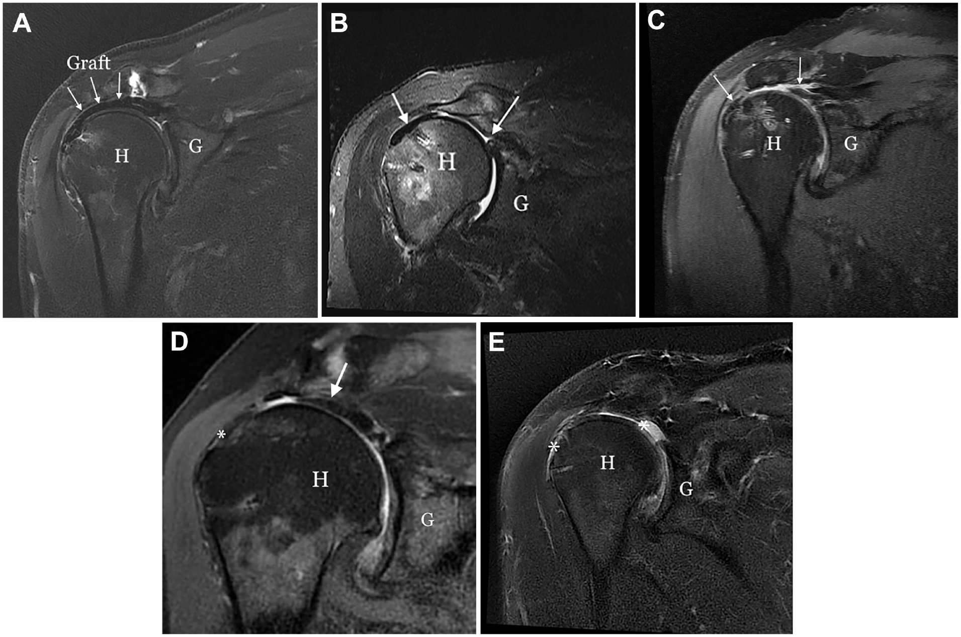
Postoperative magnetic resonance imaging. G, glenoid; H, humeral head. (A) Intact graft (arrows). (B) Graft tear from glenoid (G) (arrow pointing to graft healed to tuberosity). (C) Midsubstance tear (arrow pointing to graft ends, laterally healed to the tuberosity and portion of graft medially healed to glenoid). (D) Graft tear from the tuberosity with balled-up graft on glenoid (arrow) with a bare tuberosity (asterisk). (E) Dissolved or absent graft from the tuberosity and glenoid (asterisks).
On the basis of the previously described classification, 44 we assigned patients to 3 groups: group 1, intact (healed) graft; group 2, tuberosity covered (graft tear from glenoid or midsubstance); or group 3, tuberosity bare (graft tear from tuberosity or dissolved) (Table 1). There was no difference between the 3 groups in patient age (P = .54) and sex (P = .69), preoperative AHD (P = .76), Goutallier stage (P = .05), and Hamada grade (P = .18).
Patient Characteristics Between 3 Groups a

Group 1, healed graft. Group 2, tuberosity covered. Group 3, tuberosity bare. AHD, acromiohumeral distance; ASES, American Shoulder and Elbow Surgeons; Oxford, Oxford Shoulder Score; Post, postoperative; Pre, preoperative; VAS, visual analog scale; SANE, Single Assessment Numeric Evaluation.
Kruskal-Wallis P.
Fisher exact P.
Functional Outcomes Based on Graft Tear Location
The mean clinical follow-up was 53.3 ± 25 months (range, 14-98 months). Overall, there was significant improvement in ASES (36.3 ± 14.1 to 76.6 ± 22; P < .001), Oxford (26.2 ± 9.4 to 41.8 ± 7; P < .001), and VAS (7.1 ± 1.8 to 2.2 ± 2.6; P < .001) scores. There was significant correlation between graft integrity with the final outcome. Patients in groups 1 and 2 had significant improvement in ASES (P < .001) and Oxford (P < .001) scores as well as reduction in VAS (P < .001) (Table 2). In groups 1 and 2, 100% of patients achieved MCID and SCB for the ASES score. In group 1, 93% achieved PASS and in group 2, 87% achieved PASS for the ASES score. Patients in group 3 did not have improvement in ASES (P = .50) or Oxford (P = .81) scores but did have significant improvement in VAS (P =.031). However, the postoperative VAS in group 3 (5.4 ± 1.6) was significantly higher than group 1 (0.9 ± 1.9) and 2 (1.3 ± 1.7) (P < .001). In group 3, 40% of patients achieved MCID and SCB, and 10% achieved PASS for the ASES score.
Pre- and Postoperative Functional Outcome Scores Based on Graft Integrity and Tear Location a

Group 1, healed graft. Group 2, tuberosity covered. Group 3, tuberosity bare. AHD, acromiohumeral distance; ASES, American Shoulder and Elbow Surgeons; Oxford, Oxford Shoulder Score; VAS, visual analog scale.
Student t test.
Table 3 demonstrates that there was no significant difference between group 1 and group 2 in postoperative ASES (88.5 ± 11.5 to 86.1 ± 11.2; P = .59), Oxford (46.2 ± 2.3 to 44.2 ± 3.8; P = .07), VAS (0.9 ± 1.9 to 1.3 ± 1.7; P = .42), and SANE (85.4 ± 9.9 to 83.2 ± 13; P = .92), respectively. The postoperative SANE of groups 1 and 2 was significantly higher compared with group 3 (45.4 ± 20.7) (P < .001). In groups 1 and 2, 93% of patients achieved PASS, while in group 3 only 10% achieved PASS for SANE.
Differences Between Group 1 and Group 2 a

Group 1, healed graft. Group 2, tuberosity covered. ASES, American Shoulder and Elbow Surgeons; Oxford, Oxford Shoulder Score; Post, postoperative; Pre, preoperative; VAS, visual analog scale; SANE, Single Assessment Numeric Evaluation.
Kruskal-Wallis P.
Association Between Sex, Preoperative AHD, Preoperative Goutallier Stage, and Previous Surgery With Successful Outcome
As explained in the Methods section, we considered an ASES score >70 a successful outcome18,22,47,49 and evaluated whether sex, previous surgery, preoperative AHD, and preoperative Goutallier stage had an effect on a successful outcome. There was no difference in the mean postoperative ASES score for women (74 ± 22.3) and that of men (77.9 ± 22.2) (P = .40). Women and men had similar improvement in mean ASES of 40 points. The mean postoperative Oxford score for women was 42 ± 7 and that of men was 42.3 ± 5.4 (P = .91). Women had a mean improvement in Oxford of 16, while men had a mean improvement of 15.6. There was no difference in the percentage of women (30.8% versus 38.5%; P = .73) in patients with a successful outcome compared with those with a poor outcome, respectively (Table 4).
Effect of Sex, Previous Surgery, Preoperative AHD, and Preoperative Goutallier on a Successful Outcome a

ASES, American Shoulder and Elbow Surgeons; AHD, acromiohumeral distance.
Fisher exact P.
Kruskal-Wallis P.
There was no change in preoperative to postoperative AHD, respectively, in group 1 (7.7 ± 2.4 vs 8.1 ± 2.1; P = .73), group 2 (7.4 ± 2.5 vs 7.9 ± 3.1; P = .77), or group 3 (6.2 ± 2.5 vs 5.4 ± 2.5; P = .44) (Table 2). There was no difference in the preoperative AHD (7.7 ± 2.3 mm) in patients with ASES scores >70 and the preoperative AHD (6.6 ± 2.8 mm) of those with ASES scores <70 (P = .57) (Table 4).
Pre- and postoperative Goutallier stage for each group are summarized in Table 1. Although the percentage of lower Goutallier stages (73.1%) was higher in patients with a successful outcome compared with those with a poor outcome (46.2%), it did not reach significance (P = .16) (Table 4).
Although patients without previous surgery had a higher mean post ASES score (81.8 ± 19.2) compared with those who had a previous operation (73.8 ± 25.4), this difference was not statistically significant (P = .24) and did not meet the MCID (11.2). There was no difference between having had a previous surgery and no previous surgery in achieving an ASES >70 (P = .06).
Discussion
The primary finding of our study was that patients who underwent SCR with a dermal allograft and developed a graft tear leaving the tuberosity covered had significant and equivalent functional outcomes and pain scores as those with an intact graft. Those patients who had a graft tear leaving the tuberosity bare did not have significant functional improvements. This raises the question as to whether a Biologic Tuberoplasty42,58 (fixation of an acellular dermal allograft to cover the greater tuberosity) is sufficient, rather than performing SCR in patients with MRCT, as it is less time-consuming, less expensive, and technically less demanding. 43 We found no other statistically significant associations between age, sex, previous surgeries, preoperative AHD, preoperative Hamada grade, preoperative Goutallier stage of the supraspinatus on functional outcomes.
Mihata et al39,40 have reported significant clinical improvement after SCR with TFL. In the United States, ADM, or dermal allograft, has gained popularity.16,21,29,50,59 The majority of studies in the literature have reported good and excellent outcomes after SCR with a dermal allograft.13,16,29,50,55,67 Several studies have reported graft tears after SCR, but none have correlated functional outcomes with location of graft tear. The first study to report SCR graft tears was by Denard et al, 16 who reported on 59 patients. Their indication for obtaining postoperative MRI was “patients who were willing to undergo an MRI,” unlike our study in which MRIs were routinely obtained on all patients regardless of symptoms. This biased symptomatic patients to undergo an MRI. Only 20 of 59 patients (34%) underwent a postoperative MRI, with 11 (55%) revealing a tear: 1 (9.1%) from the glenoid, 3 (27.3%%) from the midsubstance, and 7 (63.6%) from the tuberosity. The “tear” group had a significantly worse postoperative VAS and ASES scores compared with the intact group. Because the majority of the tears were from the tuberosity, the tear group, as a whole, fared worse. Lim et al 38 reported outcomes of SCR using TFL in 31 patients who underwent routine postoperative MRI. The authors reported 9 re-tears: 7 (77.8%) with the tuberosity covered and 2 (22.2%) with the tuberosity bare. There were no significant differences in postoperative ASES, Constant, VAS, range of motion, and external rotation strength between the intact and the “graft tear” groups, although the study may have been underpowered. Because most (77.8%) of the tears in that study left the tuberosity covered, no differences were appreciated between the intact and tear groups. Pennington et al. 50 reported on 88 patients who underwent an SCR with an ADM, with only 4 patients (4.5%) undergoing an MRI. Of those, 3 (75%) demonstrated a tear from the tuberosity. In the Methods section, the authors stated that “postoperative advanced imaging with MRI was only performed on those patients who expressed dissatisfaction with their level of pain, or who had insufficient functional improvement in terms of strength and range of motion,” which is consistent with symptomatic patients having tears leaving the tuberosity bare.
Two studies have specifically reported on the location of graft tears but did not correlate them with functional outcomes. Lacheta et al 35 obtained postoperative MRI scans of SCR in 22 patients at a mean of 2.5 months. They found that there were no tears from the tuberosity, 5 midsubstance tears, and 4 tears from the glenoid side. In this series, the tear group is equivalent to our group 2, where the graft tears left the tuberosity covered. The authors found no difference in ASES and SANE scores between the intact and tear groups. Lee et al 36 reported the clinical and MRI results of 46 patients who underwent SCR (30 TFL and 16 ADM). Postoperative MRI scans of all shoulders were performed and revealed a 35% graft tear rate. Location of the graft tear was 10 from the tuberosity, 2 from the glenoid, 3 midsubstance, and 1 at both sites (floating graft). Only patients with lateral-sided graft tears did poorly and required a revision, which is consistent with our findings. Badman et al, 3 in a cohort of 10 patients, diagnosed graft failure in 7 patients (70%) with MRI at 1 year. Of the 7 failures, 4 failed from the glenoid, 2 from the midsubstance, and 1 from the tuberosity. The majority of failures in this cohort would be in our group 2; therefore, the author reported no difference in ASES score between the intact group (3 patients) and tear group (7 patients, with 6 being in tuberosity covered category). Shin et al 54 obtained postoperative MRI scans in 21 patients with SCR and found intact grafts in 14 patients (66.7%). Of the 7 patients with graft tears, 3 (42.9%) were from the glenoid, 3 (42.9%) from the tuberosity, and 1 (14.3%) from both sides. Therefore, 4 of 7 patients in the tear group had a bare tuberosity. The authors did not find a difference in postoperative ASES between the 2 groups. Campbell et al 12 performed postoperative MRI scans on 24 shoulders after SCR and noted 12 torn grafts, most commonly at the glenoid side (8 of 12). They reported no difference in ASES scores between the healed and the tear groups because the majority of the tears left the tuberosity covered. In a study by LaBelle et al, 34 postoperative MRI scans revealed graft failure in 13 of 21 shoulders (62%), with 6 from the tuberosity, 4 from the glenoid side, and 3 midsubstance graft failures. Thus, 7 of 13 tears in their cohort left the tuberosity covered, leading to slightly lower ASES score in the tear group, but this was not significant. Ohta et al 48 reviewed MRI scans of 49 patients who underwent SCR with TFL and found that of the 5 graft tears, all were from the tuberosity. Those patients had no significant improvement in the UCLA score. Yoon et al 66 reviewed the postoperative MRI scans of 30 patients who underwent SCR with a hybrid graft (TFL and dermal allograft) and found that all 5 of graft tears were from the tuberosity. The intact group had higher postoperative ASES score, but this was not found to be significant.
Mirzayan et al 45 have also reported on 25 patients who had a mixture of bridging 64 and SCR procedures in shoulders with MRCTs. They found similar results as in this current study; those patients who had a graft tear leaving the tuberosity covered had similar functional outcomes as those with intact grafts. The authors theorized that one of the sources of pain is from bone-to-bone contact between the greater tuberosity and the acromion. In a shoulder with an MRCT, this contact occurs in a dynamic fashion when the deltoid muscle contracts without the presence of the supraspinatus tendon, raising the center of rotation of the humeral head and leading to rubbing of the greater tuberosity with the undersurface of the acromion. Acromiohumeral distance is a static measurement on a plain radiograph with the arm at rest in an adducted position. As an example, a shoulder that has Hamada grade 1 with an AHD of 10 mm, can still have bone-to bone contact with deltoid contraction. An intact graft, or a graft tear leaving the tuberosity covered, acts as an interpositional biologic tissue preventing contact between the tuberosity and the acromion. The authors proposed the term “biologic tuberoplasty” to describe this phenomenon.
The precise mechanism by which SCR and other joint-preserving techniques improve pain and function after MRCT remains debatable. Our work supports that for SCR, while there may be a contribution of the ADM to humeral head depression, there is a positive clinical outcome from the biologic tuberoplasty or spacer effect that prevents the humeral head from coming in contact with the acromion. It is plausible that the SCR functions as a spacer, 31 keeping the humeral head down, while tendon transfers dynamically or statically pull the humeral head inferiorly. This explains why the outcomes of these techniques degrade with Hamada grade 3 changes (fixed apposition of the greater tuberosity on the acromion).1,16,19
Acromiohumeral distance has been used as both a prognostic indicator, as well as a way to measure the radiographic success of SCR. Most studies have reported a significant increase in AHD after SCR,29,37,38,40,50 with only one study showing no significant increase. 16 Our findings are consistent with the latter in that we did not find improvement in the AHD after SCR. This may be because all the shoulders in our series were Hamada grade 1 or 2, with no or minimal superior migration. Lee et al 37 identified AHD as a significant prognostic factor for graft failure in SCR, noting that patients with graft failure had significantly decreased immediate and 1-year postoperative AHD, as well as smaller improvements in AHD from the preoperative to the postoperative period compared with patients with intact grafts. Our findings contradict theirs, as there was no change in AHD in shoulders regardless of Hamada grade. This may be because the majority (11 of 13) of graft tears in their study were from the humeral side, while the majority of tears in our study were from the glenoid side.
Strengths and Limitations
The strengths of this study are an adequate sample size with postoperative MRI obtained in each symptomatic and asymptomatic patient, consistently applied surgical technique, patient-reported outcomes, and pain scores.
Our study also has several limitations. The most significant limitation is that there was a large gap in the timing of when the MRI was performed and when the clinical data were obtained. The final patient-reported outcomes were obtained many months or years after the postoperative MRI, so it is possible that some tears that came later may have been missed and may have thrown off the correlation between graft integrity and clinical outcome. However, Lee et al 36 demonstrated in their study that dermal allograft tears were noted at 4.3 months after SCR. Therefore, we believe that a minimum of 6 months for obtaining the MRI was adequate to capture graft tears. Other limitations include that it was retrospective, without a control group. The sample size may have been too small to capture small differences between group 1 and group 2. There were no range of motion or strength measurements. However, we believe that ASES and Oxford scores are a satisfactory record of patient functional range of motion. In addition, patients ultimately decided whether to have an MRI, which may have biased the sample.
The ultimate question remains as to whether, in patients with an MRCT, covering the tuberosity with a dermal allograft (Biologic Tuberoplasty) may be sufficient instead performing an SCR, which is technically demanding, time-consuming, and costly. 43 Some early preliminary studies are indicating that may be the case.42,57 Leaving the tuberosity bare leads to bone-to-bone contact between the acromion and greater tuberosity when the deltoid is activated and the rotator cuff is absent.
Conclusion
Patients who underwent SCR and developed a graft tear leaving the tuberosity covered had equivalent functional outcomes at 4 years to those with an intact graft.
Footnotes
Submitted March 15, 2024; accepted July 17, 2024.
One or more of the authors has declared the following potential conflict of interest or source of funding: R.M. has received honoraria from Arthrex not related to this manuscript, consulting fees from Arthrex, and speaking fees from Empire Medical. A.S. has received compensation from Arthrex for services other than consulting, support for education from Arthrex, and support for education from ImpactOrtho. AOSSM checks author disclosures against the Open Payments Database (OPD). AOSSM has not conducted an independent investigation on the OPD and disclaims any liability or responsibility relating thereto. This study was approved by the Kaiser Permanente Southern California institutional review board (No. 11147).