






Abstract
Background:
The insufficient regeneration of fibrocartilage at the tendon enthesis is the primary cause of retearing after surgical reattachment of the rotator cuff. Exosomes derived from bone marrow–derived mesenchymal stem cells (BMSC-Exos) and kartogenin (KGN) have been demonstrated to induce fibrocartilage formation. Loading drugs into exosomes may lead to a synergistic effect, significantly enhancing the inherent activity of both components. However, further investigation is necessary to determine whether loading KGN into BMSC-Exos could yield superior efficacy in promoting tendon enthesis healing.
Purpose:
To study the effect and mechanism of KGN-loaded BMSC-Exos (Kl-BMSC-Exos) on tendon enthesis repair and biomechanical properties in a rat rotator cuff injury (RCI) model.
Study Design:
Controlled laboratory study.
Methods:
The characteristics and in vivo retention of exosomes were demonstrated using nanoflow cytometry, transmission electron microscopy, and in vivo imaging of a small animal. The differentiation markers of BMSCs were assessed through quantitative polymerase chain reaction and immunofluorescence assays. Unilateral supraspinatus tenotomy and repair were performed in rats to establish the RCI model. Gelatin sponges were utilized to contain and deliver exosomes. In total, 44 rats were randomly assigned to 4 groups: sham, RCI, BMSC-Exos, and Kl-BMSC-Exos. Tendon enthesis regeneration and biomechanical properties were evaluated 8 weeks after surgery. RNA sequencing of BMSCs was performed to elucidate the underlying mechanism through which Kl-BMSC-Exos enhance tendon enthesis healing.
Results:
No discernible disparities in fundamental characteristics were evident between BMSC-Exos and Kl-BMSC-Exos. Incorporating exosomes into a gelatin sponge extended the in vivo retention time from 7 to 14 days. Kl-BMSC-Exos were more effective in inducing differentiation markers of BMSCs, improving fibrocartilage regeneration, organizing collagen fiber arrangement, and enhancing the biomechanical properties of tendon enthesis. Furthermore, transcriptomics suggested that Mospd1 was involved in Kl-BMSC-Exos–mediated tendon enthesis healing by enhancing fibrocartilage regeneration.
Conclusion:
The incorporation of exosomes into a gelatin sponge significantly enhances their in vivo retention time. Kl-BMSC-Exos can expedite the healing of RCI by enhancing chondrogenesis and fibrocartilage regeneration, providing more organized collagen fiber arrangement and superior biomechanical properties of the rotator cuff enthesis. The promotion of rotator cuff enthesis regeneration may contribute to enhancing the chondrogenic potential in BMSCs through Kl-BMSC-Exos–mediated upregulation of Mospd1.
Clinical Relevance:
As a cell-free therapeutic approach, Kl-BMSC-Exos displayed a better therapeutic effect on tendon enthesis healing than BMSC-Exos did, and these can be used as a biologic augmentation to enhance the healing of rotator cuff enthesis.
Rotator cuff injury (RCI) is a prevalent dysfunction that leads to chronic pain and restricted movement, significantly impairing patients’ quality of life.4,21 Although advances in surgical skills, techniques, technology, and equipment have significantly facilitated the arthroscopic repair of full-thickness rotator cuff tears, failed repairs are frequently reported.22,36 The formation of fibrovascular scar tissue between the tendon and bone may account for the biomechanically inferior structure as compared with the native enthesis, as it lacks a natural arrangement of collagen fibers, thereby hindering enthesis regeneration.24,32,36 In light of this perspective, exploring effective augmentative treatment strategies could decrease the rate of rotator cuff repair failure.
Exosomes are a population of extracellular vesicles based on bilayer lipid membranes, which not only encapsulate various bioactive molecules but also serve as natural carriers to facilitate intercellular communication.6,12 Given their inherent membrane penetrability and nanoscale dimensions, exosomes can efficiently release encapsulated drugs at the desired site in a controlled manner, thereby significantly enhancing their therapeutic efficacy.19,40 For these reasons, exosome-based interventions have been investigated in various diseases, including rotator cuff tearing, 31 osteoarthritis, 33 and osteochondral defects. 17
In tendon enthesis healing, fibrocartilage may fail to reform as expected. Instead, scar-like tissue forms, which is mechanically inferior to normal tissue.30,37,42 Kartogenin (KGN) is a novel inducer of chondrogenic tissue formation and is widely studied in cell-free therapy in the field of regeneration for cartilage regeneration and tendon-bone healing. 11 Exosomes derived from bone marrow–derived mesenchymal stem cells (BMSC-Exos) 8 and KGN 42 have shown favorable results in rotator cuff healing by enhancing fibrocartilage regeneration. The use of exosomes alone for tendon enthesis repair, however, may exhibit limited effectiveness in recovering biomechanical properties.3,16,27,38 Meanwhile, in situ KGN injection leads to excessive fibrocartilage-like tissue in unintended areas, which is not favorable for optimal tendon enthesis healing. 39 Here, we orchestrated KGN-loaded exosomes and gelatin sponges to investigate the effect and mechanism of KGN-loaded BMSC-Exos (Kl-BMSC-Exos) on tendon enthesis repair and biomechanical properties in a rat RCI model.
Methods
BMSC Culture and Trilineage Differentiation Assay
The extraction of BMSCs was conducted as previously described. 38 Briefly, the femurs and tibias of neonatal rats were aseptically excised. The bone marrow was subsequently flushed out and homogenized using a syringe. Subsequently, the bone marrow cells were filtered, concentrated, resuspended, and finally seeded into 100-mm culture dishes. The cells were cultured in DMEM/F12 (Hyclone) medium supplemented with 10% fetal bovine serum (Gibco) and a combination of 100-μg/mL penicillin and streptomycin (Hyclone) at a temperature of 37°C under a CO2 concentration of 5%. Upon reaching a confluency of 95%, the cells were enzymatically dissociated for subsequent passaging or differentiation assays. Trilineage differentiation was assessed using commercially available kits (Cyagen Biosciences) following established protocols.
Isolation of Exosomes
Exosomes were collected from the BMSCs medium after ultracentrifugation as previously described. 5 Briefly, the conditioned medium was centrifuged at 500g for 15 minutes, at 3000g for 15 minutes, and at 12,000g for 30 minutes at 4°C to remove cells and debris. Exosomes were collected by centrifuging at 140,000g for 90 minutes at 4°C. The condensation was purified twice. Purified exosomes were resuspended with (1) phosphate-buffered saline (PBS) for transmission electron microscopy (TEM; Zeiss), (2) radioimmunoprecipitation assay buffer for Western blot analysis, and (3) fetal bovine serum–free medium for in vivo and in vitro experiments.
Animal and Surgical Procedure
Fifty male Sprague-Dawley rats were purchased from the laboratory animal center of Xiamen University. All rats were kept in a specific pathogen–free environment with 3 animals per cage to minimize potential confounders. Animals (16 weeks of age, 460-520 g) were randomly divided into 4 groups: sham, RCI, BMSC-Exos, and Kl-BMSC-Exos. At 8 weeks postoperatively, 5 rats in each group were sacrificed for biomechanical testing and 6 rats for histologic evaluation (Figure 1). All animal experiments in this study were approved by the Institutional Animal Care and Use Committee of Xiamen University. Supraspinatus repair surgery was utilized to establish the RCI model. 23 In brief, rats were anesthetized with pentobarbital (40 mg/kg) and then underwent depilation and sterilization. They were secured in the lateral decubitus position before surgery. After a skin incision on the lateral aspect of the left shoulder, the anterior and lateral deltoid muscles were longitudinally incised, allowing subsequent access to the proximal humerus through blunt dissection. Before a supraspinatus tendon tenotomy was performed, the acromioclavicular ligament was incised to expose the insertion of the supraspinatus tendon. After that, the tendon was resected at the insertion and the stump was refreshed. Two 0.8-mm bone tunnels were created at the edge of the greater tuberosity. Two gelatin sponges with a size of 6 × 2 × 0.5 cm (Jingling) were cut into 2.5 × 5.0 × 5.0–mm pieces and immersed into the exosome solution to completely absorb 1 mL of KGN-loaded exosome suspension. Then the half-dry and half-wet exosome-soaking sponge was tucked under the tendon of the supraspinatus, and a transosseous suture bridge technique was employed to compress the tendon and the gelatin sponge underneath onto the footprint area for gelatin sponge fixation and rotator cuff repair (see Appendix Figure A4, available in the online version of this article). Finally, the deltoid muscle and skin incisions were closed in layers. For sham surgery, we only surgically exposed the left supraspinatus and closed the incision.
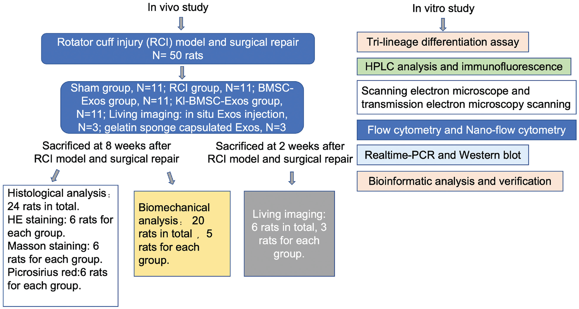
Flowchart illustrating the study design. BMSC-Exos, bone marrow mesenchymal stem cell–derived exosomes; HE, hematoxylin and eosin; HPLC, high-performance liquid chromatography; Kl-BMSC-Exos, kartogenin-loaded bone marrow mesenchymal stem cell–derived exosomes; PCR, polymerase chain reaction.
Exosome Uptaking and Visualization
Exosome isolation was performed as previously described. 5 Purified exosomes were resuspended with 5μM CM-DiI (Yeasen) and incubated at 37°C for 20 minutes. The CM-DiI–stained exosomes were then diluted with 20 mL of PBS and centrifuged at 140,000g for 70 minutes at 4°C. The condensation was washed twice to remove surplus dye. After resuspension in PBS, the quantity of CM-DiI–labeled exosomes was determined by nanoflow cytometry, and 1011 CM-DiI–labeled exosomes were utilized to incubate with BMSCs for 4 hours. For visualization, the skeleton of the cell was stained with phalloidin at 37°C for 30 minutes, and the nucleus was stained with DAPI at 37°C for 1 minute. The internalization of exosomes was visualized by using LSM880 confocal microscopy (Zeiss). For flow cytometry analysis, the fluorescence intensity was detected at an excitation wavelength of 570 nm (Thermo Fisher).
KGN Loading and High-Performance Liquid Chromatography Analysis
Incubation and sonication were utilized to load KGN into BMSC-Exos. 14 For incubation, 30 μg of KGN was mixed with 1011 BMSC-Exos and incubated at 37°C for 60 minutes. An equivalent quantity of drug and exosomes was employed to conduct the sonication loading. Briefly, 1011 exosomes were mixed with 30 μg of KGN in 1 mL of PBS. The KGN-exosome mixture was then subjected to sonication using a model 505 Sonic Dismembrator equipped with a 0.25 tip under the following conditions: 20% amplitude and 6 cycles of 30 seconds, on/off for 3 minutes, with a 2-minute cooling period between cycles. After sonication, the solution containing KGN-loaded exosomes was incubated at 37°C for 60 minutes to facilitate recovery of the exosomal membrane. The excess KGN was removed from the mixture through subsequent dilution and additional ultracentrifugation at 4°C for 70 minutes. Acetonitrile was employed to release the drug from the exosomes, followed by high-performance liquid chromatography analysis (Dionex) to determine the loading efficiency of KGN. 14 The loading efficiency was calculated as follows: mass of loaded KGN/total KGN dosage.
Scanning Electron Microscope
To visualize the exosomes loaded in a gelatin sponge, a 3 × 3 × 10–mm gelatin was immersed into PBS with 1011 exosomes at 4°C for 12 hours. After washing with PBS 3 times, the gelatin sponge was allowed to air-dry. The samples were sprayed with metal and observed under scanning electron microscopy (Zeiss).
Small Animal In Vivo Imaging
The retention time of exosomes in a gelatin sponge was measured by using an IVIS spectral imaging system (PerkinElmer). Exosomes were stained by 5-μm DiR at 37°C for 20 minutes and recollected as previously described. 5 In total, 1 × 10¹¹ particles of DiR-labeled exosomes were loaded into the gelatin sponge, and the squeezed sponge was surgically implanted beneath the rotator cuff. Images of the rotator cuff were recorded by an IVIS Spectrum imaging system at 1, 7, and 14 days after implantation.
Biomechanical Measurement
At 8 weeks after surgery, the tendon-humeri from 5 rats in each experimental group were harvested to conduct biomechanical evaluations using established methodologies described in a previous study. 34 Briefly, the tendon samples were tested on a mechanical testing machine (Instron ITW) with a loading sensor of 100 N using the following protocols: (1) A steel fixture with a 3-mm hole was used to block the humeral head. (2) The prepared tendon was passed through the hole and clamped by a needle holder (See Figure 9, red dotted line). (3) The fixture plate was securely fastened in the biomechanical test machine using sutures and 2 additional needle holders. (4) The humerus was lodged just below the middle hole of the fixture to provide an opposing force. (5) The specimen was preloaded with a force of 0.1 N, followed by cyclic elongation ranging from 0 to 0.5 mm for 20 cycles at a constant speed of 5mm/min. (6) The load-displacement curve was recorded at a constant elongation rate of 5mm/min.
Real-time Polymerase Chain Reaction
Total RNA was extracted from exosome-treated BMSCs by using a fast isolation kit (Bioflux). All procedures were conducted following the protocol provided by the kit. The primer sequences are listed in Table A1 (available in the online version).
Immunofluorescence Analysis
A total of 1011 purified exosomes were utilized to incubate with BMSCs for 12 hours in each group. Cells were fixed with 10% neutral buffered formalin, penetrated by 0.2% Triton X-100, blocked by 5% bovine serum albumin, and washed with PBS before being incubated with primary antibodies overnight. After incubation with the corresponding secondary antibodies at room temperature for 4 hours, the cells were washed 3 times by PBS, and the images were captured using LSM880 confocal microscopy (Zeiss).
Western Blot
Western blot was performed as previously described. 5 The antibodies are listed in Table A2 (available in the online version).
Histologic Analysis
The explanted humeral head with the repaired supraspinatus tendon was fixed in 10% neutral buffered formalin for 24 hours. Subsequently, it underwent decalcification for another 24 hours (Solarbio) and then embedding in paraffin. Coronal 3 mm–thick sections of tendon enthesis were placed on slides, and 1 representative section of the tendon insertion from each shoulder was stained with hematoxylin and eosin, Masson trichrome, and picrosirius red. Picrosirius red staining was observed by using polarized light microscopy. The general morphologic characteristics of the maturation of the tendon enthesis were quantitatively determined using a histologic scoring system that has been described.1,9 Two investigators scored all the slides in a blinded fashion. A perfect score in this scoring system was 28 points, and a higher score indicated better healing of tendon enthesis.1,9 Furthermore, the area ratio of new fibrocartilage formation at the repair site and the collagen birefringence was quantitatively determined by using Image J software.26,34
Bioinformatic Analysis
BMSCs were incubated with PBS, BMSC-Exos, and Kl-BMSC-Exos for 7 days. Three samples in each group were used for RNA sequencing. The quality of the resulting complementary DNA libraries was assessed using the Bioanalyzer 2100 (Agilent). Sequencing was performed on an Illumina Hiseq 4000 platform (LC Bio). The differentially expressed genes (DEGs) at a transcriptional level were identified with log2(fold change) >1 and with P < .05 by using R studio. The coexpression genes in BMSCs treated with BMSC-Exos and Kl-BMSC-Exos were sorted by using Draw Venn diagram online tools, and the volcano plot was displayed by using R studio.
siRNA Synthesis and Transfection
siRNA was designed and synthesized by Beijing Tsingke Biotech. siRNA stocking solution (100 μM) was diluted to 50 nM by using the opti-MEM medium (Gibco) and transfection reagent (Tsingke). The mixture was then incubated with BMSCs for 6 hours at 37°C. Afterward, the culture medium was replaced, and the cells were continued to be cultured in the differentiation medium for another 7 days. The siRNA sequences are as follows: Mospd1-siRNA-1, TGGTGCCTCTCTACCTCCACTTAAG; Mospd1-siRNA-2, TACCTCCACTTAAGTGTGAATCAAA.
Statistics
All data were presented as mean and standard deviation (mean ± SD) from at least 3 independent experiments in each cell experiment. The normality test and Brown-Forsythe test were conducted before the parametric test. Unpaired t tests were performed to analyze the differences between 2 groups. The statistical analysis for comparing differences among ≥3 groups was performed utilizing either a 1-way analysis of variance followed by Tukey post hoc test or a 2-way analysis of variance incorporating a Tukey multiple-comparisons test (for assessing variations at different time points). All data were utilized to perform further analysis. The testing for normality and homogeneity of variances, as well as all statistical analyses, was conducted using GraphPad software version 9. Significance was set at P < .05 in all experiments.
Results
Isolation and Identification of BMSCs
The BMSCs used in our study were evaluated with the surface markers and the ability of trilineage differentiation. The BMSCs successfully differentiated into chondrocytes, osteocytes, or adipocytes under conditioned medium in vitro (see Appendix Figure A1A). Flow cytometry showed that CD29 and CD90 were positive in isolated cells (100% and 99.2%); meanwhile, CD34 and CD45, markers of vascular and immune cells, were expressed at quite low levels (0.34% and 0.47%, respectively) (Figure A1B).
Characteristics of BMSC-Exos and Kl-BMSC-Exos
The typical characteristics of exosomes include a size range of 40 to 150 nm, a cup-shaped morphology, and specific surface biomarkers. 10 TEM, nanoflow cytometry, and Western blot were performed to identify the extracted Exos in this study. The vesicles in both groups exhibited a cup-shaped morphology under TEM observation (Figure 2A). Meanwhile, the nanoflow cytometry showed that the particle size distribution of BMSC-Exos and Kl-BMSC-Exos ranged from 51.75 ± 2.29 to 123.25 ± 13.00 nm and from 53.25 ± 1.00 to 123.25 ± 13.43 nm, respectively (Figure 2B). BMSC-Exos and Kl-BMSC-Exos positively expressed CD9, CD63, CD81, and TSG101 (Figure 2C), which indicates that the exosomes used in this study were consistent with the classic identification. 10 Overall, BMSC-Exos and Kl-BMSC-Exos showed similar morphology, particle size distribution, and surface markers, indicating that KGN loading by the sonication method has no effect on the basic characteristics of exosomes.
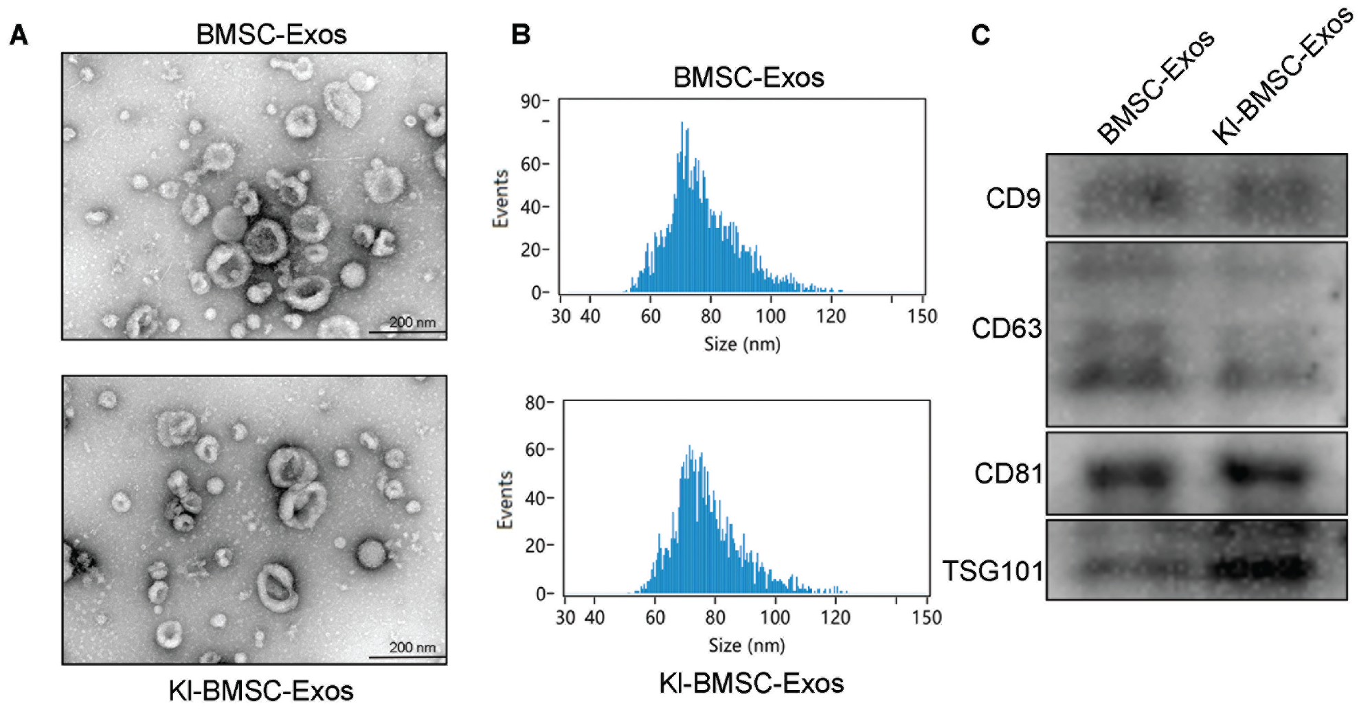
Characteristics of BMSC-Exos and Kl-BMSC-Exos. (A) Transmission electron microscopy revealed the morphology of BMSC-Exos and Kl-BMSC-Exos. (B) Nanoflow cytometry displayed the particle size distribution of BMSC-Exos and Kl-BMSC-Exos. (C) Western blot assay confirmed typical exosomal markers. BMSC-Exos, bone marrow mesenchymal stem cell–derived exosomes; Kl-BMSC-Exos, kartogenin-loaded bone marrow mesenchymal stem cell–derived exosomes.
KGN Loading Efficiency of Exosomes
High-performance liquid chromatography was carried out to calculate the loading efficiency of KGN. The results demonstrated the successful incorporation of KGN into exosomes using a sonication assay (Appendix Figure A2B), achieving a loading efficiency of 27.2% ± 2.5%, which exhibited an increase >3-fold as compared with that achieved using an incubation assay (6.4% ± 0.1%; P = .0074). Our results suggest that the ultrasonic method is more effective and can maintain a relatively high loading efficiency.
Internalization of Exosomes
To investigate whether the modified exosomes could enter into the cytoplasm of BMSCs, CM-DiI–labeled exosomes were incubated with BMSCs for 4 hours. Confocal microscope analysis showed that a large number of exosomes were uptaken by BMSCs in the BMSC-Exos group and Kl-BMSC-Exos group and mainly distributed around the nucleus (Figure 3A), which indicates that exosomes can successfully enter the target cell. Flow cytometry was employed to quantify the uptake efficiency of exosomes by BMSCs. The uptake rates of the BMSC-Exos group and the Kl-BMSC-Exos group were 21.9% ± 1.5% and 22.1% ± 1.3% (P = .8887), respectively, indicating that KGN loading does not affect exosome internalization (Figure 3B).
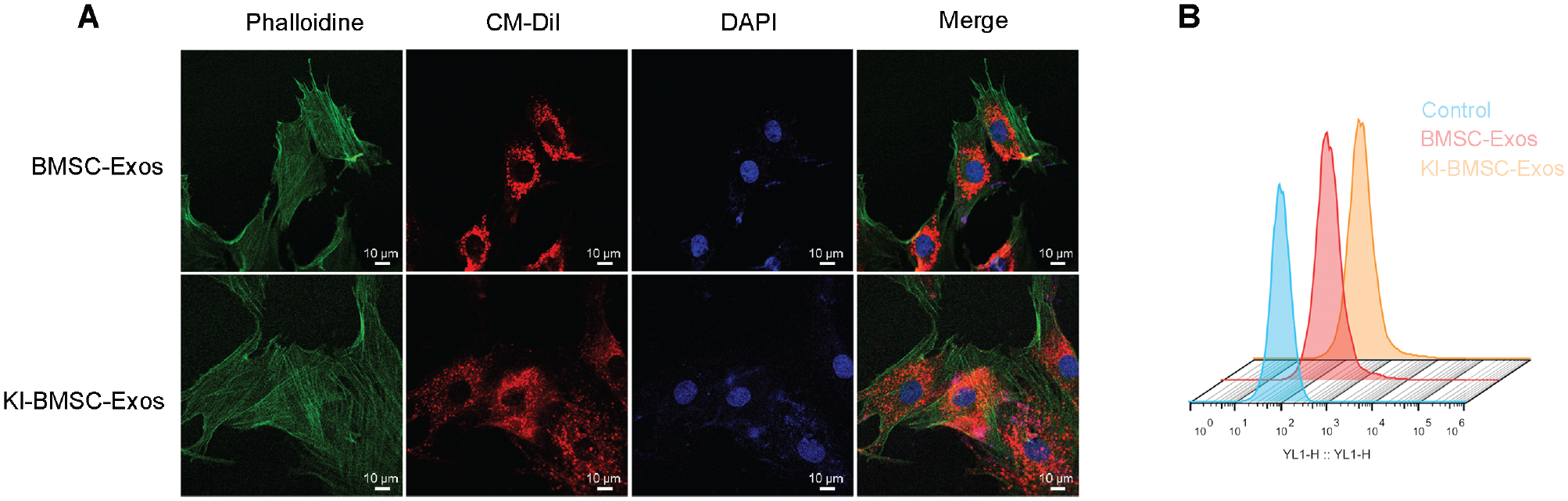
Uptaking and visualization of BMSC-Exos and KI-BMSC-Exos. (A) Immunofluorescence visualized the internalization of exosomes. Scale bar = 10 μm. (B) Flow cytometry was utilized to quantify the internalization of exosomes. BMSC-Exos, bone marrow mesenchymal stem cell–derived exosomes; Kl-BMSC-Exos, kartogenin-loaded bone marrow mesenchymal stem cell–derived exosomes.
Cellular Response to BMSC-Exos and Kl-BMSC-Exos
After 7 days of different exosomal interventions, the mRNA expression of Col2 (vs control, P = .0003; vs BMSC-Exos, P = .0004), Sox9 (vs control, P < .001; vs BMSC-Exos, P < .001), and Runx1 (vs control, P < .001; vs BMSC-Exos, P < .001) in Kl-BMSC-Exos–treated BMSCs revealed a statistically increasing trend when compared with those incubated with BMSC-Exos or PBS (Figure 4B). Although a 2.01-fold (vs control, P < .001) elevation of Runx1 was observed in BMSC-Exos–treated cells, the degree of increase was statistically less than that in the group treated with Kl-BMSC-Exos (P < .001). Similar results were seen in tendon-generic markers and osteogenic-related indicators (Figure 4, A and C). BMSC-Exos and Kl-BMSC-Exos upregulated the mRNA expression level of Tnmd (P < .001), Mkx (P = .0081; P < .001), Col1 (P < .001), Alp (P =.0735; P < .001), Ocn (P < .001), and Runx2 (P = .9846; P < .001) to varying degrees in coincubated BMSCs. However, the Kl-BMSC-Exos group exhibited a superior elevation in the mRNA expression of Tnmd (1.92- vs 2.76-fold, P = .0011), Mkx (1.67- vs 2.46-fold, P = .0026), Col1 (1.98- vs 3.43-fold, P < .001), Alp (1.78- vs 2.97-fold, P = .0043), Ocn (1.78- vs 2.97-fold, P = .0101) and Runx2 (0.97- vs 1.48-fold, P < .001) than that incubated with BMSC-Exos alone.
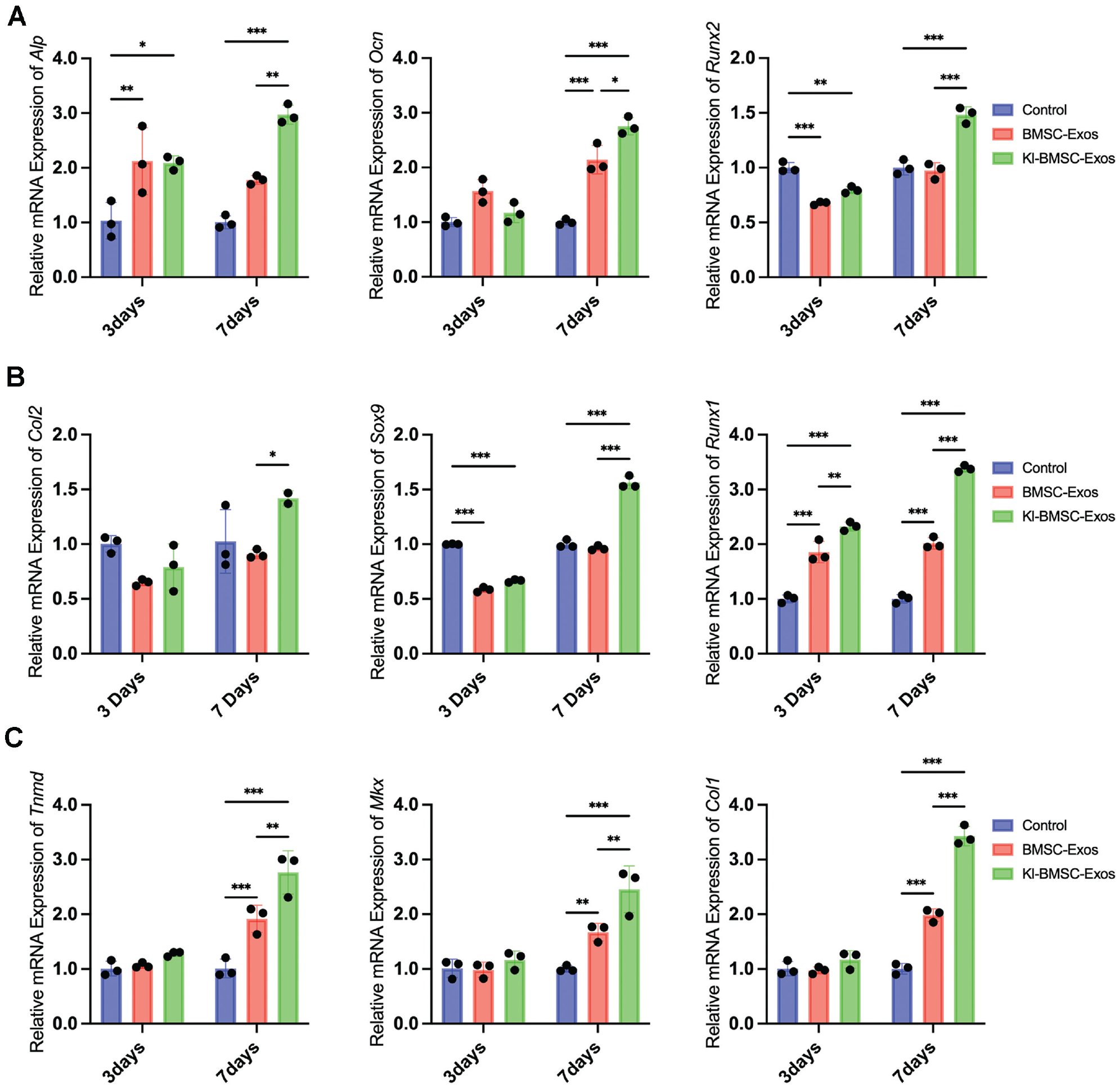
(A-C) Quantitative polymerase chain reaction analysis revealed the relative gene expression of BMSCs supplemented with BMSC-Exos or Kl-BMSC-Exos. Data are presented as mean ± SD (3 samples per group). *P < .05. **P < .01. ***P < .001. BMSC-Exos, bone marrow mesenchymal stem cell–derived exosomes; Kl-BMSC-Exos, kartogenin-loaded bone marrow mesenchymal stem cell–derived exosomes.
The protein expression of collagen 2 and aggrecan was provoked by BMSC-Exos and Kl-BMSC-Exos intervention. The increase was more pronounced in the Kl-BMSC-Exos–treated group, indicating that Kl-BMSC-Exos effectively enhanced the chondrogenesis of BMSCs as compared with using BMSC-Exos alone (Figure 5).
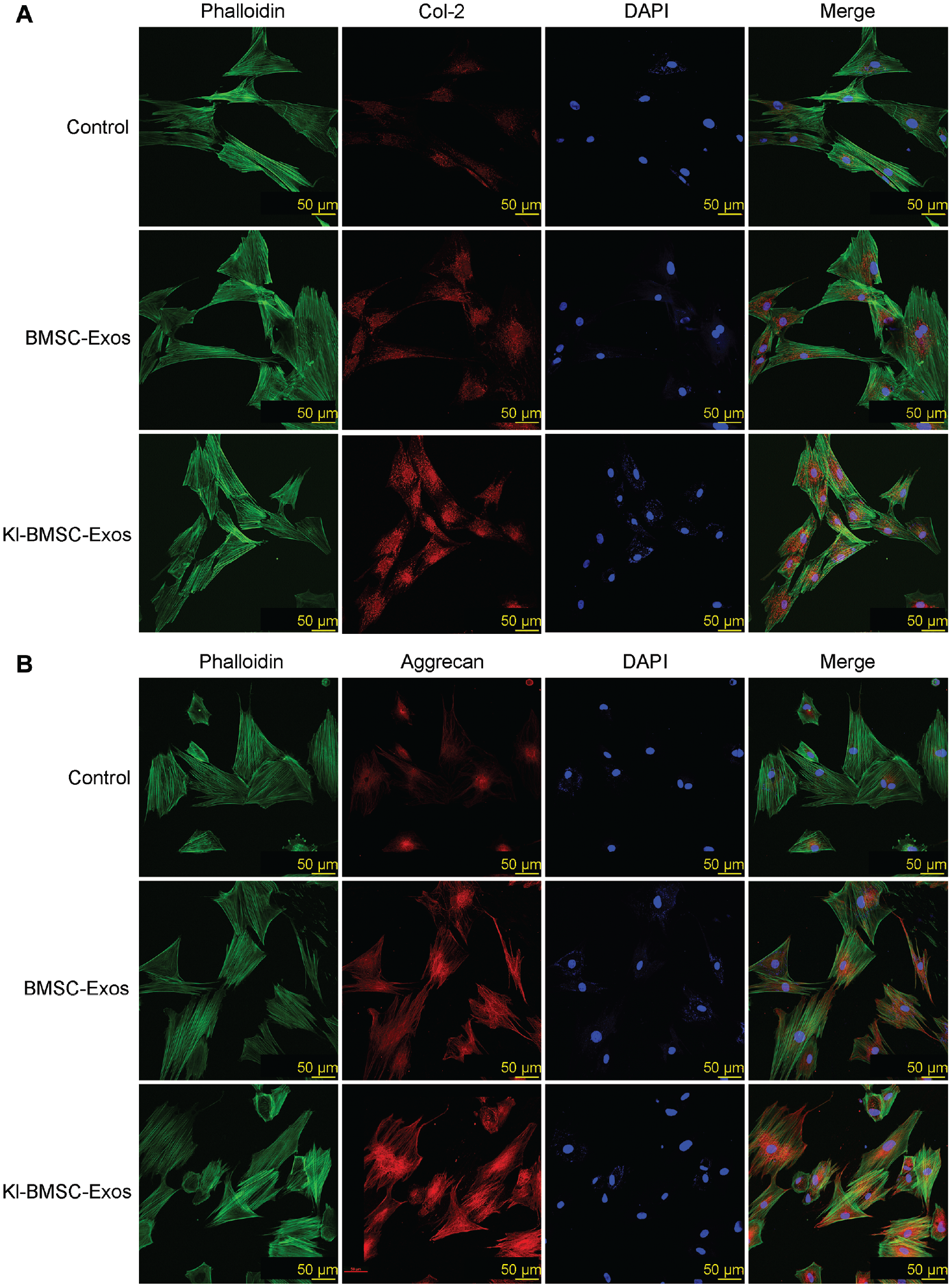
Immunofluorescence visualized the relative expression level of cartilage-specific protein (A) Col-2 and (B) aggrecan in BMSCs supplemented with BMSC-Exos or Kl-BMSC-Exos. Scale bar = 50 μm. BMSC-Exos, bone marrow mesenchymal stem cell–derived exosomes; Col-2, collagen type 2; Kl-BMSC-Exos, kartogenin-loaded bone marrow mesenchymal stem cell–derived exosomes.
Loading and Controlled Releasing of Exosomes
Easy degradation is a challenge that constrains the practical application of exosomes. To avoid rapid degradation, we loaded exosomes into a gelatin sponge to extend the retention time. In vitro, the preservation of exosomes on a gelatin sponge was demonstrated by TEM detection (Figure 6A). Additionally, to assess the in vivo retention duration of exosomes, we established an RCI model and implanted the exosome-loaded gelatin sponge beneath the footprint area of supraspinatus. The DiR tracking assay revealed that the fluorescence intensity of exosomes that were loaded into gelatin was maintained for 14 days (Figure 6B). In contrast, in the group without a gelatin sponge, the fluorescence intensity dramatically decreased on the third day after implantation. The average radiant efficiency reflects the precise quantity of the fluorescence intensity within the selected region of interest, providing an accurate indication of the quantity and intensity of stained exosomes. The in situ injection group demonstrated a significantly lower average radiant efficiency of 6.88 × 10⁷± 1.30 × 10⁷ (mean ± SD) on day 7, as compared with the gelatin-capsulated exosome group with 1.48 × 10⁸± 2.19 × 10⁷ (mean ± SD) (P = .0292) (Figure 6C). The average radiant efficiency of the in situ injection group rapidly decreased from 6.88 × 10⁷± 1.30 × 10⁷ (mean ± SD) to 3.10 × 10⁷± 6.06 × 10⁶ (mean ± SD) on day 14, while the gelatin-capsulated group exhibited a controlled decline, ranging from 1.48 × 10⁸± 2.19 × 10⁷ to 8.56 × 10⁷± 5.69 × 10⁶, which was approximately 2.1 to 2.7 times higher than that of the in situ injection group (P = .001). These results suggest that loading exosomes into a gelatin sponge could significantly prolong their retention time, which is beneficial for exerting a long-term therapeutic effect.
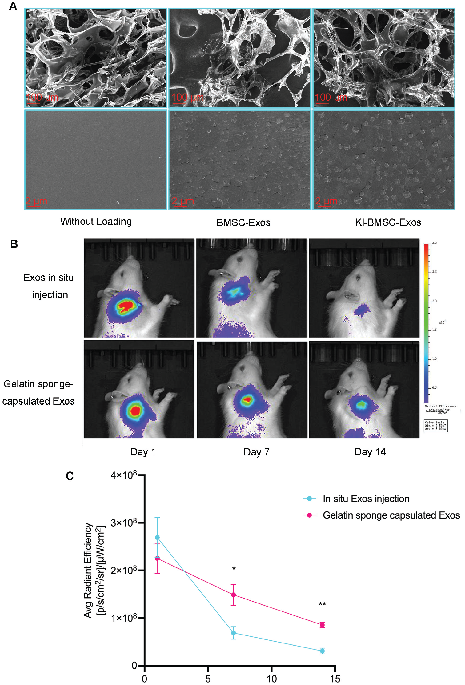
(A) Scanning electron microscopy showed the loading of exosomes on gelatin sponges.
Histologic Analysis
Hematoxylin and eosin staining revealed that the continuity between the tendon and the bone was complete in all model groups, with the repaired tendon better integrated into the bone in the BMSC-Exos group and the Kl-BMSC-Exos group (Figure 7A). In the RCI group, sparse newly formed fibrocartilage was found in the RCI group, and abundant scar-like fibrous tissue was observed at the tendon enthesis, which exhibited a disorganized pattern and showed limited integration within the bone. In contrast, fibers showed better arrangement in the BMSC-Exos group and the Kl-BMSC-Exos groups. When compared with the BMSC-Exos group, the Kl-BMSC-Exos group showed enhanced enthesis regeneration, as shown by the morphologic characteristics of the tendon enthesis. Specifically, in the Kl-BMSC-Exos group, a large amount of fibrocartilage regeneration was observed, and new fibers were organized along the longitudinal axis of the tensile force. Meanwhile, the native transitional 4-layer structure of the tendon enthesis was partially restored in the Kl-BMSC-Exos group. In terms of the BMSC-Exos group, a limited amount of fibrous ingrowth was observed at the tendon enthesis, with collagen fibers displaying a crimp pattern (Figures 7A and 8A). However, the collagen fiber arrangement was not as regularly organized as that in the Kl-BMSC-Exos group. Less neofibrocartilage was formed in the BMSC-Exos group and the RCI group. In addition, the Kl-BMSC-Exos group exhibited the highest histologic score (vs RCI, P < .001; vs BMSC-Exos, P = .0183) (Figure 7B) and collagen birefringence (vs RCI, P < .001; vs BMSC-Exos, P = .0190) (Figure 8B) among the 3 surgically repaired groups. The BMSC-Exos group had a higher histologic score than the RCI group (P = .0075). Yet, there was no significance between the BMSC-Exos group and the RCI group in collagen birefringence (P = .0776). Masson trichrome staining was employed to evaluate the regenerated collagen fibers in the supraspinatus enthesis. The Kl-BMSC-Exos group displayed more organized collagen fiber than the BMSC-Exos group and the RCI group (Figures 7A, 7C, and 8A). Similar trends were observed in the fibrocartilage area ratio. The Kl-BMSC-Exos group demonstrated the largest amount of neofibrocartilage formation (vs RCI, P < .001; vs BMSC-Exos, P = .0063), followed by the BMSC-Exos group and then the RCI group (RCI vs BMSC-Exos, P = .002) (Figure 7D).
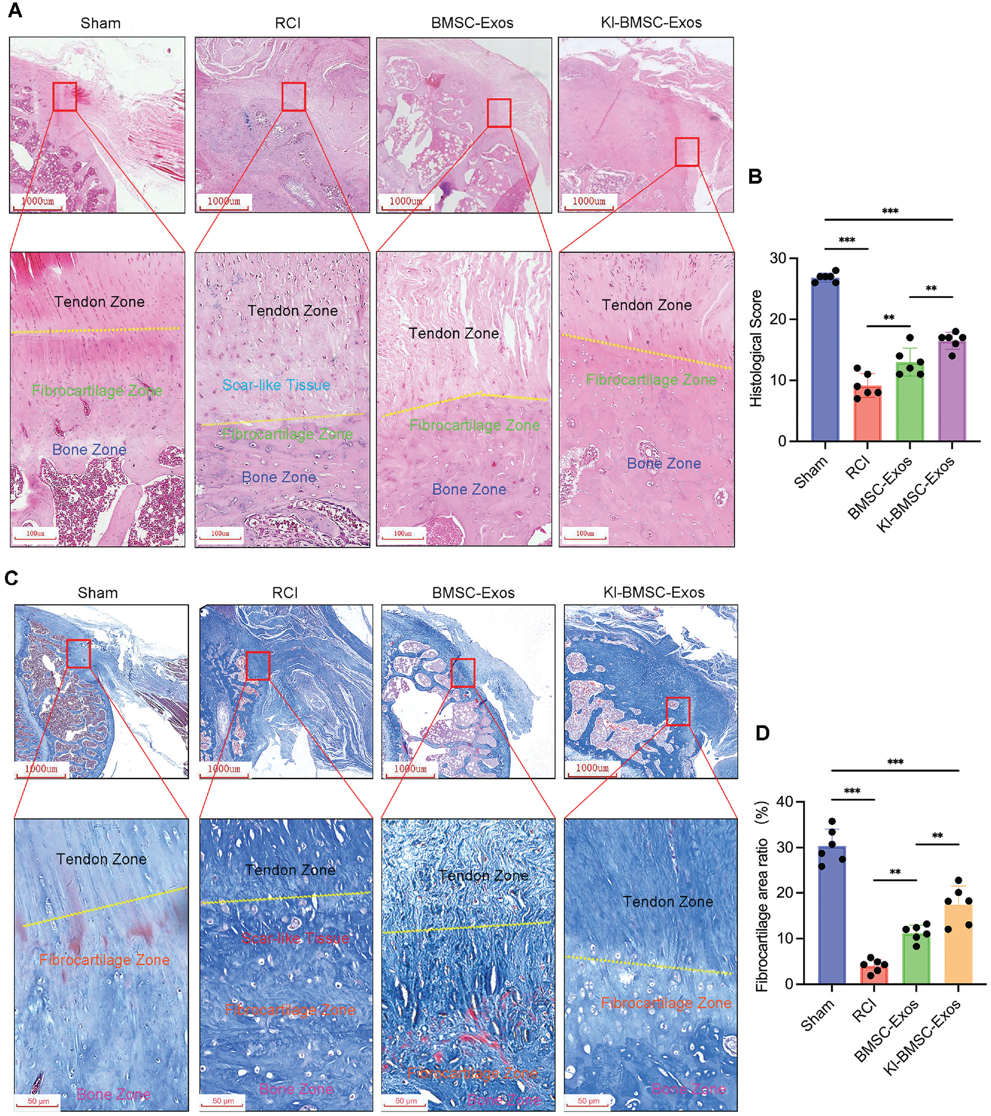
(A) Hematoxylin and eosin staining, (B) histologic score, (C) Masson trichrome staining, and (D) metachromasia ratio were used to evaluate the tendon enthesis regeneration of various groups at 8 weeks postoperatively. Data are expressed as mean ± SD (6 rats per group). **P < .01. ***P < .001. Scale bar = 1000 μm and 50 μm. BMSC-Exos, bone marrow mesenchymal stem cell–derived exosomes; Kl-BMSC-Exos, kartogenin-loaded bone marrow mesenchymal stem cell–derived exosomes; RCI, rotator cuff injury.
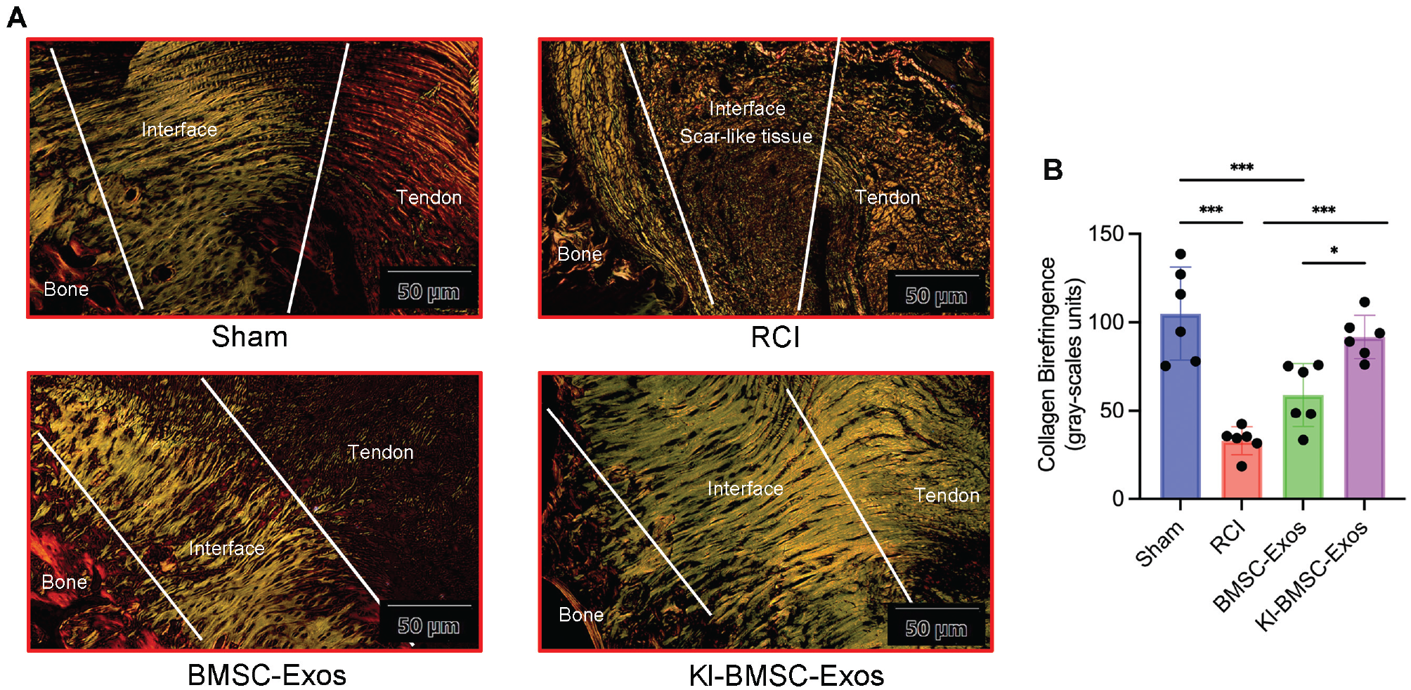
(A) Picrosirius red staining and (B) collagen birefringence were used to evaluate the tendon enthesis regeneration of various groups at 8 weeks postoperatively. Data are expressed as mean ± SD (6 rats per group). *P < .05. ***P < .001. Scale bar = 50 μm. BMSC-Exos, bone marrow mesenchymal stem cell–derived exosomes; Kl-BMSC-Exos, kartogenin-loaded bone marrow mesenchymal stem cell–derived exosomes; RCI, rotator cuff injury.
Biomechanical Testing
To evaluate the biomechanical properties of the regenerated tendon enthesis, we developed a fixture to securely fasten the specimen onto the testing apparatus (Figure 9A). The biomechanical testing results revealed that the native tendon enthesis demonstrated the highest failure loading of 39.95 ± 4.76 N among all experimental groups (P < .001) (Figure 9B). At 8 weeks postoperatively, the Kl-BMSC-Exos group demonstrated a significantly higher failure load with an ultimate loading capacity of 28.79 ± 2.70 N as compared with the BMSC-Exos group (18.52 ± 3.23 N; P = .002) and the RCI group (13.17 ± 3.56 N; P < .001). However, there was no statistical significance between the RCI group and the BMSC-Exos group (P = .1345). Similar results were seen in stiffness analysis. Kl-BMSC-Exos exhibited a higher value of stiffness than the RCI group (P < .001) and the BMSC-Exos group (P = .048) (Figure 9C). Meanwhile, no statistical difference was observed between the RCI group and the BMSC-Exos group (P = .0673).
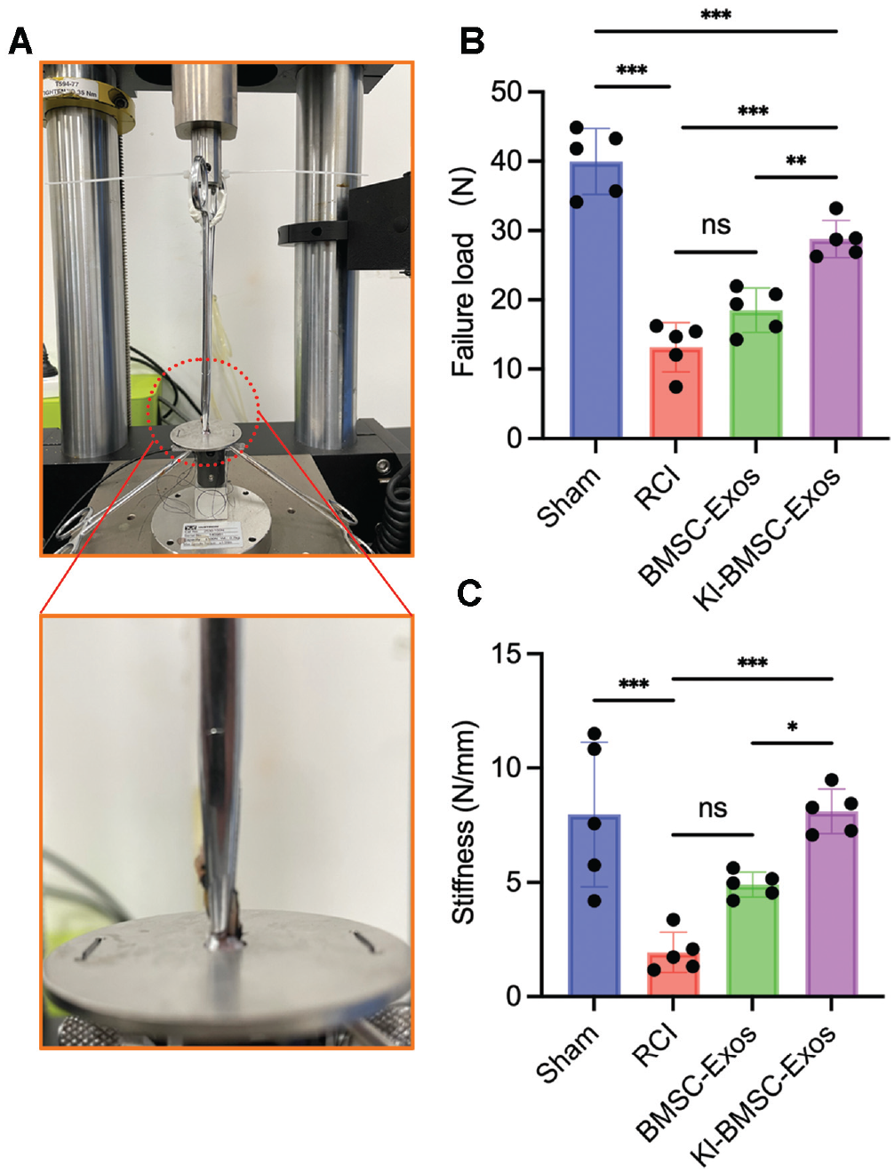
(A) Photographs demonstrated the biomechanical test setting for rotator cuff enthesis. (B) Failure load and (C) stiffness analysis revealed the biomechanical properties of tendon enthesis in various groups. Data are expressed as mean ± SD (5 rats per group). ns, not significant (P > .05). *P < .05. **P < .01. ***P < .001. BMSC-Exos, bone marrow mesenchymal stem cell–derived exosomes; Kl-BMSC-Exos, kartogenin-loaded bone marrow mesenchymal stem cell–derived exosomes; RCI, rotator cuff injury.
Bioinformatic Analysis and Verification
By employing the RNA sequencing methodology, we investigated alterations in mRNA expression within exosome-treated BMSCs (Figure 10).

Heat maps of differentially expressed genes: (A) Ctrl versus BMSC-Exos, (B) BMSC-Exos versus Kl-BMSC-Exos, and (C) Ctrl versus Kl-BMSC-Exos. BMSC-Exos, bone marrow mesenchymal stem cell–derived exosomes; Ctrl, control; Kl-BMSC-Exos, kartogenin-loaded bone marrow mesenchymal stem cell–derived exosomes.
Notably, all samples exhibited 3 distinct clustering patterns, indicating that our treatment induced diverse categories and levels of mRNA expression in BMSCs (Figure 11A). In total, we identified 112 DEGs in control versus BMSC-Exos, 125 DEGs in control versus Kl-BMSC-Exo, and 108 DEGs in BMSC-Exos versus Kl-MSC-Exos (Figure 11B). When compared with the control group and the BMSC-Exos group, 64 and 60 upregulated mRNAs were observed in Kl-BMSC-Exos–treated BMSCs, respectively (Figure 11, B-D). Among these upregulated DEGs, there were 9 coexpression genes (Figure 11E). Among them, Mospd1 ranked the highest and may have the potential to regulate the chondrodifferentiation of BMSCs. The expression of Mospd1 in BMSCs treated by BMSC-Exos or Kl-MSC-Exos was verified using quantitative polymerase chain reaction (qPCR) analysis. The expression levels were significantly increased by approximately 3.7-fold (P < .001) and 4.6-fold (P < .001) after 3 or 7 days of Kl-MSC-Exos intervention, as compared with the control group (Appendix Figure A3, available online). To evaluate the potential role of Mospd1 in BMSC differentiation, Mospd1-specific siRNA was synthesized and transfected into BMSCs. Interference efficiency was demonstrated by qPCR analysis (P < .001) (Figure 11F). Furthermore, qPCR analysis indicated that Kl-BMSC-Exos may initiate the chondrogenesis of BMSCs through a Mospd1-dependent pathway, as evidenced by (1) the upregulation of early chondrogenesis markers Sox9 and Runx1 (P < .001) (Figure 11G) and (2) the effect of siMospd1 transfection in abolishing the promoting effect of Kl-BMSC-Exos. (P < .001) (Figure 11G). Altogether, these results suggest that Mospd1 plays a role in the enhanced chondrogenesis of BMSCs in vitro. Further study using Mospd1 knock-in and knockout mice could clarify the specific role of Mospd1 in rotator cuff enthesis healing.

(A) PCA revealed distinct gene expression patterns among the 3 groups. (B) The DEG analysis revealed upregulation and downregulation of gene expression across the 3 experimental groups. (C, D) Volcano plots reveal upregulation and downregulation of gene expression across the experimental groups. (E) The Venn diagram displays the coexpression of upregulated genes: Ctrl versus Kl-BMSC-Exos group and BMSC-Exos versus Kl-BMSC-Exos group. (F, G) Relative mRNA expression of Mospd1 and chondrogenic markers in BMSCs. Data are expressed as mean ± SD (3 rats per group). *P < .05. **P < .01. ***P < .001. BMSC-Exos, bone marrow mesenchymal stem cell–derived exosomes; Ctrl, control; DEG, differentially expressed gene; Kl-BMSC-Exos, kartogenin-loaded bone marrow mesenchymal stem cell–derived exosomes; PCA, principal component analysis.
Discussion
Rotator cuff enthesis healing is an intractable clinical challenge because of the complex tissue organization. 21 Several biologic augmentation strategies have demonstrated that application of KGN, 39 platelet-rich plasma, 41 BMSC implantation, 15 exosomes derived from platelet-rich plasma, 23 and BMSC-Exos 18 could accelerate tendon enthesis healing by restoring the functional fibrocartilage. In this study, a cell-free tissue-engineered gelatin sponge sustainably releasing Kl-BMSC-Exos enhanced the healing process of the rotator cuff enthesis by promoting fibrous arrangement, fibrocartilage regeneration, histologic evaluation, and biomechanical properties potentially through Mospd1 pathway–induced chondrogenesis.
Numerous studies have shown that BMSCs exert their regulatory function in enthesis healing by releasing exosomes.18,27,38 However, undesired loss of exosomes attributed to fluid flow and joint movements should be considered when BMSC-Exos are to be applied in tendon enthesis healing.38,39 In our study, instead of in situ injection, we utilized a commercially available gelatin sponge to construct a scaffold capable of retaining the exosomes during joint movements, thereby extending the retention time of exosomes from 7 to 14 days.
Although BMSC-Exos exhibited reparative efficacy in tendon enthesis healing, application of exosomes alone may not yield optimal biomechanical recovery.26,38 Thus, KGN, a chondrogenesis initiator, has been incorporated into exosomes because in situ injection of KGN could lead to excessive formation of fibrocartilage-like tissues in unexpected areas.25,39,42 Drug loading and delivery using exosomes as vehicles is an emerging field and has attracted growing interest. A study conducted by Xu et al 35 demonstrated that encapsulation of KGN into exosomes could serve better fibrocartilage repair in an OA model. However, KGN dosages vary in the literature. Xu et al used protein concentration to quantify exosomes and incorporated 12 μg of exosomes with 12.69 ng of KGN. They reported that this dosage was sufficient for in vivo exosomal visualization and effective chondrogenesis. Yet, in our pilot study, we failed to detect sufficient fluorescence in the in vivo and in vitro experiments using that dosage of exosomes. In another study, Kim et al 14 reported that a larger number of exosomes could be retained for a longer period and was more suitable for in vivo tracking. Therefore, we decided to administer a dosage of 1011 exosomes. In our study, we confirmed that loading KGN into exosomes enhanced the chondroinductive potency of BMSC-Exos, as shown by significantly upregulated expression of early markers of chondrogenesis: Sox9 and Runx1 of BMSCs. The enhanced chondrogenesis of BMSCs may have led to abundant fibrocartilage regeneration in the RCI rat model.
More important, in addition to increasing fibrocartilage regeneration, Kl-BMSC-Exos induce a more organized arrangement of collagen fibers and higher collagen birefringence as compared with BMSC-Exos (Figures 7 and 8), thereby imparting adequate mechanical strength for the regenerated rotator cuff enthesis. Disorganized and abundant scar-like tissue was observed at the tendon-bone interfaces in the RCI group. The Kl-BMSC-Exos group demonstrated a more organized cell alignment and higher collagen birefringence at 8 weeks postoperatively, in contrast to the RCI group and the BMSC-Exos group. Meanwhile, parallel collagen fibers and a 4-layer continuous enthesis structure were observed in the KGN-loaded group. These findings indicate that Kl-BMSC-Exos facilitate fibrocartilage regeneration, which may lead to a microscopically near-native enthesis repair with organized and parallel collagen fibers. However, the specific mechanism of how Kl-BMSC-Exos induce fibrocartilage regeneration needs investigation.
The failure load and stiffness in the Kl-BMSC-Exos group were higher than those obtained from BMSC-Exos alone in our study. However, when compared with the native rotator cuff, Kl-BMSC-Exos treatment still cannot achieve a perfect histologic score and biomechanical properties. The inherent difficulty of enthesis healing and the imperfect dosage of Kl-BMSC-Exos may account for this issue; incorporating more biomimetic adjustments may help our system achieve optimal healing.
Cai et al 2 performed a well-designed and convincing study about exosomes derived from KGN-preconditioned BMSCs in enthesis regeneration, demonstrating that exosomes derived from KGN-preconditioned BMSCs effectively promote cartilage formation as well as collagen maturation and organization in enthesis regeneration, contributing to enhanced biomechanical properties after rotator cuff repair. The exosomes that Cai et al used were derived from KGN-pretreated BMSCs. In their study, the constituents of exosomes derived from primed BMSCs may play a role in enthesis regeneration. In comparison, our study utilized BMSC-derived exosomes as proregeneration biologics and a natural delivery system. In other words, this method delivered the BMSC-derived exosomes and KGN at the same time, which is distinct from delivering only exosomes derived from KGN-primed BMSCs. However, in our future investigations, this method could be improved by engineering the exosomes with peptides to achieve a more pronounced and precise therapeutic effect. 20
Clarifying the mechanisms of Kl-BMSC-Exos promoting tendon enthesis healing is essential for potential application and future improvement of this biologic augmentation. Mospd1 has been reported to play a pivotal role in the proliferation and differentiation of BMSCs.13,29 In our study, the RNA sequencing revealed that the most significant upregulation of Mospd1 was found in the Kl-BMSC-Exos group, suggesting that Mospd1 may play a pivotal role in enhanced tendon enthesis healing. This conclusion is supported by 2 previous studies, which reported that Mospd1 acts as a novel contributor to the regulation of mesenchymal stem cell proliferation and differentiation.7,13 Furthermore, in our study, inhibition of Mospd1 mRNA expression using siRNA hindered chondrogenesis in BMSCs and abolished the positive effects induced by Kl-BMSC-Exos, suggesting that tendon enthesis healing mediated by Kl-MSC-Exos may rely on upregulation of Mospd1.
Our study also has some limitations. First, sex as a biological variable was not examined. 28 Second, we utilized an acute rotator cuff repair model rather than a chronic degenerative rotator cuff model, which is more clinically relevant. Last, additional trials using large animal models in preclinical studies are indispensable before clinical conclusions.
Conclusion
This study demonstrates that gelatin sponge impregnated with Kl-BMSC-Exos serves to provide sufficient in vivo retention time and can improve tendon enthesis healing in the rat RCI model by enhancing chondrogenesis of BMSCs and fibrocartilage regeneration, as well as by providing more organized collagen fiber arrangement and superior biomechanical properties, as compared with using BMSC-Exos alone. The RNA sequencing revealed that Kl-BMSC-Exos promote tendon enthesis healing, which may be mediated through a Mospd1-dependent pathway. The present study offers novel perspectives on expediting tendon enthesis healing using biologics, which is promising in clinical application.
Supplemental Material
sj-pdf-1-ajs-10.1177_03635465241296141 – Supplemental material for Kartogenin-Loaded Exosomes Derived From Bone Marrow Mesenchymal Stem Cells Enhance Chondrogenesis and Expedite Tendon Enthesis Healing in a Rat Model of Rotator Cuff Injury
Supplemental material, sj-pdf-1-ajs-10.1177_03635465241296141 for Kartogenin-Loaded Exosomes Derived From Bone Marrow Mesenchymal Stem Cells Enhance Chondrogenesis and Expedite Tendon Enthesis Healing in a Rat Model of Rotator Cuff Injury by Yue Wang, Ji-Zheng Qin, Chao-Yu Xie, Xin-Zhou Peng, Jian-Hua Wang and Shao-Jie Wang in The American Journal of Sports Medicine
Footnotes
Acknowledgements
The authors thank Yang Xu and Yong-Kang Ma for their help with exosome isolation. They also acknowledge Jing Shang and Gui-Xia Li for their help with cell transfection.
Submitted February 8, 2024; accepted August 26, 2024.
One or more of the authors has declared the following potential conflict of interest or source of funding: This work was supported by the National Natural Science Foundation of China (82072404 to S.-J.W.), National Natural Science Foundation of China Youth Fund Project (82002266 to J.-Z.Q.), Science Fund for Distinguished Young Scholars of Fujian Province (2020D030 to S.-J.W.), Fujian Eyas Project for Young Top-notch Talent (to S.-J.W.), Natural Science Foundation of Fujian Province (2020J011211 to J.-Z.Q.), and Key Projects for Healthcare in Xiamen (3502Z20234011 to S.-J.W.). AOSSM checks author disclosures against the Open Payments Database (OPD). AOSSM has not conducted an independent investigation on the OPD and disclaims any liability or responsibility relating thereto.

References
Supplementary Material
Please find the following supplemental material available below.
For Open Access articles published under a Creative Commons License, all supplemental material carries the same license as the article it is associated with.
For non-Open Access articles published, all supplemental material carries a non-exclusive license, and permission requests for re-use of supplemental material or any part of supplemental material shall be sent directly to the copyright owner as specified in the copyright notice associated with the article.
