
Abstract
Background:
Gluteus medius tears are an increasingly recognized cause of lateral hip pain in active individuals, yet no gold standard repair technique has been established, and failure and retear rates remain high. Iliotibial band (ITB) graft augmentation is a validated option in rotator cuff and hip labral repairs for stronger repair construct, but its biomechanical utility in gluteus medius repairs has not been investigated.
Hypothesis:
It was hypothesized that ITB-augmented gluteus medius repairs would demonstrate superior ultimate load to failure and increased repair stiffness as compared with nonaugmented repairs.
Study Design:
Controlled laboratory study.
Methods:
Fourteen unpaired fresh-frozen cadaveric hemipelvises with intact gluteus medius tendons were randomized into ITB-augmented and nonaugmented groups and tested biomechanically with a tensile testing machine. Native stiffness was assessed as follows: 10-N preload for 2 minutes, 150 cycles at 0.8 Hz from 10 to 125 N to simulate early partial weightbearing, followed by a quasistatic load to 60 N at 1 mm/s. Tendons were then elevated from their anatomic footprint on the greater trochanter and repaired using double-row suture bridge configurations, with augmented repair incorporating ITB graft fixation. After preconditioning (10 N for 2 minutes, 150 cycles at 0.8 Hz from 10 to 125 N to simulate early partial weightbearing), specimens were loaded to failure at a constant displacement rate of 31 mm/min. Failure load, repair stiffness, and mode of failure were recorded. Data analysis included Shapiro-Wilk test, independent samples 2-tailed t tests, and paired t tests.
Results:
ITB-augmented repairs demonstrated a 98% stronger failure load (mean ± SD, 294.6 ± 87.9 N) than nonaugmented repairs (149.0 ± 40.4 N; P = .002) and were significantly less stiff to their native state (28.1 ± 6.7 vs 137.6 ± 98.1 N/mm; P = .023). The stiffnesses of the native tendons were not different from each other (P = .773).
Conclusion:
Gluteus medius repairs augmented with ITB grafts led to greater failure load than nonaugmented repairs and were less stiff than the native state.
Clinical Relevance:
Utilization of ITB-augmented double-row repairs when treating tears of the gluteus medius tendon can potentially reduce high failure rates.
The gluteus medius is the primary hip abductor, critical for pelvic stability during single-leg stance and gait by controlling transverse and frontal plane hip and femur motion.3,17,39 Tears are increasingly identified in active individuals with lateral hip pain, abductor weakness, and gait disturbances, reflecting improved diagnostic accuracy and awareness.12,13,36,43,46 Minimally invasive surgical techniques, particularly endoscopic repair, have expanded surgical indications.12,13,36,43,46 Newer strategies, including tendon compression bridge, double-row suture anchor repair, and single-row repair with trochanteric microfracture, show promise, although clinical and biomechanical support remains limited.10,26,36 Thus, no gold standard exists for surgical repair.
Full-thickness gluteus medius repairs are challenging given the tendon’s short, broad structure, limited substance for multiple suture passes or locking constructs, and a musculotendinous junction near the bony insertion. 44 Chronic tears worsen these constraints with retraction, degeneration, poor tissue quality, and fatty infiltration.16,27,31,38,40,43 Biomechanical demands further compromise repair, as single-leg stance and ambulation generate compressive forces of 2.5 to 3 times the body weight concentrated at the musculotendinous junction and insertion.38,44 Biomechanical studies show failure occurring predominantly at these sites.25,37 Clinically, retear rates range from 0% to 35.7%, with up to 21.1% failing at 2 years.1,2
Double-row repair provides superior footprint coverage, tendon-to-bone contact, and construct strength as compared with single-row techniques.25,38 To further reduce repair failure or retear, patch augmentation strategies have been introduced.16,19,33 Dermal allografts offer high initial strength but are biomechanically inert and have not shown clear clinical superiority.5,6,33 Collagen- and bovine-based patches provide strength and cell support but are limited by nonstructural properties, hyperinflammatory and allergic risk, and xenograft-specific issues.6,19,21,30,41 Biocomposite scaffolds are promising but remain limited by their novelty and lack of long-term and clinical data.19,23,28
Iliotibial band (ITB) grafts are well established for reconstruction and augmentation in rotator cuff and hip labral repairs, with favorable outcomes.9,10,32 The ITB contributes to hip abduction and lateral stability via its connections with the gluteus maximus and tensor fascia latae.22,24 By transmitting muscular forces, it facilitates abduction and reinforces the lateral hip joint, supporting its use as an augmentation graft.7,15,34 However, ITB grafts have not been studied for primary gluteus medius repair. Autografts generally demonstrate superior incorporation and durability, immediate availability, and avoidance of immunologic complications, potentially overcoming limitations of dermal allografts, collagen-based patches, and biocomposite scaffolds.4,5,19,30
Existing biomechanical studies on gluteus medius repairs have focused on repair technique (single vs double row), anchor fixation, surgical approach (open vs endoscopic), and the effects of bone quality and decortication on construct strength and failure.11,18,25,29,37 These studies established baseline metrics, including footprint coverage, gap formation, load to failure, and the influence of technique and bone quality on repair integrity. To our knowledge, no prior studies have evaluated the biomechanical properties of full-thickness gluteus medius repairs augmented with ITB graft.
The purpose of this study was to compare the biomechanical performance of gluteus medius repairs with and without ITB graft augmentation regarding failure load, stiffness, and mode of failure. We hypothesized that ITB-augmented repairs would demonstrate superior biomechanical characteristics at time zero as compared with nonaugmented repairs.
Methods
Power Analysis and Study Design
Power analysis based on prior literature indicated a sample size of 14 unpaired specimens (mean difference, 145.7 N; SD of differences, 84.2 N; power, 0.8), with 7 specimens per test group. 18 Thus, this study employed an unpaired design with 14 specimens randomly assigned to 2 even groups: with and without ITB augmentation.
Specimen Preparation
A total of 14 unpaired, fresh-frozen, human cadaveric hemipelvises were included (mean age, 63 years [range, 42-75]; 8 male, 6 female). Specimens were randomly assigned to either an ITB augmentation repair group (n = 7; mean age, 63.4 years; 3 female, 4 male) or a no-augmentation group (n = 7; mean age, 62 years; 3 female, 4 male). The deidentified cadaveric specimens used were donated to a tissue bank designated for medical research and purchased by the authors’ institution. The use of cadaveric specimens donated for medical research is institutional review board exempt. Specimens were selected per the following inclusion criteria: age <75 years and body mass index between 20 and 30. Specimens were excluded if they had any prior surgery to the hip, osteoarthritis, osteoporosis, sepsis, degenerative joint disease, HIV I/II, hepatitis B, or hepatitis C.
Specimens were thawed at room temperature for 24 hours before dissection and testing. Skin and subcutaneous tissue were dissected. Fatty infiltration of the gluteus medius tendon was qualitatively assessed by 2 fellowship-trained orthopaedic surgeons based on gross visual inspection of the muscle and tendon. Assessment was informed by the general criteria of the Goutallier classification (ie, relative proportion of muscle to viable intramuscular fat). Specimens with advanced fatty degeneration (equivalent to Goutallier grade ≥3) were excluded. The femur was transversely cut with a bone saw, leaving 130 mm distal from the greater trochanter intact. The femur was potted with polymethyl methacrylate (Fricke Dental) in cylindrical potting, and horizontal screws were placed in the femur to augment fixation within the construct. The gluteus medius muscle was cut so that ~5 cm of muscle was left intact. The cut ends of the muscle were whipstitched with a No. 2 nonabsorbable braided suture (Ethibond Excel; Ethicon) through high-absorbency, nonwoven sponges (McKennson) attached to the muscle edges. After whipstitching, additional reinforcement with figure-of-8 sutures was used to unitize the muscle belly for clamping efficiency. Sutures were passed through the musculotendinous junction and the sponge to prevent slippage during testing. ITB grafts were harvested from each specimen as single-layer strips measuring 30 × 30 mm from the region just distal and slightly posterior to the greater trochanter. This location was selected because it provides adequate tissue thickness and is readily accessible during open gluteus medius repair. The harvested graft was kept moist with saline-soaked gauze until fixation. Consistency was maintained by harvesting from the same anatomic region in all specimens.
Biomechanical Testing: Native State
Before the repairs, the native state was tested. The native gluteus medius tendon was mounted to a dynamic tensile testing machine (Bionix Tabletop System; MTS Systems) with the muscular portion of the gluteus medius fixed within a custom clamping fixture on the end effector and the femur secured to a fixture secured to the base of the testing machine (Figure 1). The base fixture was adjusted such that the line of action of the end effector was aligned to the line of action of the gluteus medius in a neutral anatomic stance, with the muscle positioned 20° lateral to the greater trochanter. The tendon was clamped such that the direction of loading was in the physiologic direction of the pull of the muscle. Each specimen underwent the following testing sequence: (1) 10-N preload for 2 minutes, (2) 150 cycles from 10 to 125 N at 0.8 Hz, and (3) load to 60 N at 1 mm/s to record the force-displacement curve. In this way, native stiffness was calculated while avoiding specimen damage. The cyclic loading protocol was chosen to represent submaximal physiologic tensile forces experienced by the gluteus medius during the early postoperative period with a 125-N upper limit consistent with prior biomechanical studies of abductor repairs that simulated early functional loads.18,25 Native stiffness was calculated as the slope of the linear portion of the force-displacement curve. The specimen was then removed from the testing machine.
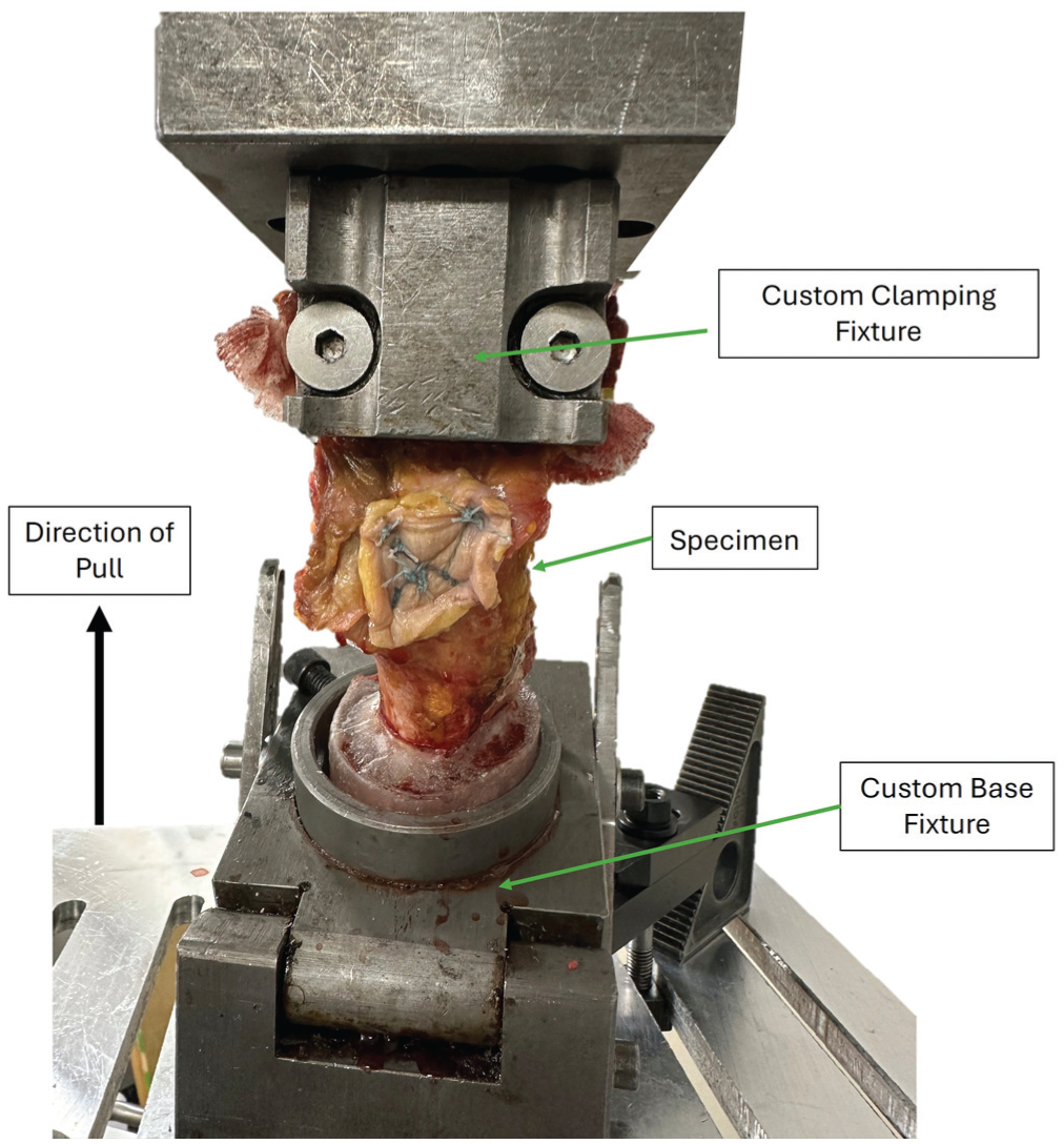
Mechanical testing setup.
Nonaugmented Gluteus Medius Repair
After native state testing, the gluteus medius tendon was carefully elevated from the bone by a No. 10 blade scalpel (Figure 2A). The anterior medial row anchor was placed in the center of the anterior facet, and the posterior anchor was placed in the geometric center of the posterior facet. The lateral row anchors were placed in line with the medial row anchors, 1 cm distal to the lateral edge of the anatomic footprint of the gluteus medius tendon. The medial row anchors were single loaded with suture (No. 4 FiberWire; Arthrex), and the 2 limbs were brought through the tendon in a single pass. These were then tied to the lateral row anchors in a standard “X” suture bridge configuration and were tied by 5 alternating half hitches on reversing posts.
ITB-Augmented Gluteus Medius Repair
A similar construct was used for the augmented repair (Figure 2B). Suture anchors were placed via the same anatomic landmarks, and identical anchors were utilized for the nonaugmented and ITB-augmented constructs. The 2 free suture limbs from the medial row were brought through the tendon and the ITB in a single pass. To achieve constant graft tensioning, an 8-mm offset technique was employed: sutures were passed through the gluteus medius tendon approximately 8 mm proximal to the corresponding graft pass, resulting in an estimated 8 mm of proximal graft advancement upon tying. This method provided a reproducible approach to tensioning based on controlled advancement rather than applied force. The sutures from the lateral row anchors were then brought through the ITB graft and tied in a horizontal mattress configuration, thereby anchoring the ITB laterally to bone. These sutures were then tied to the medial row sutures to create an “X” pattern suture bridge. After completion of the suture bridge, 2 additional proximal figure-of-8 sutures were placed into the ITB graft and the proximal gluteus medius tendon to unitize the ITB graft to the gluteus medius tendon, creating a load-sharing construct.
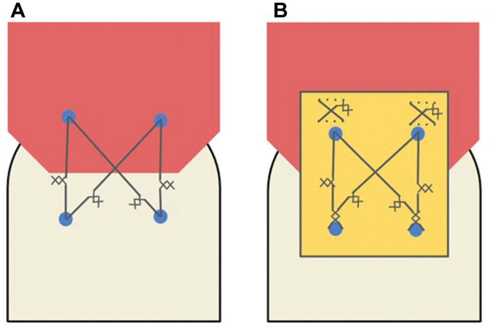
Schematic of the gluteus medius repair (A) without and (B) with iliotibial band augmentation.
Biomechanical Testing: Repair State
Specimens were remounted to the dynamic materials testing machine using the same protocol as the native state. Each specimen underwent the following testing sequence: (1) 10-N preload for 2 minutes, (2) 150 cycles from 10 to 125 N at 0.8 Hz, and (3) load to failure at a constant ramp displacement of 31 mm/min. The cyclic loading protocol was chosen to represent submaximal physiologic tensile forces experienced by the gluteus medius during the early postoperative period with a 125-N upper limit, consistent with prior biomechanical studies of abductor repairs that simulated early functional loads.18,25 Force-displacement data were recorded, and repair stiffness was calculated as the slope of the linear portion of the force-displacement curve. Failure load and mode of failure were recorded.
Statistical Analysis
Statistical analyses were performed in commercial software (MATLAB; MathWorks), and data were expressed as mean and standard deviation. Three biomechanical outcomes are reported in this study: failure load, native stiffness, and repair stiffness. Shapiro-Wilk tests were used to verify normal distribution of the data. Paired t tests with a Bonferroni correction were used to compare within-specimen stiffness data between the repair groups and between the repair state and the native state within each repair group as these data were normally distributed. Between-group comparisons (no augmentation vs ITB augmentation) for failure load and repair stiffness were conducted with independent samples 2-tailed t tests. Statistical significance was set at α = .05, with β error at .80.
Results
The repair with ITB augmentation was 98% stronger than the repair without augmentation (Figure 3). The repair without ITB augmentation was not different to its native state. The repair with ITB augmentation was significantly less stiff than its native state. Mode of failure was visually confirmed for each specimen during load-to-failure testing and defined as the first structural rupture or <10% loss in load-carrying capacity. All specimens failed via suture pull-through of the tendon except for 1 specimen in the ITB augmentation group, which failed at the myotendinous junction.
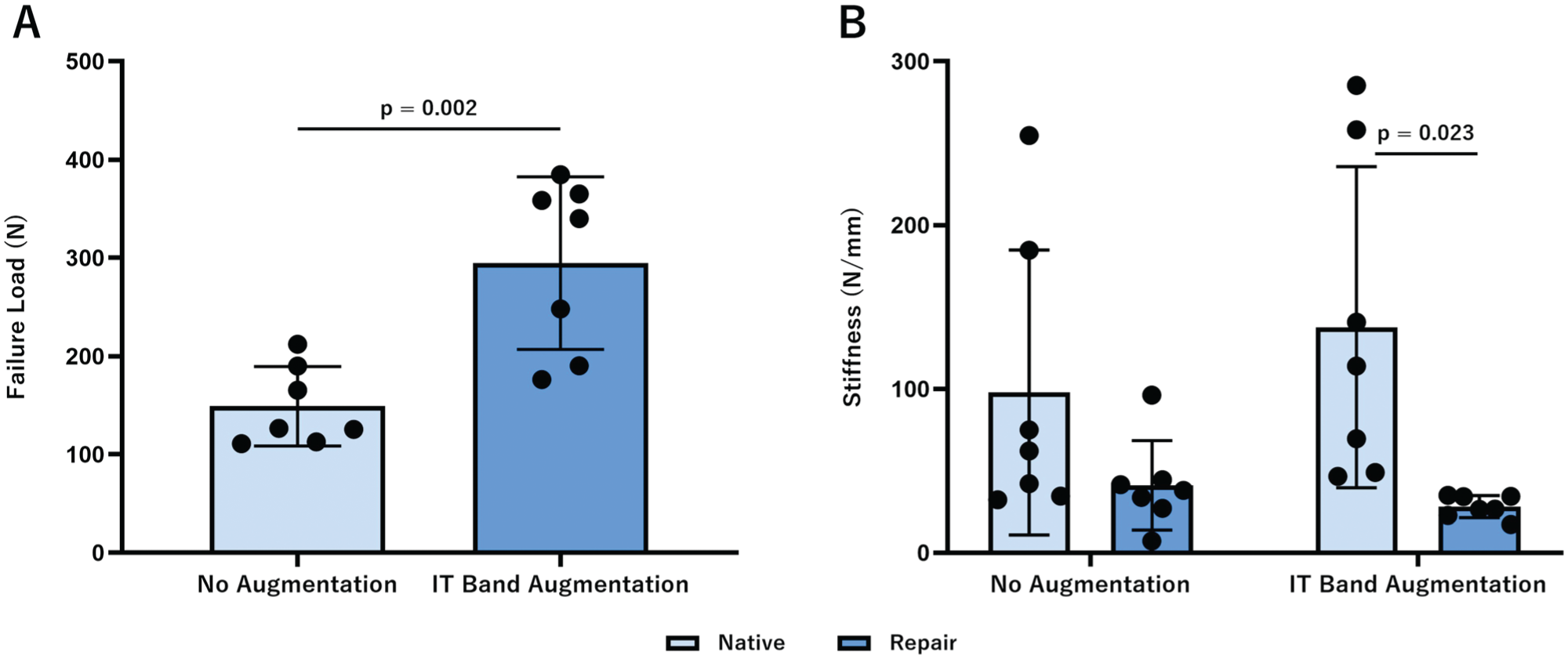
(A) Failure load and (B) stiffness of the gluteus medius repair with and without iliotibial band augmentation. Data are presented as mean (SD). P < .05 was considered statistically significant.
Discussion
The most important finding of this study was that repair of full-thickness gluteus medius tears augmented with ITB grafts resulted in significantly greater failure load as compared with repairs without ITB augmentation, confirming the initial hypothesis. Repairs with ITB augmentation were 98% stronger than repairs without ITB augmentation, indicating substantial improvement in the construct strength relative to established techniques. Interestingly, neither repair configuration restored the tendon to its native stiffness. In fact, repair with ITB augmentation significantly reduced stiffness by 20.4% as compared with the native state, suggesting that while ITB augmentation enhances load-bearing capacity, it may not fully restore native tendon function and elasticity.
Previous cadaveric studies investigating repair techniques for gluteus medius tears have primarily focused on the configurations of the repair construct. Double-row suture repair with knotless lateral anchors has demonstrated superior mechanical characteristics to single-row constructs, while comparison of open versus endoscopic approaches has yielded no significant differences in biomechanical outcomes.11,18,25 In our study, the maximum load to failure (249 ± 87.9 N with ITB augmentation; 149.0 ± 40.4 N without) was lower than previously reported values for double-row repairs (348-454 N), while repair stiffness (28.1-41.2 N/mm) was comparable to prior reports (43-51 N/mm).11,25 Although our study did not reproduce the higher loads reported in previous literature, it is the first to show a significant difference between the gluteus medius repair constructs. Additionally, bone mineral density and greater trochanteric decortication have shown to influence suture anchor pullout strength after abductor tendon repairs.11,37 These studies highlight the mechanical advantage of using the double-row repair construct to maximize footprint coverage and failure load, while emphasizing how poor bone quality and increased decortication have detrimental effects on construct stability.11,18,25,37
Consistent with these prior cadaveric investigations, our study observed failure in the ITB augmented and nonaugmented groups occurring predominantly at the musculotendinous junction and bone-anchor-tendon interface, with suture pull-through being the most common mode of failure.25,37 Gluteus medius repairs with ITB grafts improved failure load as compared with the traditional double-row construct without augmentation. These results are consistent with existing literature on ITB augmentation in hip labral and rotator cuff reconstruction techniques, where the graft has been shown to enhance mechanical strength in soft tissue repairs.9,10,32,35
Repair of full-thickness gluteus medius tears is often challenging, particularly when nonoperative management fails long-term, as this can lead to progressive tendon retraction, separation from its anatomic footprint, and fatty infiltration.4,31,42,43,45 When compounded by poor tendon quality, degeneration, or insufficient residual tendon at the musculotendinous junction, primary repair may place excessive tension on the construct that significantly increases the risk of failure.40,45 The results of this study suggest that ITB autograft augmentation may enhance initial repair strength, providing a mechanical advantage that could improve outcomes in compromised tissue. The ITB is readily accessible for autograft harvest and has demonstrated success in hip labral and rotator cuff reconstruction, further supporting its potential role in gluteus medius repair.9,10,32,35 However, clinical application of ITB autograft augmentation must consider potential drawbacks, including donor site morbidity, increased surgical time, and the possible effects of lateral knee stability, given the ITB’s role as a stabilizer of the frontal plane.8,14,20 These trade-offs should be weighed against the potential biomechanical benefits demonstrated in this study when considering translation to clinical practice. While our findings support the biomechanical benefit of ITB augmentation, future investigation is needed to assess long-term healing, graft incorporation, and clinical outcomes.
This study was not without limitations. As a cadaveric model, it does not account for biologic healing, graft integration, and the benefits of postoperative rehabilitation. While ITB augmentation demonstrated improved biomechanical strength at the time of repair, these results may not directly translate into clinical outcomes and warrant further investigation. Additionally, all soft tissue aside from the gluteus medius was removed, which may not be entirely representative of active hip biomechanics or tendon load sharing. The applied tensile loads were also unidirectional and therefore did not fully reproduce the multidirectional forces experienced by the gluteus medius during functional activities. As a result, all repairs were performed using an open approach, which may differ from the endoscopic techniques most commonly used in clinical practice. Additionally, anatomic differences such as femoral offset, neck-shaft angle, gluteus medius moment arm, and tendon cross-sectional area could influence estimated hip loads, load distribution, and repair mechanics. While all specimens were positioned in a standardized orientation to minimize variability, these inherent differences between unpaired specimens may have introduced unaccounted biomechanical variability. Finally, the small sample size and use of unpaired specimens reduce the power to detect small to moderate effects. Nevertheless, failure load exhibited a large observed effect with high achieved power, supporting the robustness of that comparison within the constraints of a cadaveric time-zero model.
Conclusion
Gluteus medius repairs augmented with ITB grafts led to greater failure load than nonaugmented repairs and were less stiff than the native state.
Footnotes
Submitted September 4, 2025; accepted December 20, 2025.
One or more of the authors has declared the following potential conflict of interest or source of funding: M.J.P. reports the following outside the submitted work: royalties from Smith & Nephew, Inc, Arthrosurface, Bledsoe, ConMed Linvatec, DJO, SLACK Inc, and Elsevier; owner/shareholder in Arthrosurface, MJP Innovations LLC, Vail Valley Surgery Center, Vail MSO Holdings LLC; shareholder in MIS, EFFRx, Olatec, iBalance (Arthrex), Manna Tree Partners, Stryker, Trimble, 3M, Bristol Myers, Squibb, Pfizer, AbbVie, Johnson & Johnson; board member for the International Society of Hip Arthroscopy and Vail Health Services; and co-chair for the Steadman Philippon Research Institute. M.J.P. has also received education support from Smith+Nephew, Inc, ConMed Linvatec, Ossur, Arthrex, and Siemens Medical Solutions; speaking fees and consulting from Smith & Nephew, Inc, MIS, Olatec, and NICE Recovery Systems; and hospitality payments from Siemens Medical Solutions and Synthes GmbH. J.A.G. declares consulting fees from Bioventus and research funding from Arthrex, Inc. AOSSM checks author disclosures against the Open Payments Database (OPD). AOSSM has not conducted an independent investigation on the OPD and disclaims any liability or responsibility relating thereto.