
Abstract
Background:
Sufficient blood vessel formation in bioengineered tissues is essential in order to keep the viability of the organs. Impaired development of blood vasculatures results in failure of the implanted tissue. The cellular source which is seeded in the scaffold is one of the crucial factors involved in tissue engineering methods.
Materials and methods:
Considering the notable competence of Bone Marrow derived Mesenchymal Stem Cell aggregates for tissue engineering purposes, in this study BM-aggregates and expanded BM-MSCs were applied without any inductive agent or co-cultured cells, in order to investigate their own angiogenesis potency in vivo. BM-aggregates and BM-MSC were seeded in Poly-L Lactic acid (PLLA) scaffold and implanted in the peritoneal cavity of mice.
Result:
Immunohistochemistry results indicated that there was a significant difference (p < 0.050) in CD31+ cells between PLLA scaffolds contained cultured BM-MSC; PLLA scaffolds contained BM-aggregates and empty PLLA. According to morphological evidence, obvious connections with recipient vasculature and acceptable integration with surroundings were established in MSC and aggregate-seeded scaffolds.
Conclusion:
Our findings revealed cultured BM-MSC and BM-aggregates, capacity in order to develop numerous connections between PLLA scaffold and recipient’s vasculature which is crucial to the survival of tissues, and considerable tendency to develop constructs containing CD31+ endothelial cells which can contribute in vessel’s tube formation.
Keywords
Introduction
Ageing, disease and trauma as the major causes of tissue damage and injury impose enormous operational and financial burden on the health care system and individuals. 1 Researchers in two main areas of regenerative medicine; cell therapy, and tissue engineering are exploring to find efficient replacements and treatments for damaged tissues. 2 Despite all efforts, the clinical achievements have been limited due to some obstacles. One of the most important problems is the insufficient angiogenesis which prevents the efficient integration and viability of implanted tissue. Features of cell source, characteristics of the scaffold, and the interaction of cell source and the scaffold are the most critical aspects of tissue engineering.3–5 Mesenchymal Stem Cells (MSCs) are one of the most prevalent types of adult stem cells which are used in several studies.6–8 Furthermore several completed clinical trials are using MSCs for the treatment of bone and tissues disease, for example knee osteoarthritis, dental implants, maxillary bone cysts, etc.9–11 Based on our previous studies MSCs are not isolated in a niche; in fact, they are in the form of cell aggregates. We have extracted MSC’s preserved in their native niche-like environment assumed as the stromal component of haematopoietic stem cell niches.12,13 In combination with 3-D scaffold subsequent to transplanting into the mice, native BM structure was observed in transplanted bioengineered tissue. 14 In addition to the remarkable competence of BM-aggregates they are available in urgent cases and no expansion or passage is needed, in contrary with expanded MSCs; furthermore, in the form of cell aggregates, haematopoietic and mesenchymal stem cells reside in their original and their behaviour remains much the same as their in vivo behaviour. Mentioned characteristics of BM-aggregates make them appealing for tissue regeneration purposes so we assessed osteogenic differentiation potential and feasibility of their attachment to scaffold which revealed BM-aggregate’s faster osteogenic differentiation than expanded MSCs. 15 Promising properties of BM-aggregate considering the necessity of angiogenesis in bone tissue engineering and other transplanted tissues led us to investigate the angiogenesis potential of BM-aggregates. This research was conducted to evaluate the angiogenesis potential of BM-aggregate versus cultured BM-MSC and their interaction in contact with Poly L-Lactic Acid (PLLA) scaffold comparing with an empty scaffold. PLLA is a degradable, accessible, biocompatible scaffold with favourable mechanical attributes.16,17 In vivo researches would be essential to explore the practical findings to promote angiogenesis in transplanted tissues.
Materials and methods
Results
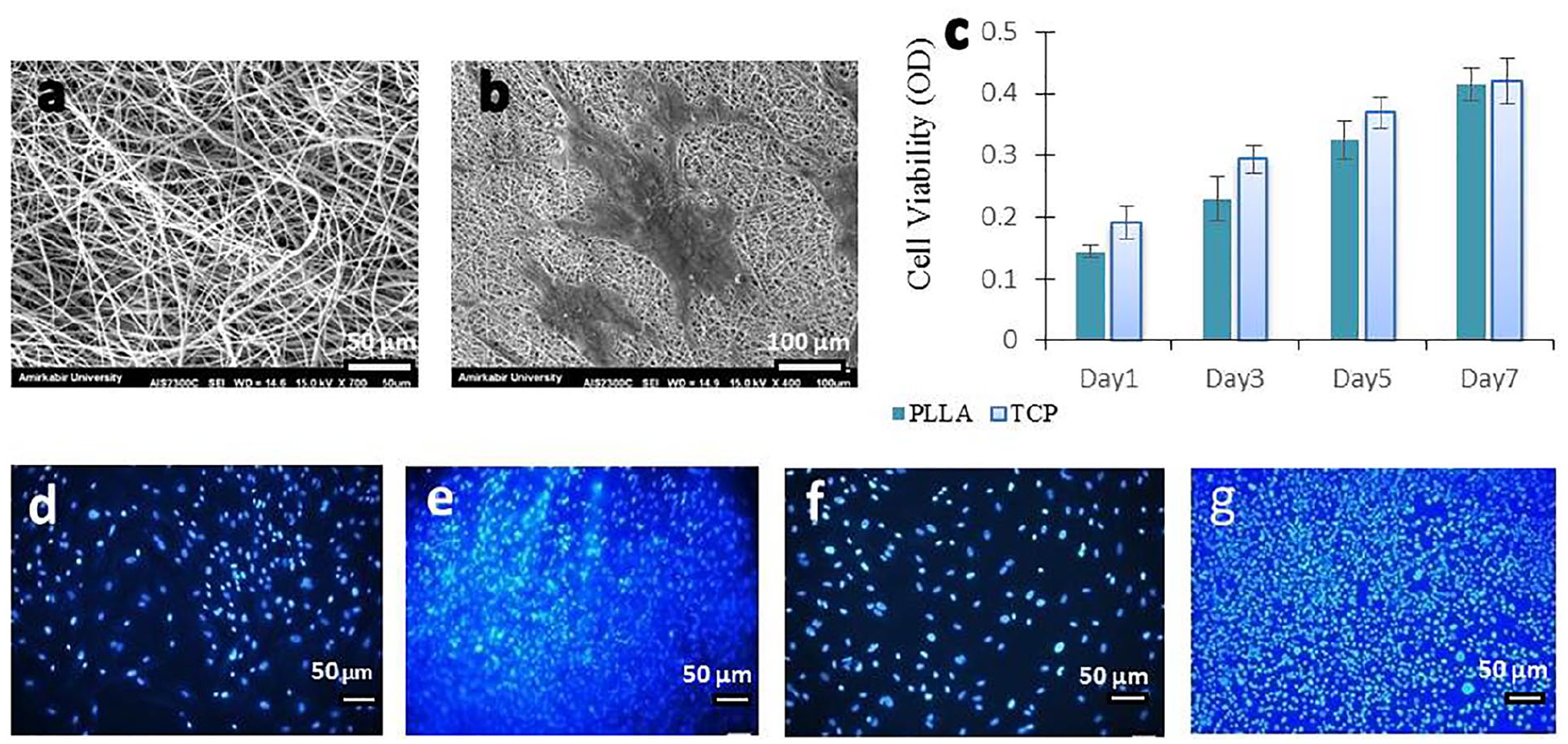
Scaffold characterization. (a) SEM revealed the scaffold’s porous structure, fibre’s diameter was estimated 994 ± 0.4 nm (scale bar: 50 μm). (b) MSC strongly attached and spread among the fibres (scale bar: 100 μm). (c) MTT illustrated the proliferation rate of cells on 1, 3, 5 and 7 days. No significant difference observed between PLLA attached cells and tissue cultured plate (TCP), applying an independent T-test (p < 0.05). Error bars represent the Standard Deviation of mean. (d, e) MSC cultivated on scaffold and plate, treated by DAPI after 24 h, (f, g) and 7 days demonstrated an increased number of nucleus in both groups (scale bars: 50 μm).
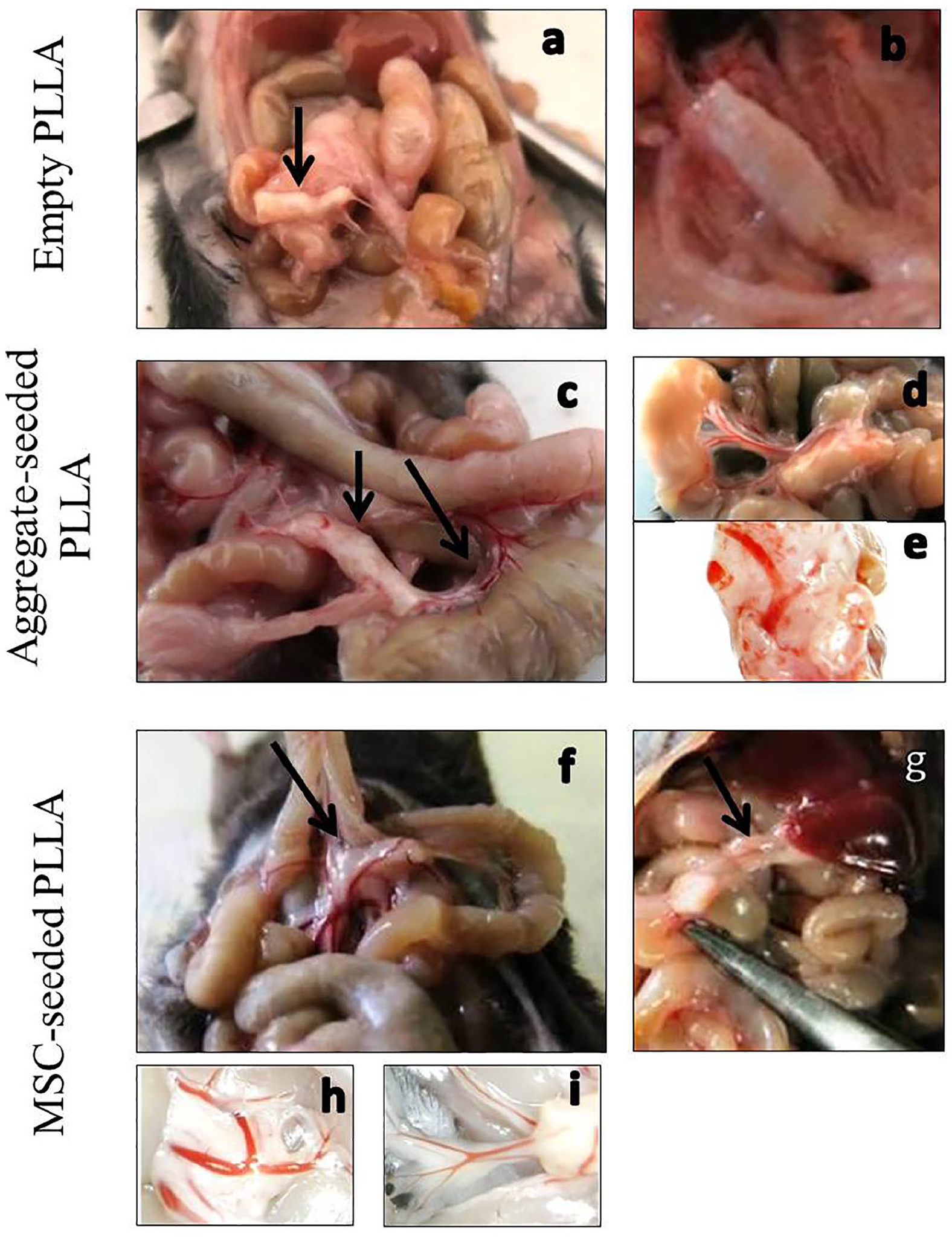
Morphological description of harvested transplanted scaffolds after 18 days. (a, b) Empty PLLA was poorly connected to other abdominal organs. (c, d) Aggregate-seeded scaffolds were connected to the organs and some vessels surrounded the structure from different sides especially the penetration of some vessels from holes in sides of the rolled scaffold was observed, (e) these vessels were attached to the scaffold and penetrated the construct. (f, g) MSC-seeded scaffolds were in good interaction with omentum vasculature and organs (h, i) many vessels penetrated and integrated in to them.
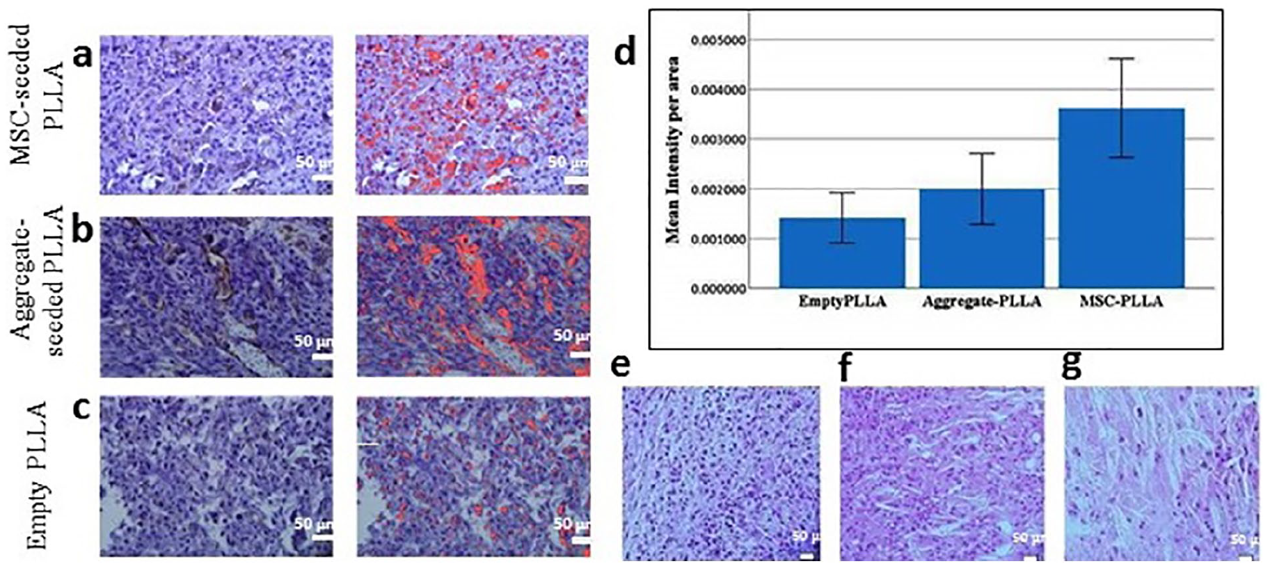
IHC, H&E, image analysis and statistical process. (a, b) More CD31+ cells (marked in red) comparing with empty scaffold (the scaffolds which transplanted empty with no seeded cells) were detected in MSC and aggregate seeded scaffolds. The structure of vessels and their CD31+ endothelial cells were apparent. (c) CD31+ areas of empty scaffolds (marked in red) were slight; (all scale bars: 50 μm). (d) The ratio of dyed area to the total area of image in percent, were analysed. The difference between per area means of MSC and aggregate-PLLA with empty PLLA were significant. p-Values less than 0.05 were appointed significant. Error bars represent Standard Deviation of mean. H&E staining demonstrated cell population spread within the scaffold fibres in (e) BM-aggregate seeded scaffolds (f) MSC seeded ones and (g) the empty scaffold which contained lower cell density in comparison with two other groups.
Discussion
According to morphological evidence, MSC and aggregate-seeded scaffolds contain abundant cellularity. Obvious connections with recipient vasculature and acceptable integration with surroundings were established that were not the same with empty PLLA. This apparent morphological difference besides the significant difference in CD31+ cells would indicate the angiogenesis potency of these cell sources and their efficient features to keep the viability of the implanted tissue. Our previous study agrees with these results, BM haematopoietic stem and progenitor cells niche-like units seeded in scaffold which existed 1 month in recipient showed native BM structure and efficient connection to the mouse vascular system was established while empty scaffolds were not connected to the recipient tissues and vasculature. 14 The existence of cell populations in our empty scaffolds insists on the acceptable features of the scaffold and might be relevant to the appointed duration that scaffolds existed in recipients. Considering the undeniable necessity of angiogenesis and effectual connection between implanted tissue and recipient’s vasculature in order to keep the viability of the implant and cell population it contains; it would be expected that empty PLLA with poor connections become ablated during longest periods as well as the cited study’s evidence. More studies with longer durations are recommended.
MSCs have been applied in many angiogenesis investigations and several aspects of this kind of cells have been evaluated; researchers have co-cultured MSCs with different kinds of cells, for example, endothelial outgrowth cells or human umbilical vein endothelial cells, in order to analyse their effect on network formation or vasculogenesis ability of those cells26–28; also they have been induced to differentiate to endothelial cells which are able to establish vessels. 22 MSCs have been used with special inductive agents or scaffolds in vitro and in vivo in order to promote vasculogenesis in many tissues. In this study we applied expanded BM-MSC and BM-aggregates without any inductive agent or co-cultured cells, to investigate their own angiogenesis potency in vivo. In a study Matrigel-embedded cultured-MSCs were implanted subcutaneously into mice; elicited a robust angiogenic reaction, leading to a remarkable increase in vessel density. Researchers stated the in vivo vascular differentiation of MSCs might be related to the interaction between the cells and matrigel. It should be considered that matrigel contained a wide variety of elements, any of which might provoke signals promoting endothelial differentiation. 17 In our study, we applied PLLA to make suitable matrices for cells and we hadn’t utilized differentiation-inductive agents.
Both aggregate-seeded and MSC-loaded scaffolds demonstrated angiogenesis tendency in order to establish numerous connections between PLLA scaffold and recipient’s vasculature, and considerable propensity to develop constructs containing CD31+ endothelial cells which can contribute to vessel’s tube formation. MSCs are multipotent adult stem cells that were used in tremendous researches and nominated as one of the most practical cell sources in many studies of regenerative medicine.29,30 MSC Cultivation and in vitro expansion are essential in order to achieve a sufficient amount of cells which is needed in most clinical and research protocols,12,31 while BM-aggregate is available in urgent cases with no need for expansion and passage which keep it acquitted from controversial subsequent of several expansions.32,33 Also its interaction with the microenvironment and other niche components may prevent unfavourable changes.12,13 Although the existence of several distinguishable vessel structures with CD31+ endothelial cells in aggregate contained scaffolds as well as the MSC ones, CD31+ percent area of the expanded MSC was higher. The amount of BM-aggregate which was seeded in the scaffold might modify this score and this issue should be involved, so we recommend applying BM-aggregates in different types of bioengineered scaffold furthermore optimizing the required concentration for further researches. Due to the notable osteogenic properties of BM-aggregates, we have introduced them as a suitable source for bone tissue engineering, 15 according to angiogenesis potency which they have shown in this study and for the aforementioned reasons, BM-aggregate would be introduced as an applicable and noteworthy cell source in bone tissue engineering and other fields of regenerative medicine. It seems more studies are needed to investigate various characteristics of BM-aggregate.
Footnotes
Declaration of conflicting
The author(s) declared no potential conflicts of interest with respect to the research, authorship, and/or publication of this article.
Funding
The author(s) disclosed receipt of the following financial support for the research, authorship, and/or publication of this article: Isfahan University of Medical Sciences, Isfahan, Iran (grant number: 394768).