
Abstract
Despite technological advances in mechanical circulatory support devices to treat end-stage heart failure, blood damage induced by non-physiological shear stress in operation often triggered clinical hemocompatibility complications. The loss of high molecular weight von Willebrand Factor (HMW-VWF) has been considered as an essential cause of gastrointestinal bleeding. In addition to the mechanics factors, interface factors may also affect blood damage, especially the surface characteristics. In this study, the effect of surface roughness on VWF damage under flow condition was investigated. A roller pump circulation experimental platform with a roughness embedded sample chamber was constructed to provide blood shearing flow condition. VWF molecular weight analysis, VWF antigen (VWF-Ag) concentration assay, and VWF ristocetin cofactor activity (VWF-Rico) assay were performed on the sheared blood samples. These variables are the main functional indicators of VWF. It was found that the surface roughness induced VWF damage is mainly caused by the loss of HMW-VWF rather than reducing the total amount of VWF. The threshold value of surface roughness for a rapid increase in the degradation of HMW-VWF under low flow rate was obtained between Ra 0.4 and 0.6 μm, which was smaller than the threshold for hemolysis. Our findings indicated that VWF is more sensitive to the interface factor of surface roughness than red blood cells, thus has a higher requirement for blood pump design. It could provide reference for the material design and processing in developing mechanical circulatory support devices.
Introduction
Heart failure (HF) affects nearly 26 million people worldwide, poses a growing burden on public health.1–3 Although mechanical circulatory support devices could help patients with HF to recover by providing hemodynamic support,4,5 it is often associated with clinical hemocompatibility complications, including hemolysis and thrombosis. The causes of these two complications have been confirmed as the damage of red blood cells and platelets, respectively. 6 With the development of blood pumps and the application of magnetic levitation technology, these two blood damage associated complications have been greatly improved. 7
However, in recent years, the gastrointestinal bleeding complication in the clinical use of blood pumps have been paid more attention. 8 Studies have shown that the degradation of high molecular weight von Willebrand Factor (HMW-VWF) induced by high mechanical shear stress in blood pumps is the main trigger of this syndrome.9,10 Acquired von Willebrand syndrome (AVWS), characterized by the loss in HMW-VWF, has recently been considered as one of the causes of gastrointestinal bleeding. 11 VWF is a large multimeric glycoprotein, which plays an essential role in the clotting process. When the blood vessel ruptures, the HMW-VWF adheres to the wound and expands into a chain, and then the chain captures platelets. A large number of platelets are mediated by VWF and eventually adhere to the collagen fiber to form thrombus, which could stem bleeding. Under the high mechanical shear stress, the HMW-VWF will expand into chains and be degraded into small molecules; thus the maximum coagulation efficiency cannot be exerted.12–14
In addition to mechanics factors, blood damage may also be affected by interface factors, including properties of bulk material and surface characteristics. At present, researchers mainly focus on topics that the blood damage caused by surface roughness and the effect of surface microstructure on platelet adhesion. Surface roughness is an irregular material surface microstructure that has been found to increase hemolysis and platelet adhesion. 15 Takami et al. 16 found that hemolysis was not affected when the surface roughness was less than Ra 2 μm under flow condition. They hypothesized that 2 μm is the minimum thickness of red blood cells. Maruyama et al. 17 investigated the influence of surface roughness on hemolysis by changing the rotor roughness using a blood-shearing platform of a rotational shear stressor. They concluded that when the surface roughness Ra reached 0.8 μm, the hemolysis value would increase sharply. Linneweber et al. 18 showed that the ratio of platelet adhesion increased by 40% and 76% respectively when the surface roughness increased from 0.05 to 0.2 and 0.4 μm.
Previous studies mainly focused on the damage of red blood cells and platelet. Besides, it was found that blood damage under static and flow condition was obviously different. Since blood will flow through the inner surface of working blood pump, investigating the influence of surface roughness on VWF under flow condition was necessary. In this study, a roller pump circulation experimental platform with a roughness embedded sample chamber was constructed to provide blood shearing flow condition. VWF molecular weight analysis, VWF antigen (VWF-Ag) concentration assay, and VWF ristocetin cofactor activity (VWF-Rico) assay were then performed on the sheared blood samples. The results of this study are expected to provide guidance and reference for the surface roughness design of blood pumps.
Materials and methods
Blood collection
Porcine blood was collected from Taihu white pig (Changshu, China) at the Experimental Animal Center of Soochow University, which had passed the ethical review of animal experiments of Soochow University. One unit of blood (400 mL) was drawn from the artery and directly collected into a sterilized blood bag (HnG Medical Incorporated, Ontario, Canada) mixed with 0.4% heparin sodium (Yuanye Bio-Technology, Shanghai, China) at a ratio of 1:20. The blood bag was gently shaken to mix the blood with anticoagulant fully. To prevent coagulation, the activated coagulation time (ACT) of blood was controlled to be >300 s. 19
Experimental circuit
A custom-made roller pump was designed to satisfy the experimental requirements, which were mainly composed of pump body and pump pipe (Figure 1(a)). An experimental circulation platform was constructed with the roller pump, 1 m tubing with the inside diameter of 3/8 inch, and a blood bag (Figure 1(c)). Flat grinding was used to manufacture small pieces with different roughness on material surfaces. The grinding material was made of stainless steel. The piece was embedded with its processed face inwards into a 3D printed tubing connector (Figure 1(b)), then connected to the circulation loop. The diameter of tubing connector was the same as tubing. When the roller pump was turned on, blood flowed continuously through the surfaces of roughness samples. According to the results of calculation and simulation, the flow state in the roughness sample chamber was strict laminar flow.
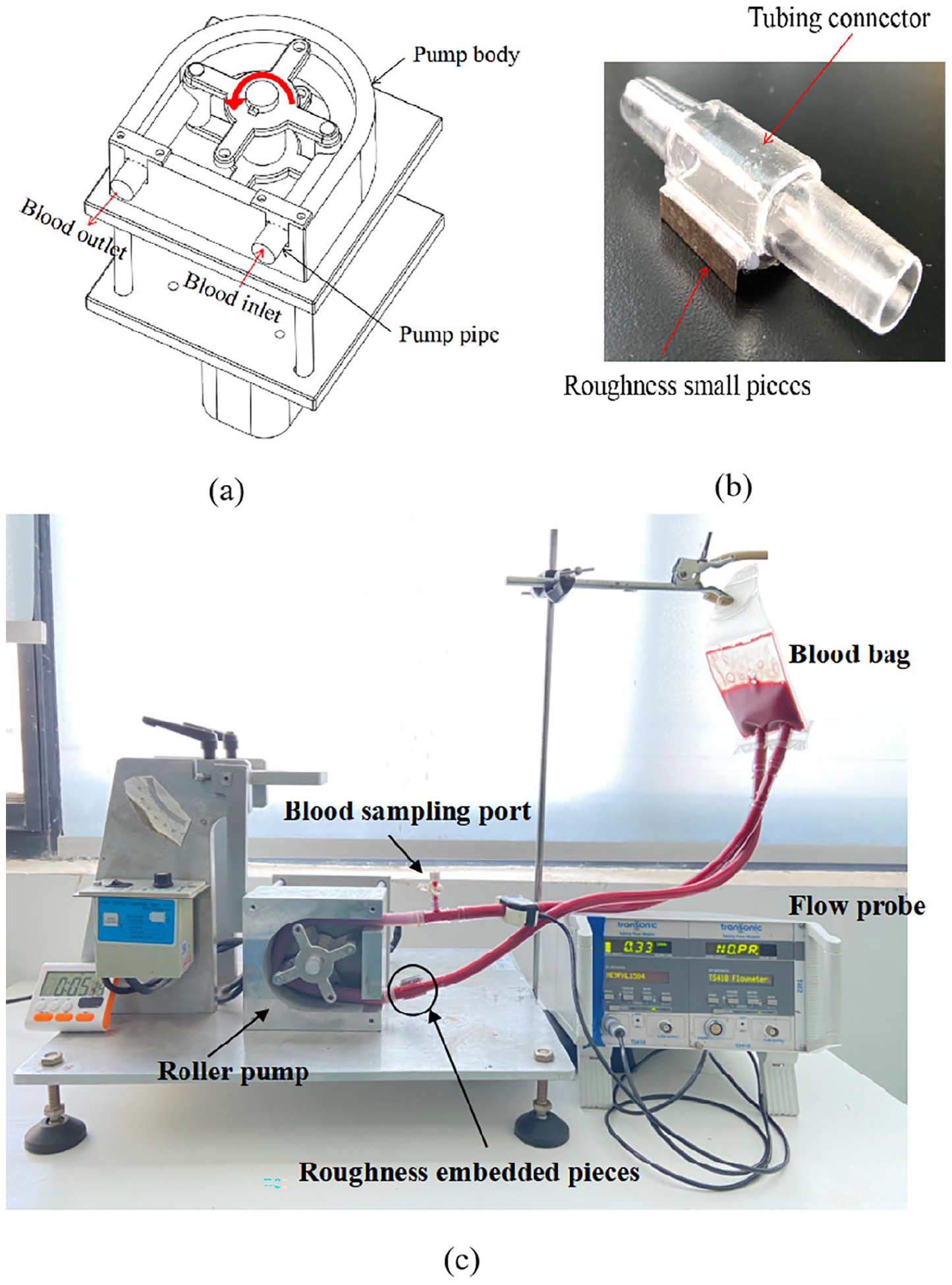
Assembly drawing of the custom-made roller pump (a), roughness embedded sample chamber (b), and roller pump circulation experimental platform (c).
During the experiment, 150 mL porcine blood was primed into the circuits. The hematocrit (HCT) of blood was 31%. 20 All air bubbles were carefully removed before circulation. The blood bag was hung upside down so that the blood could be mixed well by gravity. The flow rate was set as 0.35 ± 0.02 L/min, monitored in real-time by a flow probe (Transonic System, NY, USA). The roughness sample was arranged at the inlet of roller pump, and the blood sampling port was placed at the outlet of roller pump. The blood in the circuits was circulated continuously for 1 h at room temperature. Blood samples were collected through the sample port at time zero and every 10 min of pumping. In this research, four groups of circulating experiments were conducted, in which four small piece samples with different surface roughness were embedded into the loop respectively. Blood samples collected were then centrifuged at 4000 rpm for 10 min. The supernatant was taken out and stored at −20°C for subsequent experiments.
HMW-VWF analysis
Collected platelet-poor plasma was mixed with sample loading buffer (1:20). SDS-agarose gel electrophoresis was used for separating VWF multimers for 0.5 h at a current of 30 mA and then 3 h at a current of 50 mA. After electrophoresis, the protein ladders were transferred onto a 0.45 μm polyvinylidene difluoride membrane (Immobilon-P; Millipore Corporation, Bedford, MA, USA) overnight at a current of 70 mA. Then, primary antibody (Polyclonal Rabbit Anit-Human Von Willebrand Factor, Cell Signaling Technology, Boston, USA) (1:4000 dilution) and secondary antibody (Polyclonal Goat Anti-rabbit lgG HRP-linked Antibody, Massachusetts, USA) (1:2000 dilution) were used to detect VWF multimers. Luminous substrate (20 × LumiGLO Reagent and 20 × Peroxide, Cell Signaling Technology, Boston, USA) was added to the membrane, making the film sensitive to light. Finally, through development and fixing, bands can be displayed on the film. Image-J software was used to process the VWF bands image obtained from the Kodak film (Rayco Medical Products Company Limited for Carestream Health; Xiamen, China) development. Western blot bands from 1 (lowest molecular weight band) to 8 were classified as low to medium molecular weight VWF, while all the bands above 8 as HMW-VWF. 21 The section of HMW-VWF was selected to acquire the gray value corresponding to each sample. The degradation of HMW-VWF was expressed as,
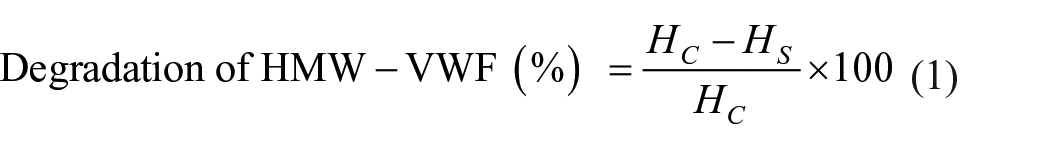
Where Hs is the gray value of HMW bands corresponding to the blood sample at each time point, and Hc is the gray value of HMW bands corresponding to the initial sample at time zero.
VWF antigen and ristocetin cofactor activity assays
The leading functional indicators of VWF include VWF antigen (VWF-Ag), VWF ristocetin cofactor activity (VWF-Rico), and VWF ristocetin activity: antigen ratio (VWF-Rico: VWF-Ag). VWF-Ag and VWF-Rico were quantified by the enzyme-linked immunosorbent assay (ELISA) (ELISA; von Willebrand Factor Test Kit; Nanjingjianchengbio, China). VWF-Ag was used for measuring the level of total VWF in plasma. VWF-Rico was used for measuring the ability of plasma to agglutinate platelets in the presence of the antibiotic ristocetin. VWF-Rico: VWF-Ag is a comparison of platelet binding activity to the concentration of VWF, which demonstrates a change of functional activity compared to antigen concentration. The whole process of ELISA included five main steps: sample addition, incubation, HRP-conjugated antibodies application, color development, and terminated liquid addition.18,19 After these steps, the absorbance was measured using a microplate reader (Molecular Devices, California, USA) at a wavelength of 450 nm. The standard curve linear regression equations between standard liquid concentration and absorbance for the VWF-Ag and VWF-Rico were established by ELISAcalc software. Then the VWF-Ag and VWF-Rico in the plasma samples were calculated according to the regression equation respectively, followed by the VWF-Rico: VWF-Ag.
Statistical analysis
Three valid measurements were obtained from repeated tests of all the surface roughness groups. The averages and standard deviations were calculated using a data analysis software (Origin version 9, OriginLab, Northampton, MA, USA). Due to multiple measurements taken from each surface roughness group, statistical analysis was carried out using Student t-test. A value of p < 0.05 was considered to be statistically significant.
Results
Roughness profiles and Ra values
The surface roughness (Ra values) of small pieces obtained by flat grinding were measured with a surface texture-measuring instrument (Tokyo Seimitsu Co., Ltd., Tokyo, Japan). The measurement length of the small piece sample was 0.25 mm. The roughness profiles for the surface were shown in Figure 2. In this research, four kinds of small piece samples with different roughness were obtained, namely Ra 0.1, Ra 0.2, Ra 0.4, and Ra 0.6 μm.
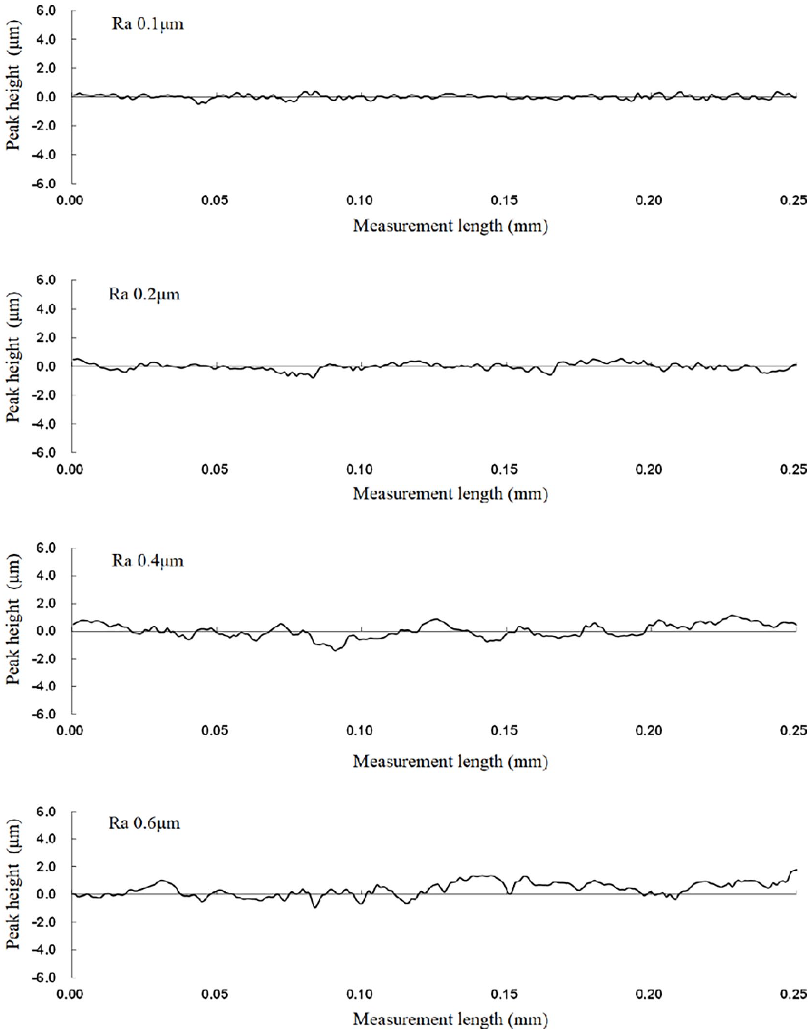
Roughness profiles for surface roughness ranging from Ra 0.1 to 0.6 µm.
HMW-VWF analysis
The images of VWF multimer bands for different surface roughness obtained by immunoblotting were shown in Figure 3. The section in the dotted box represented the HMW-VWF. It demonstrated that the HMW-VWF degradation did not change obviously with time except for Ra 0.6 μm sample (p > 0.05). According to the images, the exact values of HMW-VWF degradation were calculated and shown in Figure 4. The highest degradation of HMW-VWF in Ra 0.1, Ra 0.2, and Ra 0.4 μm groups was below 5%, which was at a shallow level. Besides, the HMW-VWF degradation of these three groups fluctuated with time during 1-h circulation, indicating that the loss of HMW-VWF did not increase significantly with time. The situation that HMW-VWF degradation was negative at 60 min for Ra 0.4 μm group may be due to the lower image resolution, which did not affect the overall outcome. On the contrary, the degradation of HMW-VWF for Ra 0.6 μm group significantly increased with time. It was also obviously higher than other groups at each time point, which had already reached 11.4% in 20 min. The highest degradation of HMW-VWF reached 27.7% at 60 min, which was even more than seven times higher than that of other groups. To sum up, when the surface roughness increased to Ra 0.6 μm, the degradation of HMW-VWF became significantly higher and increased with time.
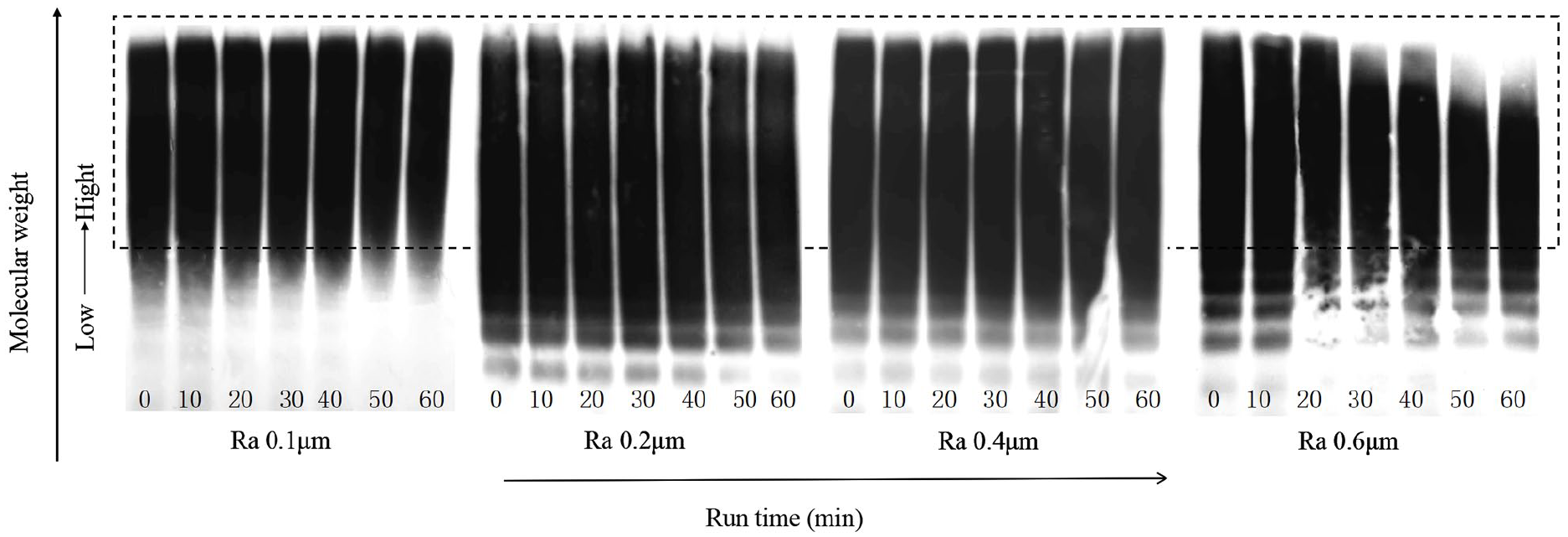
The image of VWF multimers for different surface roughness.
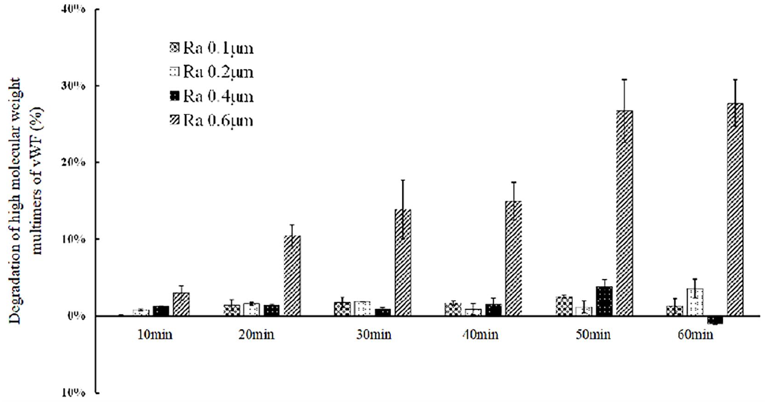
Degradation of HMW-VWF with time under different surface roughness.
VWF antigen and ristocetin cofactor activity assays
The VWF-Ag concentration of each sample was shown in Figure 5(a). It can be clearly seen that the VWF-Ag concentration of each group did not change significantly with time. For all the samples, the concentration of VWF-Ag fluctuated with time, with a change of ±100 U/L during the whole circulating process. The result of VWF-Rico activity was shown in Figure 5(b). In general, the VWF-Rico activity in all the groups showed a decreasing trend over time. At all time points, level of VWF-Rico activity in group Ra 0.6 μm was >15% lower compared to other groups. Then, the ratio of VWF-Rico activity and VWF-Ag concentration was calculated throughout the entire experiment, the results of VWF-Rico: VWF-Ag were shown in Figure 5(c). It demonstrated that VWF-Rico: VWF-Ag did not change significantly with time except for the Ra 0.6 μm group. The reduction of VWF-Rico: VWF-Ag in Ra 0.1, Ra 0.2, and Ra 0.4 μm groups were <5% throughout the entire circulation process. On the contrary, the VWF-Rico: VWF-Ag in Ra 0.6 μm group decreased >13% during the whole 60 min. Furthermore, it was also obviously lower than other groups at each time point, and this disparity reached the maximum (12.3%) at 60 min.
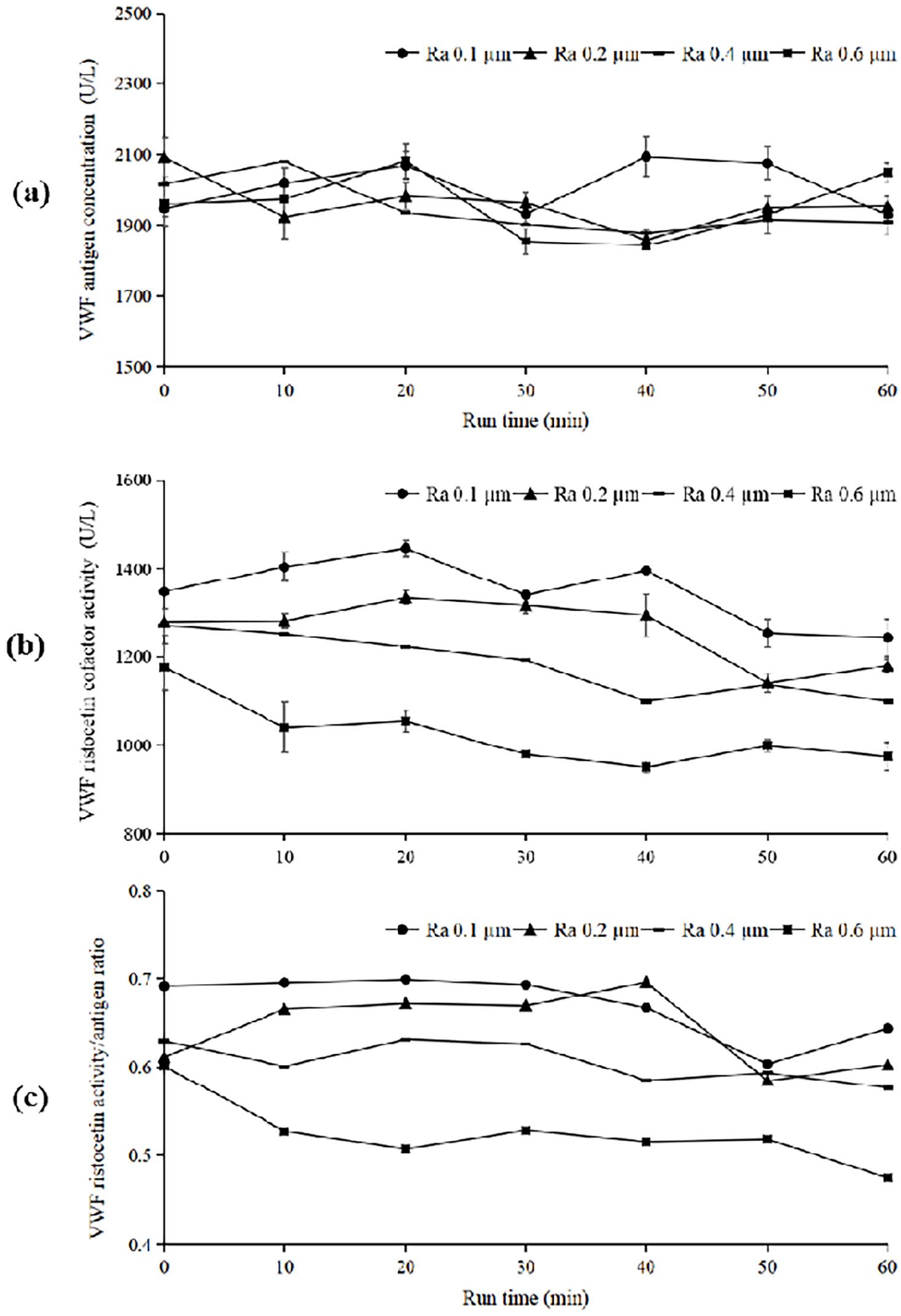
Serial outcomes in functional assays of VWF-Ag (a), VWF-Rico (b), and the ratio of VWF-Rico to VWF-Ag (c).
Discussion
A roller pump circulation platform was designed to investigate the damage of VWF resulting from surface roughness under flow condition in this study. Related literature 22 proposed that roller pump was not suitable for the study of hemolysis, because the hemolysis caused by roller pump itself was so serious that the hemolysis caused by surface roughness can be ignored. However, according to our previous study, the custom-made roller pump did not cause obvious damage to VWF. We let the roller pump drive the blood to circulate in the loop without roughness sample for 60 min, and found no significant degradation of HMW-VWF (Supplemental Figure 1S). Therefore, the roller pump circulation platform could be used to study the law of VWF damage caused by surface roughness under flow condition. This may be due to the different mechanism of red blood cells and VWF damage. The red blood cell damage is caused by the rupture of cell membrane, while the damage principle of VWF is that the HMW-VWF was unfolded, then cleaved by ADAMTS13 (von Willebrand factor-cleaving protease) into VWF fragments. 23 Therefore, continuously squeezing of the pump pipe in the roller pump will rupture the red blood cells, but may not unfold the HMW-VWF chain to further cause cleavage.
Certain surface roughness can cause damage of VWF under flow condition. The reduction of VWF-Rico: VWF-Ag in Ra 0.1, Ra 0.2, and Ra 0.4 μm groups were less than that in Ra 0.6 μm group throughout the entire circulation process. The degradation of HMW-VWF in Ra 0.6 μm group increased significantly with time, while other groups of smaller surface roughness did not. It indicated that the reduction of VWF-Rico: VWF-Ag was consistent with the degradation of HMW-VWF. This may be due to the fact that only the HMW-VWF plays a role in coagulation process, and the loss of HMW-VWF means the reduction of coagulation efficiency.
The results of HMW-VWF degradation showed that there was a threshold value of surface roughness for VWF damage, which was between Ra 0.4 and Ra 0.6 μm. In the literature of hemolysis, there was also a threshold for red blood cell damage caused by surface roughness (between Ra 0.6 and Ra 0.8 μm). 17 It indicated that VWF is more sensitive to the interface factor of surface roughness than red blood cells. The reason may be that a rough surface not only brings higher shear stress, but also makes it easier for VWF to adhere to the irregular grooves on surface. Especially, since the flow rate was 0.35 ± 0.02 L/min in this study, which was much smaller than other kinds of circulation platforms, the threshold revealed may not be the threshold of VWF damage caused by shear stress but the threshold of VWF adhesion. Therefore, higher surface roughness could increase the adhesion of VWF, which allows HMW-VWF to expand longer at lower shear stress and thus more easily be cleaved into small segments. In order to minimize the degradation of HMW-VWF, the surface roughness of mechanical circulatory support devices should be controlled less than Ra 0.4 μm.
Limitations and future directions
There were also some study limitations and future directions. VWF damage could be related to the profile of roughened surface. For the surfaces with same Ra, the maximum height of profile (Rz) and the maximum profile peak height (Rp) of them may be different. Therefore, the Rz and Rp may also be involved to evaluate surface roughness in the future. Besides, surface roughness is an irregular surface microstructure that is randomly distributed. Hence, we planned to design regular microstructures on surface to accurately control the near-surface flow field and study the influence on blood damage. In addition, the flow rate provided by the roller pump in this study was 0.35 ± 0.02 L/min. We planned to study the damage of VWF under larger flow rates, so as to more comprehensively illustrate the mechanism of surface roughness affecting VWF damage.
Conclusion
This study illustrated that the roller pump circulation platform can be used to investigate the damage of surface roughness on VWF under flow condition. The results shown here indicated that there was a threshold of surface roughness between Ra 0.4 and Ra 0.6 μm for VWF damage under low flow rate, which was smaller than the threshold for hemolysis. In addition, there was a strong Positive correlation between the degradation of HMW-VWF and the decrease of VWF-Rico. This work could provide guidance and reference for the surface design and improvement of mechanical circulatory support devices. It is highly recommended to pay attention to surface roughness of the devices while conventionally focusing on structural optimization.
Supplemental Material
sj-tif-1-jao-10.1177_03913988211056961 – Supplemental material for The influence of surface roughness on the damage of von Willebrand Factor under shear flow condition
Supplemental material, sj-tif-1-jao-10.1177_03913988211056961 for The influence of surface roughness on the damage of von Willebrand Factor under shear flow condition by Xu Mei, Bin Lu, Min Zhong, Yuxin Zhu, Liudi Zhang and Wanning Ge in The International Journal of Artificial Organs
Footnotes
Declaration of conflicting interests
The author(s) declared no potential conflicts of interest with respect to the research, authorship, and/or publication of this article.
Funding
The author(s) disclosed receipt of the following financial support for the research, authorship, and/or publication of this article: This work was supported by the National Natural Science Foundation of China (grant number 31700817); the China Postdoctoral Science Foundation (grant number 2017M620224) and the Natural Science Foundation of Jiangsu Province (grant number BK20170352).
Supplemental material
Supplemental material for this article is available online.
References
Supplementary Material
Please find the following supplemental material available below.
For Open Access articles published under a Creative Commons License, all supplemental material carries the same license as the article it is associated with.
For non-Open Access articles published, all supplemental material carries a non-exclusive license, and permission requests for re-use of supplemental material or any part of supplemental material shall be sent directly to the copyright owner as specified in the copyright notice associated with the article.