
Abstract
The development of natural-based wound dressings is of great interest in the field of skin tissue engineering. Herein, different bioactive molecules such as gelatin (GEL), chitosan (CH) and mucilage (MU) were used to prepare a wound dressing. The physico-chemical and biological characterizations occurring after γ-irradiation were investigated. Results showed that Electron Paramagnetic Resonance (EPR) spectroscopy of un-irradiated GEL-CH-MU biomaterial showed two paramagnetic centers which correspond to g = 1.89 and g = 2.033. A generated new active center appeared at g = 2.003 at 25 kGy due to the interactions of gamma rays with the polymer chain creating signals at the absorbing functional groups. X-ray diffraction (XRD) spectra preserved the semi-crystalline structures between a range of 2θ (5° and 45°). Fourier Transform Infrared spectroscopy (FTIR) revealed that the initiation of cross linking phenomena. Moreover, γ-rays significantly increased antioxidant activity (9.1 ± 0.07%, p < 0.05) and exhibited a high anti-inflammatory activity (70%) at 25 kGy. Significant antibacterial activities in vitro liquid medium was observed. In addition GEL-CH-MU dressing exhibited high hemocompatibility. Conducted investigations state that such innovative dressing natural-based polymers for advanced wound care may be considered as useful for biomedical purposes.
Introduction
Gelatin (GEL) is a biopolymer with interesting properties due to low antigenicity and good biocompatibility to the physiological environment. 1 GEL-based dressings act as porous materials for cell migration and are known to promote tissue epithelialisation.1,2 However, using GEL is limited due to its fast biodegradation and reduced mechanical properties. 2 Recently, the formulation of GEL-based macroporous hybrid hydrogels by the polymers incorporation such as Polyethylene glycol (PEG), Hydroxyethyl Cellulose (HEC), and chitosan (CH) proved outstanding structural permanence as well as good mechanical stability and reversibility. 3 Herein, the incorporation of CH, a glycosaminoglycan-like biodegradable polymer, into GEL-based scaffolds may help in surmounting the potential restrictions of GEL matrices. CH is considered as a bioactive substance with reactive functional groups such as amino/acetamido group, as well as hydroxyl group. 4 Due to its hemostatic, antimicrobial, nontoxic, and biocompatible properties, CH has been widely used as a versatile biopolymer for orthopedic tissue-engineering 5 as well as in dermatology as a dressing for wounds. 4 Traditional wound dressing products are dry and used for protecting the wound from contaminations. 6 However, this type of dressing must be changed frequently and it causes maceration of healthy tissues. For that, bioactive wound dressings are designed to prevent dehydration and promote healing rather than simply covering the wound. Many bioactive substances such dextran were used not only in orthopedics to activate the bone marrow human mesenchymal stromal cells 7 but also in dermatology to enhance angiogenic responses and promote complete skin regeneration during burn wound healing. 8 Herein, bioactive polysaccharide called Mucilage (MU) was used. MU of Opuntia ficus-indica a rich carbohydrate-containing polymer was chosen for dressing preparation. MU is attractive due to its high ability to maintain a moist environment and to reduce bacterial infections. 9 It has been reported to accelerate the regeneration process by promoting neovascularization, collagen synthesis, wound contraction and tensile strength. 10 Ionizing radiation caused changes at the molecular level. Therefore, it is important to evaluate radiation effects at 5–25 kGy doses, which are normally used in the sterilization of medical products. 11 To the best of our knowledge, the synthesis, characterization, and the study of γ–radiation effects on the bioactive wound dressing starting from the present renewable resources is non-investigated. The purpose of this research is to evaluate the GEL-CH-MU biomaterial as an innovative wound dressing. The adhesive nature, together with the therapeutic characteristics may make it as promising biomaterial for wound treatment.
Material and methods
Preparation of GEL-CH-MU wound dressing
The solutions of 5% w/v were prepared by dissolving 5 g of GEL from Bovine skin (powder, gel strength ~225 g Bloom, Sigma-Aldrich, USA) in 100 ml of distilled water (99% purity, conductivity <5 µS/cm) for 30 min. Then, the solution heated at 50°C for 30 min under continuous stirring. CH (DDA~90%, viscosity Sigma-Aldrich, USA). CH solutions with concentrations of 1 wt% were prepared by dissolving in 1% acetic acid (99.8%, Sigma-Aldrich, Darmstadt, Germany) and stirred overnight at room temperature. Nopal cladodes Nopal cactus (O. ficus-indica) were harvested in a single collection and extracted as described previously. 12 Ethanolic solution (purity, ⩾99.8%, Sigma-Aldrich, USA) of 1% of MU was added to the mixture. All the polymer solutions were filtered through 0.55 µm pore-size PTFE membrane filters to remove impurities.
Gamma irradiation and electron paramagnetic resonance spectroscopy (EPR)
The irradiations were performed at the Tunisian Cobalt-60 gamma irradiation facility with energies of 1.173 and 1.332 MeV at a dose rate of 36 Gy/min. The dose rate was determined using Fricke dosimeter. EPR spectra were recorded on a Bruker ER-200D spectrometer operating at 9.8 GHz X-Band frequencies with modulation amplitude of 0, 2 mT, modulation frequency of 100 kHz, sweep width of 210 mT and microwave power of 63 mW.
Fourier transform infrared (FTIR)
The measurement was recorded at room temperature by Vertex 70 infrared spectrometer from 4000 to 400 cm−1 with a resolution of 4 cm−1 at a spectral resolution of 2 cm−1 and 32 scans.
The X-ray diffraction (XRD)
The analysis was conducted using Brucker D8 advance with Cu-Kα radiation of wavelength λ = 1.541 Å in 2θ values in the range of 15° to 90° with a step size of 0.02° and counting time of 12 s per step. The results obtained were analyzed with the X’Pert High Score Plus program.
Measurement of antioxidant activity
The free radical-scavenger activity of different composites were determined by the 1,1-diphenyl-2-picrylhydrazyl (DPPH) assay, as specified by Santhosh et al. 13
Antibacterial activity test
Staphylococcus aureus (ATCC25923) and Escherichia coli ATCC25922 a positive and negative gram bacterium, respectivily. From a young culture on agar, a dense bacterial suspension superior to 105 Colony-forming unit (CFU) cells/ml) is prepared and achieved as described perviousely. 14
In vitro hemocompatibility
Hemolysis
The hemostatic performance of the composites was tested in vitro as described previously. 15 800 µl of the composites at different concentrations were incubated with 200 µl of fresh goat whole blood (whole blood, normal saline = 8: 10) at 37°C for 1 h.
Blood anticoagulant indice
Blood anticoagulant indice was investigated according to method reported by He et al. 16 with minor modification.
In vitro anti-inflammation activity
Effect of the composites on heat-induced bovine serum albumin (BSA) denaturation assay was carried out using a method described by Chandra et al. 17 with minor modifications.
Statistical analyses
All results are presented as means (n = 3) ± standard deviations (SD). Multiple comparisons were performed using analysis of variance (ANOVA) followed by Tukey’s range test. The probability value of p < 0.05 was considered significant.
Results
EPR analysis
EPR spectroscopy detects paramagnetic substances such as radicals. Signal intensity reflects the total absorbed energy of samples under resonance conditions. As shown in Figure 1, at 5 kGy, GEL-CH-MU biomaterial generated a radical induced by irradiation on left side around the sharp peak of the center field and 1 peak on the right side (high magnetic field region) was detected at g =1.89 and g = 2.033. Exposed to 10, 15, and 20 kGy, a new signal was detected at g = 2003. This signal is attributed to the presence of Mn2+. At 25 kGy, no changes, such as broadening of the line width, or modifications in the intensities, or new superimposed spectrum were observed.
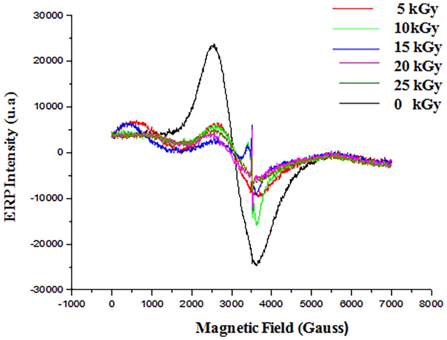
EPR spectrum of irradiated gelatine-chitosan-Mucilage (GEL-CH-MU) composite.
FTIR analysis
The functional group bands are characteristics of proteins and polysaccharides (Figure 2(a)). The signal at 1260 cm-1 is assigned to the bending vibrations of hydroxyls present in each of GEL, CH, and MU. The amide functional group combines the features of amines and ketones because it has both the N-H bond and the C=O bond. So the chemical shift demonstrated that the absorption for N–H was shifted to higher wave number 3450 cm-1 for un-irradiated composite to 3 485 cm-1 for 25 kGy dose, respectively. The absorption band at C=O stretching (amide I) for GEL-CH-MU was attributed to higher wave number at 1.638 cm-1 for the same dose. Also the biomaterial assigned a band around 2917 cm −1 to the C–H stretching vibration of the pyranose group shifted to 2950 cm −1 at 25 kGy. The bands at 1047 and 1036 cm−1 indicate the presence of monosaccharides such as mannose and glucose in pyranose ring conformations for the un-irradiated and the irradiated at 25 kGy, respectively. This increase is driven from cross linking network composed of polymer chains. The high intensity of absorption is due to the low cristallity. 18 This later behavior will be studied with XRD analysis.
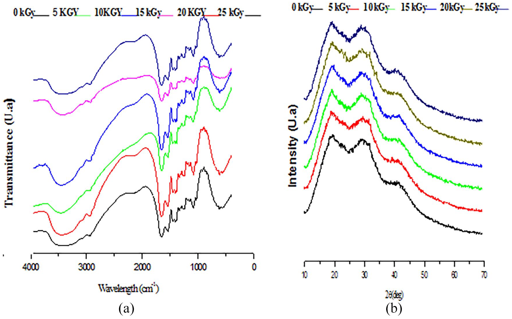
FTIR spectrum (a) and X-ray diffractograms (b) of irradiated gelatine-chitosan-Mucilage (GEL-CH-MU) composite.
XRD study
XRD pattern of GEL powder showed amorphous morphology with a characteristic broad hump in the 2θ range of 15–30 (Figure 2(b)). These characteristic peaks are usually assigned to the triplehelical crystalline structure in GEL. 19 The XRD reflection of CH revealed a sharp crystallographic peak at 2θ = 20°. 20 MU exhibited a semi-crystalline character pattern pronounced diffraction peaks at 2θ = 20.03 and 32.20. 21 After irradiation, the crystallization behavior through XRD analysis showed that the irradiation did not break the originally ordered atomic arrangement of GEL-CH-MU composite up to 25 kGy dose.
Antioxidant activity
Irradiation increased significantly antioxidant activities of the GEL-CH-MU dressing observed at doses ranging from 5 to 25 kGy (Figure 3). Particularly, data showed that all the experimented dressings treated with 25 kGy had a significant ability to reduce the stable purple colored radical DPPH into yellow-colored DPPH-H which increased up to 9.1 ± 0.07% (p < 0.05) at 5 mg/ml as compared to corresponding un-irradiated samples.
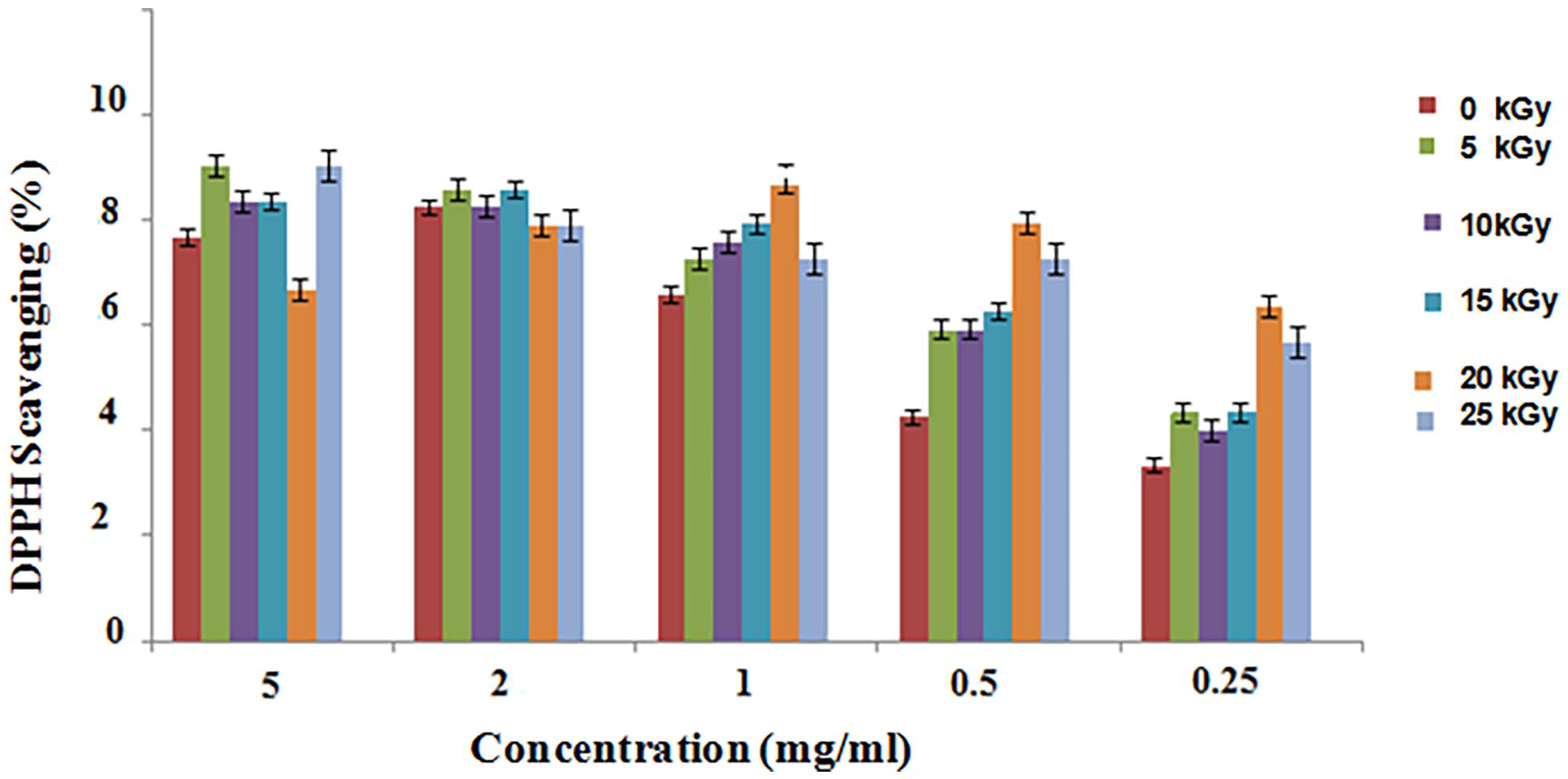
Antioxidant activity of irradiated gelatine-chitosan-Mucilage (GEL-CH-MU) composite.
Antibacterial activities
The evaluation of the antibacterial activities of GEL-CH-MU dressing on the culture medium of E. coli and S. aureus strains showed bacterial growth inhibition (Figure 4(a) and (b)). These new materials showed the best antibacterial activity at 25 kGy with an inhibition rate of 88% and, 93.33%, respectively against E. coli and S. aureus after 30 h of contact in a strong synergistic effect between CH, GEL and MU. The inhibitory effects were highly significantly different from those of un-irradiated dressings (p < 0.01).
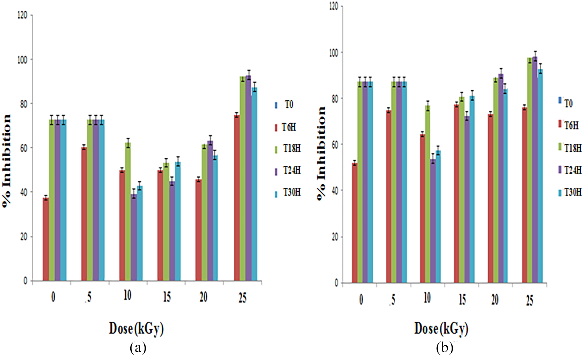
Antibacterial activities of irradiated gelatine-chitosan-Mucilage (GEL-CH-MU) composite against E. Coli (a) and S. aureus (b).
In vitro hemocompatibility
Hemolysis assay
After keeping in contact with red blood cells for 1 h, the hemolysis ratio of GEL-CH-MU material decreased from 0.185% to 0.05% when the gamma ray doses increased from 0 to 25 kGy (Figure 5(a)). At this later dose, GEL-CH-MU material revealed highly significantly lower levels of hemolysis as compared to that of GEL-CH material (p < 0.01). All the Hemolysis rates caused by GEL-CH-MU and GEL-CH dressing under different doses, indicated that dressings were not hemolytic according Standard Practice for Assessment of Hemolytic Properties of Materials F756-93 standards. 22
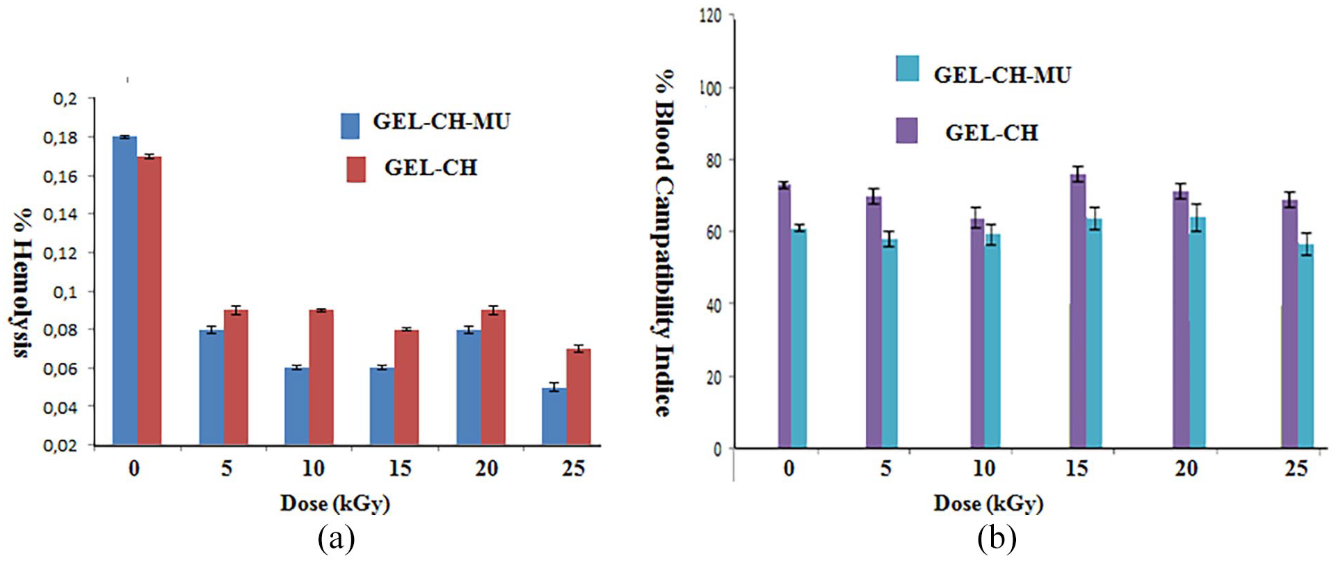
Hemolysis properties of irradiated gelatine-chitosan-Mucilage (GEL-CH-MU) (a) and Blood Coagulation Index (b).
Blood Coagulation Index (BCI)
The hemostatic potential of the newly fabricated GEL-CH-MU dressing was examined by whole blood clotting index (BCI) assay. According to Figure 5(b), at 60 min the % of BCI obtained for GEL-CH-MU revealed significantly lower BCI levels as compared to that of GEL-CH material at 25 kGy (p < 0.05). The results indicate a more desired clotting efficiency with GEL-CH-MU dressing. The incorporation of MU ameliorated blood clotting index (BCI) assay.
In vitro anti-inflammatory assays
After irradiation, GEL-CH-MU dressing significantly (p < 0.05) exhibited the highest inhibition of heat-induced BSA denaturation at 25 kGy. The inhibition % of protein denaturation was ranged from 12.0% to 70.0% at gamma ray doses range from 0 to 25 kGy. The GEL-CH-MU biomaterial exhibited a significantly higher (p < 0.01) level of anti-inflammation effect after irradiation as compared with that of GEL-CH dressing (Figure 6).
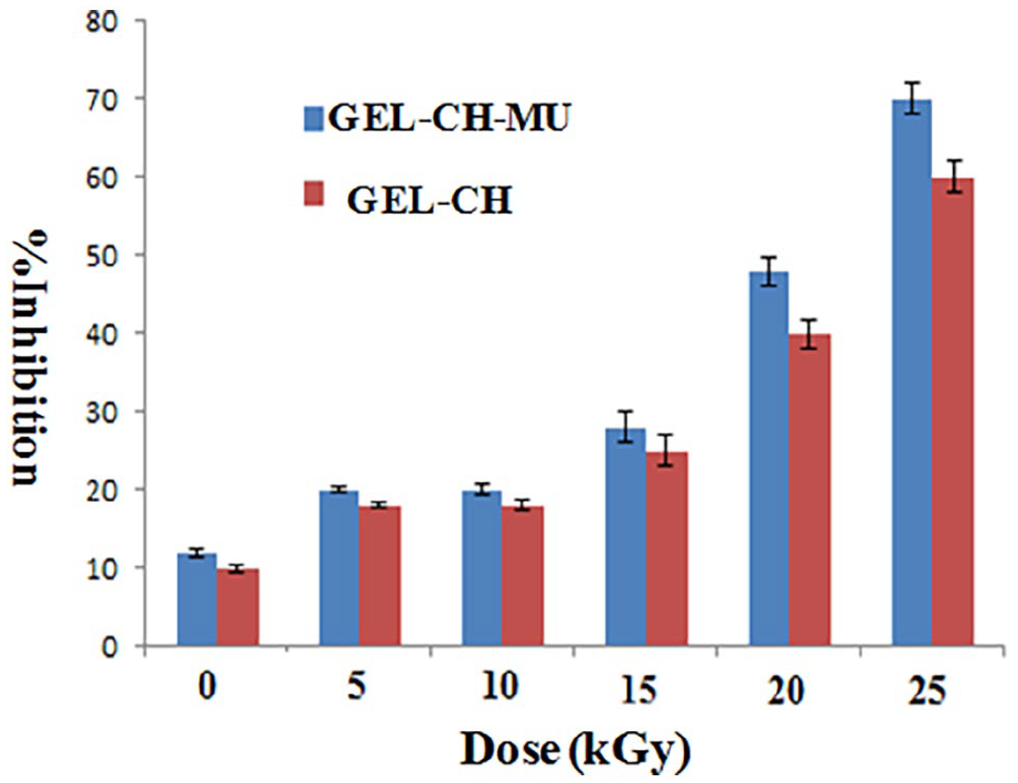
Anti-inflammatory properties of irradiated gelatine-chitosan-Mucilage (GEL-CH-MU) composite.
Discussion
A novel wound dressing based on GEL, CH, and MU as bioactive biopolymers was developed and characterized. EPR spectroscopy detects paramagnetic substances such as radicals from 5 up to 25 kGy. Signal intensity reflected the total absorbed energy of samples under resonance conditions. The Formation of these radicals can be explained by the fact that GEL-CH-MU composite contains alcohol, glycosidic, acetal oxygen, carboxyl, amino, hydroxyl and carbonyl groups. Moreover, this illustration shows that there is little amount of paramagnetic metals interfering slightly in the signal of the free radical. This signal is attributed probably to the presence of Mn2+. The minerals can be derived from sea metal incorporated in CH and also they can be contained in MU composition. 23 Herein, the amount of Mn is about 0029 and 0094 ppm in CH and MU, respectively. These amounts are remarkably inferior to the body’s tolerance level set at about 0, 1 4 mg kg-1 day-1. 24
Mn element plays a vital role in extracellular matrix synthesis and shows great potential in tissue regeneration. One study demonstrated that a composite hydrogel, which contained bioactive ions Mn2+, significantly accelerated the wound healing process and proved a significant immunomodulation.25,26 On the other hand, the FTIR spectrum proved that shift of the OH stretching peak of the GEL-CH-MU biomaterial at 25 kGy is attributed to the weakening of the hydrogen bonding interaction. Other compound such as Amide I at 1638 cm-1 band, presented a slow increase on its intensity. This modification is due to the formation of new covalent bonds because of cross linking initiation phenomena. 27 In the range 3500 to 3200 cm-1, the decrease of the band intensity between the un-irradiated and irradiated GEL-CH-MU biomaterial could be due to a lower content of free H2O, as well as, amino and hydroxyl groups due to initiation of crosslinking. This latter phenomena confers to polymers a significant improvement of their dimensional stability and so the mechanical performance. 28 In addition, it can be regarded as a good barrier against microbiological growth. 29 XRD analysis illustrated a semi-amorphous structure of GEL-CH-MU dressing. In general, the material resulting in lower crystallinity has higher tendency to enzymatic hydrolysis and biodegradation. 30 Amorphous materials absorb fluids more easily than crystalline polymers and hence degrade faster. One study using Wistar rat confirmed that the biodegradability was increased with decreasing polymer crystallinity. 31 On the other hand, the antioxidant activities showed significant increases of the DPPH radical scavenging ability. At 25 kGy, GEL-CH-MU dressing had the highest activities as compared to that of control. These results agree with previous research where it was indicated that the irradiation up to 12 kGy enhanced the DPPH scavenging activity. 32 These results might be attributed to the reaction between the free radicals and the free residual amino groups to form ammonium groups. 33 In the MU biopolymer, the presence of galacturonic is also playing a significant role in the antioxidant activities. 34 The antioxidant effects are highly dependent on efficient clearance of colonizing microorganisms, thus the antibacterial activities is a determinant factor that must be considered. Clinically, scaffold-based GEL exhibited not only efficacy in a healthy site donor patient 35 but also treatment of chronic skin ulcers. 34 However, GEL is not effective to inhibit wound infection. Herein, CH and MU were incorporated into GEL to exert an antiseptic effect to prevent bacterial colonization. Concerning antibacterial activities for CH, one proposed mechanism suggests that (1) CH can bind with microbial DNA. 36 (2) CH molecules are assumed to be able to pass through the bacterial cell wall. 36 (3) CH chelate metals and can bind to essential nutrients to microbial growth. 36 MU can reduce the amount of ATP by regulating the production of ATPase enzymes. 9 On the other hand, the blood contact with the biopolymers my induce hemolysis. So, the investigations on the interactions between the polymers and red blood cells (RBCs) can lead to a deeper understanding of the adsorption behavior. In the present study, no hemolytic activity (below 1% in all cases) was observed at the different doses. So a high hemocompatibility between the GEL-CH-MU dressing and the RBCs were observed. With increasing hydrophobicity, the polymers may enter into the cell membrane and lay the release of hemoglobin. 37 CH is a hydrophilic degradable polymer and clinically, it has been tested as a dressing at a skin-graft donor site in patients. When it comes in contact with a wound, it adheres to covering the lesion site and attracts the red blood cells, forming a seal that prevents further bleeding. GEL is an extremely hydrophilic substance and MU has abundant hydrophilic groups. Moreover, BCI reflects the coagulation effect of the GEL-CH-MU dressing. The dressing contains agents that can minimize blood loss. In fact, in the hemostatic process, metal ions, participate in several essential steps in stop bleeding properties. 38 Generally, the hemolytic process is associated to inflammatory phenomenon. Denatured proteins (DP) are considered as ones of the inflammatory mediators. DP lost its biological potency. It was determined that production of auto-antigens in certain inflammatory diseases may be due to the DP. 39 In our findings, the dressing exposition to gamma ray doses, exhibited high inhibition of heat-induced protein indicating an anti-inflammatory property. The different beneficial activities offer promising potential for various in vivo biotechnological applications.
Conclusion
A bioactive dressing based on natural polymers was developed. Significant therapeutic property was studied after γ irradiation. The EPR signals are attributed to the functional groups and the presence of manganese element. FTIR analyses showed that there was an interaction between GEL, CH, and MU molecules with accentuated absorption at 25 kGy due to cross linking phenomenon. The XRD analysis confirmed the preservation of the semicrystalline structure. The dressing enhanced antimicrobial, antioxidant, hemocompatibilty and anti-inflammatory effects. It can serve as an excellent template in skin tissue engineering application.
Footnotes
Authors Note
Keskes Hassib is now affiliated with Faculty of Medecine of Sfax, University of Sfax, Sfax, Tunisia. And author Mustpha Hidouri is now affiliated with High Institute of Applied Sciences and Technology, Gabes University, Gabes, Tunisia.
Declaration of conflicting interests
The author(s) declared no potential conflicts of interest with respect to the research, authorship, and/or publication of this article.
Funding
The author(s) received no financial support for the research, authorship, and/or publication of this article.