
Abstract
Objectives:
To evaluate the effects of combined treatment with tannic acid and ferric ions on the biomechanical and anti-calcification properties of glutaraldehyde-fixed bovine jugular veins after xenografting.
Methods:
Two-point bending test and uniaxial tensile test were used to evaluate the flexural and biomechanical properties; Subcutaneous implantation in rat and right ventricular outflow tract reconstruction of sheep were used to evaluate the anti-calcification effects; The performance of the graft in sheep models was evaluated every month after the surgery with echocardiography examination. Markers of macrophages, T lymphocytes, smooth muscle cell osteogenic differentiation and matrix metalloproteinases in sheep explants were detected by immunohistochemistry.
Results:
The flexibility of the bovine jugular veins cotreated with ferric ions-tannic acid was improved while maintaining biomechanical properties and excellent anti-calcification effects. Echocardiography results showed that the grafts functioned well in the animals without stenosis or reflux of the valve. Immunohistochemical studies showed that the osteogenic differentiation marker (Runx2) was detected in calcified regions and colocalised with the SMC marker (α-SMA). Compared to the glutaraldehyde-treated samples, T-cell marker (CD3), matrix metalloproteinase-2 and 9 expressions were reduced in the ferric ions-tannic acid treated group.
Conclusion:
Ferric ions-tannic acid treatment can give the conduits better flexibility with excellent biomechanical properties and anti-calcification effects, making it a promising bovine jugular veins processing method.
Introduction
Contegra® conduits, bovine jugular veins (BJVs) cross-linked by glutaraldehyde (GA), are clinically applied to the reconstruction of the right ventricular outflow tract (RVOT) in patients with congenital heart diseases, which has excellent haemodynamic performance and various sizes, but the long-term durability of BJVs has been limited due to structural degeneration and calcification.1,2 The extracellular matrix of BJVs contains a large amount of elastin, which cannot be crosslinked by GA, so it is prone to degradation by enzymes after transplantation and causes calcification. 3 Previously, our research group confirmed that natural polyphenols tannic acid (TA) crosslinked GA-fixed BVJs had excellent anti-calcification property due to hydrogen bond with elastin.4,5 However, TA is prone to self-association, and excessive hydrogen bond between TA and tissues will lead to reduced flexibility and increased stiffness.6,7 TA forms a stable octahedral complex with metal ions. 8 We hypothesised that the introduction of Fe3+ during TA treatment has the potential to reduce TA self-association and avoid excessive hydrogen bonds with tissues. Therefore, we conducted two-point bending test and uniaxial tensile test in vitro to evaluate the flexural and biomechanical properties of TA-Ferric chloride (FeCl3) co-treated BVJs and then conduits were subcutaneous implantation in rat and RVOT of sheep to evaluate the anti-calcification effects; we also conducted immunohistochemistry study to detected potential mechanisms responsible for calcification.
The mechanism of xenograft calcification is not fully understood. In recent years, studies9,10 have indicated that it is not just passive deposition of calcium but an active regulating process. Matrix metalloproteinases-2 (MMP-2) and 9 (MMP-9) degrade insoluble elastin to generate active peptides that can bind to the elastin receptor complex (ERC) and trigger biological pathways leading to calcification. 11 Vyavahare et al. 12 demonstrated that topical administration of synthetic MMP inhibitors significantly reduced elastin calcification in rat subdermal models, suggesting a direct link between MMPs and elastin calcification. Manji et al. 13 demonstrated a strong link between inflammatory infiltration (e.g. macrophages) and calcification of GA-fixed xenograft valves, while systemic anti-inflammatory therapy with steroids significantly alleviated calcification. Runt-related transcription factor 2-mediated osteogenic differentiation of smooth muscle cell (SMC) is an important process of vascular calcification,14,15 however, its role in xenograft calcification has not been reported. Thus, we conducted an immunohistochemical analysis of BJV explants obtained from the model of sheep RVOT reconstruction for the pathways mentioned above. We compared GA-fixed conduits with TA-FeCl3 co-treated BVJs conduits to discuss the possible causes leading to graft calcification and the potential protective effect of TA-FeCl3 treatment.
Methods
BJV collection and treatment
Fresh BJVs were harvested from a local slaughterhouse and transported in cold saline to our laboratory. After cleaning superfluous fat tissue, BJVs were treated according to the following procedures: (1) GA group: Fresh BJVs were fixed in 0.6% GA solution (50 mmol/L HEPES buffered saline, pH = 7.4) for 2 days and stored in 0.3% GA solution (same as above) for more than 7 days; (2) TA group: BJVs in the GA group were then fixed in 0.3% TA (phosphate buffer saline, PBS, pH = 5.8 ± 0.1) in the dark for 4 days and stored in 0.3% GA solution at 4 °C; (3) Fe-TA group: BJVs in the GA group were first treated with 0.9% FeCl3 solution (PBS, pH = 5.8 ± 0.1) for 6 h, then TA was added to the solution (final concentration: 0.3% m/v) and the BJVs were treated for 18 h in the dark; then, 1 M NaOH was added to adjust the pH to 8, and the treatment was continued for 8 h in the dark, the procedures were repeated 3 times, and the samples were stored in 0.3% GA solution at 4°C.
Uniaxial tensile test
An universal testing machine (Instron 3365, USA) was used for the test. Samples measuring 10 cm × 2 cm for axial stretching and 2 cm × 5 cm for circumferential stretching were isolated from the GA group (n = 5), TA group (n = 5) and Fe-TA group (n = 5). The thickness was measured at three regions using a thickness gauge, and the mean thickness of three points was entered into the testing machine. The axial tensile gauge was set at 50 mm, the circumferential stretching gauge was set at 20 mm, and the samples were then extended to failure at a rate of 100 mm/min. The stress-strain behaviour of the samples was analysed in terms of the ultimate tensile strength (in MPa), failure strain (in %), and Young’s modulus (in MPa).
Two-point bending test
The scheme was an improvement on Shrivastava’s method. 16 Samples measuring 5 cm × 0.5 cm from three groups (n = 5 for each group) were washed with saline, and the surface moisture was absorbed. Then, they were placed on a horizontal platform with the long axis parallel to the long axis of the platform. One end of 1 cm was fixed on the platform, and the remaining 4 cm part extended out of the platform, bending and dropping under gravity (Supplemental Figure S1 a photo of the device). The vertical auxiliary line in the camera was parallel to the vertical direction of the platform, and pictures were taken after 3 s of placement. ImageJ software was used to measure the droop angles relative to the horizontal.
Rat subcutaneous implantation
Male Sprague-Dawley rats (21 days old, 80 ± 5 g, n = 20) were anaesthetized by intramuscular injections of pentobarbital sodium. A small incision was made on the back of the rats, and three subdermal pouches were created. BJV samples from the GA, TA and Fe-TA groups (1 cm × 1 cm) were implanted into subdermal pouches (n = 10 per time point) and sutured with 4-0 sutures. After 21 or 60 days, the rats were euthanised by CO2 asphyxiation, and the samples were carefully collected for calcium content quantitative determination.
Quantitative calcium determination
BJV samples from the GA, TA and Fe-TA groups were carefully explanted from rats and washed twice in sterile cold PBS for 10 min. Specimens were subsequently subjected to acid hydrolysis in nitric acid. Calcium evaluation was performed in hydrolysed samples by inductively coupled plasma emission spectrometry as previously described 17 and expressed as mg/g of dry weight.
Surgical procedure of sheep RVOT reconstruction
Ordinary clean sheep (male, 40 ± 5 kg) were used for RVOT reconstruction surgery. BJV conduits from the GA group (n = 3) and the Fe-TA (n = 3) group were well washed with sterile water before implantation. The animals were fixed in the right decubitus position after anaesthesia. Valved BJV conduits were connected to the outflow tract from the right ventricle to the distal end of the main pulmonary artery under general anaesthesia at room temperature without cardiopulmonary bypass, and the autologous aortopulmonary artery was ligated. Antibiotics and warfarin were given orally after the surgery.
Ninety days (n = 1) or 180 days (n = 2) after implantation, the recipient sheep were anaesthetized, and the BJV grafts were retrieved and further processed for histology and immunohistology.
Echocardiography
The transplanted BJV grafts were evaluated using ultrasound systems (GE, Vivid E9, Boston, Mass, United States) every month after surgery. The inner diameter of the replacement pulmonary artery (BJV), the blood flow velocity and the pressure difference of the pulmonary valve (BJV valve), the size of the right ventricle and the function of the tricuspid valve were measured.
Histology and immunohistology
The BJV samples taken out of the sheep model were routinely dehydrated and embedded in paraffin, cut into 5 μm sections, and stained with HE. The von Kossa kit (Abcam, AB150687) was used to perform calcium deposition staining. For immunohistochemical (IHC) staining, paraffin blocks were cut into 4 μm sections, and high-temperature and high-pressure antigen repair was performed after conventionally dewaxing in water; 0.3% hydrogen peroxide was used to block endogenous peroxidase activity. Goat serum was used for blocking at room temperature for 1 h and then the samples were incubated overnight with diluted primary antibody at 4°C. The sections were incubated with rabbit anti-CD68 polyclonal antibody (Affinity, 1:200) to stain M1 macrophages; murine anti-CD163 monoclonal antibody (Arigo, 1:150) to stain M2 macrophages; rabbit anti-CD3 monoclonal antibody (Abcam, 1:150) to stain T lymphocytes; rabbit anti-MMP-2 polyclonal antibody (Sigma, 1:250) to stain MMP-2; rabbit anti-MMP-9 polyclonal antibody (Sigma, 1:250) to stain MMP-9; rabbit anti-alpha-SMA polyclonal antibody (Affinity, 1:250) to stain smooth muscle cells; and rabbit anti-Runx2 polyclonal antibody (Affinity, 1:200) to stain Runx2. Specimens were then treated with horseradish peroxidase-bound goat anti-rabbit or goat anti-mouse secondary antibody and diaminobenzidine (DAB) (ZSGB Biotech). Images were captured using the Panoramic Scanning Image System (3DHISTECH). To assess the staining of each antibody, a standard setting was applied for all slides and the sections were analysed by Image Pro Plus 6.0 (MEDIA CYBERNETICS), and 3 or 5 regions of interest were randomly selected for each group.
Statistical analysis
Data analysis was conducted using SPSS 23.0 software (IBM, USA). The data were expressed as the median and interquartile range (IQR) for non-normally distributed data, the differences between groups were analysed by nonparametric tests, and the Kruskall Wallis method was used for multiple comparisons. And the normally distributed data were expressed as means ± SD, the differences between groups were analysed using t-test. p value lower than 0.05 was considered statistically significant.
Results
Flexibility and biomechanical properties
The gravity drop angle of the samples in the Fe-TA group obtained from the two-point bending test was significantly increased relative to the TA group (Figure 1(a)), indicating an increase in vascular flexibility, which is consistent with the phenomenon observed with the naked eye (not shown in the picture). There were no significant differences in the axial and circumferential stress (Figure 1(b) and (e)), strain (Figure 1(c) and (f)) and elastic modulus (Figure 1(d) and (g)) of each group, indicating that the biomechanical properties of the GA-crosslinked BJV were retained after Fe3+-TA treatment.
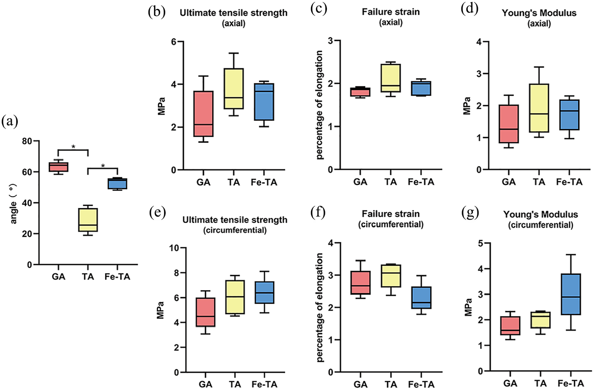
Flexibility and biomechanical parameters of BJV. (a) The gravity drop angle of the samples in the Fe-TA group, TA group and the GA group, n = 5 for each group. (b–g) The axial and circumferential stress, strain and elastic modulus of each group, n = 5 for each group. *Indicates a statistically significant difference between the two groups at a confidence level of 0.95 (GA vs TA p < 0.05, Fe-TA vs TA p < 0.05 in Figure 1(a)).
Anti-calcification effect in rat subcutaneous implantation model
After subcutaneous implantation in rat for 21 days or 60 days, the Fe-TA group showed excellent anti-calcification performance (Figure 2). The increase in calcium content in the Fe-TA group was lower than that in the TA group between the two time points, even though the difference was not statistically significant (Figure 2). Our data indicated that the addition of Fe3+ optimised the anti-calcification effect of TA.
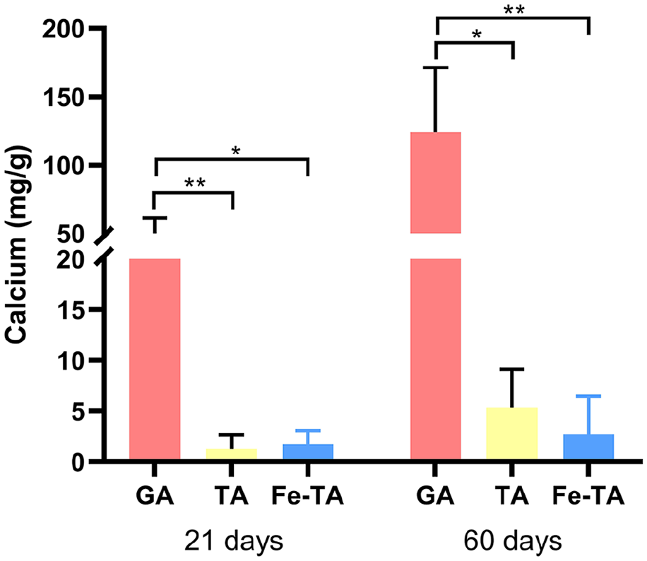
Quantitative analysis of inductively coupled plasma emission spectroscopy for calcium deposition. After 21 days or 60 days of subcutaneous implantation in rats, the calcium level in the Fe-TA group was significantly lower than that in the GA group (21 days: p < 0.05; 60 days: p < 0.001), and the increase in calcium content in the Fe-TA group was lower than that in the TA group between the two time points. *: p < 0.05, **: p < 0.001.
Echocardiography examination of graft performance
Echocardiography was performed monthly after implantation to evaluate the performance of the grafts in the sheep models (Figure 3(a) and (b)), and results show that the blood flow velocity of the bovine jugular vein valve is within 2 m/s, and the transvalvular pressure difference is within 10 mmHg (Figure 3(c)), which shows good haemodynamics and biocompatibility of BJV graft in the body, without stenosis and early valve failure.
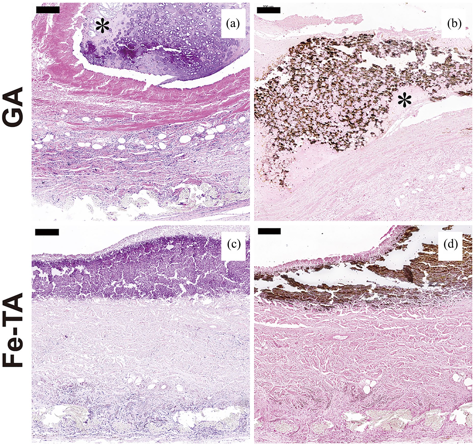
Representative image of the calcified region of the BJV in the GA group (a and b) and the Fe-TA group (c and d) 180 days after implantation in sheep (*: intimal hyperplasia). It showed calcifications in the area of intimal hyperplasia in the GA group and calcifications in the intima and medial layers in the Fe-TA group. HE (a and c) and von Kossa (b and d) staining. Scale bar = 200 μm.
Histological staining of cells infiltration and tissue calcification
HE staining showed intimal hyperplasia (indicated by an asterisk) and cells infiltrated into the medial and intimal layer in the GA group (Figure 4(a)), while cells infiltrated only in the adventitia region in the Fe-TA group (Figure 4(c)). VK staining showed that calcium deposition occurred in the area rich in elastic fibres and smooth muscle cells in the Fe-TA group (Figure 4(d)) and occurred additionally in intimal hyperplasia in the GA group (Figure 4(b)).
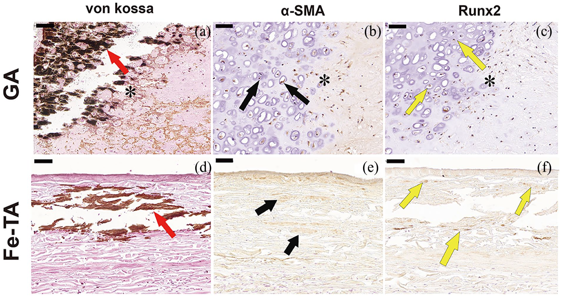
Representative image of immunohistochemical staining of α-SMA and Runx2 in the GA group and the Fe-TA group 180 days after implantation in sheep (asterisk: intimal hyperplasia). The intimal hyperplasia calcification region in the GA group ((a) indicated by red arrow) and medial calcification region in the Fe-TA group ((d) indicated by red arrow) were positive for α-SMA staining ((b and e), indicated by black arrow) and Runx2 staining ((c and f) indicated by yellow arrow). Scale bar = 50 μm.
Osteogenic differentiation in the calcified zone
Immunohistochemical staining of the calcified region was performed simultaneously with anti-α-SMA antibody and anti-Runx2 antibody. The results showed that positive staining of the SMC marker (α-SMA) (Figure 5(b) and (e)) and osteogenic marker (Runx2) (Figure 5(c) and (f)) colocalised with the calcified regions in the GA group (Figure 5(a)) and Fe-TA group (Figure 5(d)), indicating the presence of osteogenic differentiated smooth muscle cells in the calcified region.
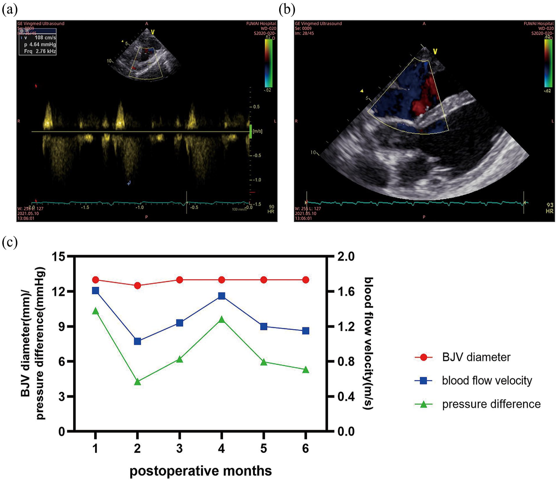
Ultrasound data acquisition and statistics: (a) pulse-doppler for the measurement of the blood flow velocity and the pressure difference of the BJV valve, (b) parasternal oblique five-chamber section for blood flow, and (c) inner diameter of the BJV, the blood flow velocity and the pressure difference of the BJV valve every month after the surgery.
Graft inflammatory cell infiltration
Both M1/M2 macrophages and T lymphocytes invaded the grafts according to the IHC staining of CD68, CD163 and CD3 after 180 days implantation in sheep (Figure 6(a)). Compared with the GA group, the number of invading T lymphocytes (CD3 staining-positive) decreased in the Fe-TA group, while the number of invading M1 macrophages (CD68 staining-positive) and M2 macrophages (CD163 staining-positive) were not significantly different as indicated by quantitative analysis of CD3, CD68, and CD163 expressions (Figure 6(b)–(d)).
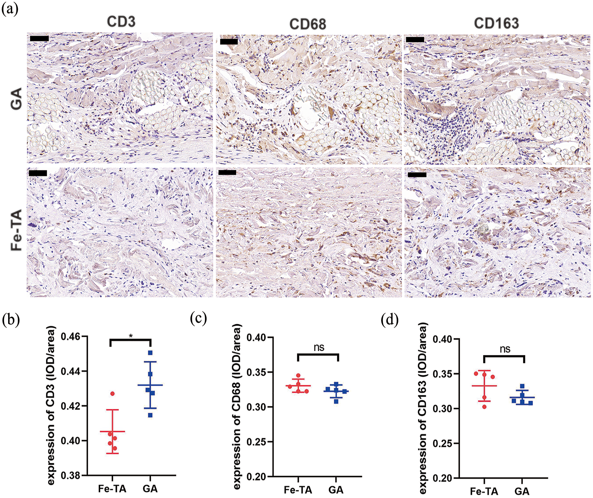
(a) Representative image of immunohistochemical staining of invading macrophages (CD68, CD163)/T lymphocytes (CD3) 180 days after implantation in sheep. Scale bar = 50 μm. (b–d) Quantitation of invading macrophages and T lymphocytes in BJV tissue sections. Data were presented as mean ± SD, n = 5. A significant difference between the groups, *p < 0.05 and ns p > 0.05.
MMP-2 and MMP-9 expression
Enzymatic hydrolysis of MMP is an important cause of elastin degradation. Immunohistochemical staining showed that MMP-2 and MMP-9 expression was lower in BJV media rich in elastic fibres in the Fe-TA group (Figure 7(a), (c), (e) and (g)) at 90 or 180 days of implantation in sheep compared with the GA group (Figure 7(b), (d), (f) and (h)) as indicated by quantitative analysis of MMP-2 and MMP-9 expressions (Figure 7(i) and (j)). These results indicated the direct role of Fe-TA treatment on MMP-2 and MMP-9 expression.
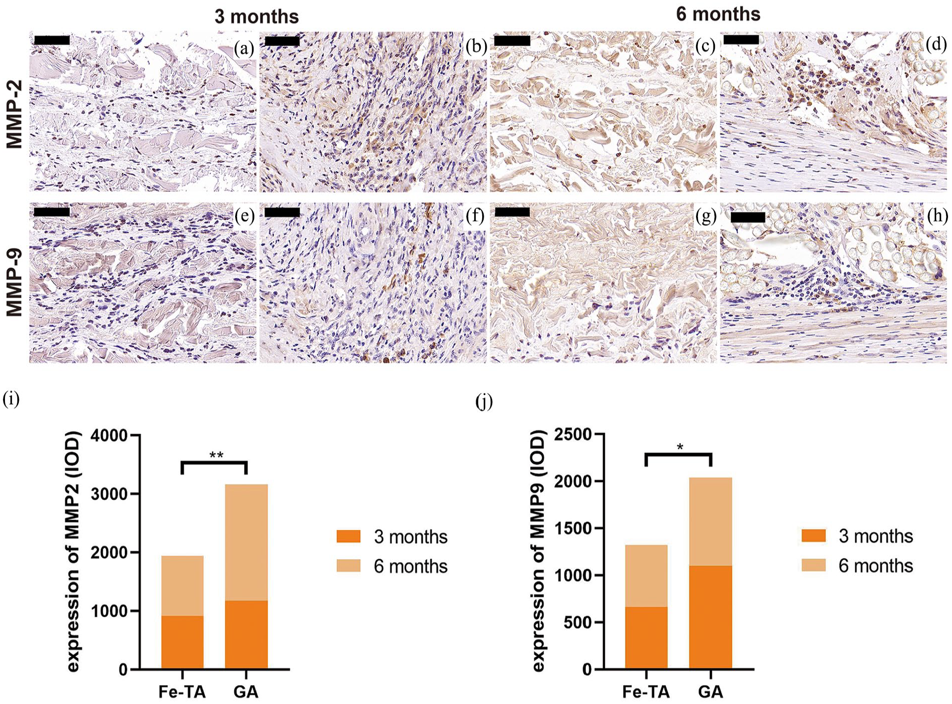
(a–h) Representative images of immunohistochemical staining of MMP-2 and MMP-9 after 90 days or 180 days implantation in sheep. Scale bar = 50 μm. (i and j) Quantitation of invading macrophages and T lymphocytes in BJV tissue sections. Data were presented as mean ± SD, n = 3. A significant difference between the groups, *p < 0.05 and ** p < 0.001.
Discussion
Our results showed that after the combined treatment of FeCl3 and TA, the flexibility of BJV tissue was improved, and the mechanical strength was preserved. However, the mechanism needs to be explored by further research. The rat subcutaneous implantation for 60 days and the reconstruction of the RVOT in sheep for 180 days (essentially no difference from the background level without implantation, data not published) showed excellent anti-calcification effects, good haemodynamics and biocompatibility, making it a good clinical application prospect.
We implanted FeCl3-TA-treated and GA-treated valved BJV conduits into the RVOT of sheep for 90 or 180 days, conducted immunohistochemical studies on key markers involved in pathways that have been reported in xenograft calcification and vascular calcification research. We found that smooth muscle cells (α-SMA staining-positive) expressing Runx2 appeared in both GA-treated and Fe3+-TA-treated calcified grafts, indicating that SMC osteogenic differentiation may be involved in the occurrence of active calcification in BJV xenografts. Runx2 is the well-known earliest and specific marker of developmental osteogenesis. Lee et al. 18 found the presence of osteoblastic cells in calcified purified porcine aortic elastin after subcutaneous implantation in rats with Reverse Transcription-Polymerase Chain Reaction (RT-PCR) and protein analysis and speculated that activated fibroblasts may undergo cell differentiation and express an osteogenic phenotype. However, the origin of osteoblastic cells in calcified elastin has not been clearly defined. In this study, we simultaneously performed immunohistochemical staining of calcified regions with anti-α-SMA and anti-Runx2 antibodies, and the results showed that the Runx2-positive cells were also α-SMA-positive cells, suggesting that SMCs may be a cellular source of osteogenic differentiation in BJV xenografts. Osteogenic differentiation of smooth muscle cells is known to be an important source of cells for vascular calcification under ageing and pathophysiological conditions, 14 and its role in xenograft calcification has not been clearly reported. In addition, there may be other possible sources of osteogenic differentiated cells, and more experiments are needed for validation.
Chekhoeva et al. 19 thought that implant deterioration was predominantly driven by invading autologous cells that induced pro-degenerative cascades, and therefore, preventive measures during the repopulation process can effectively inhibit graft degeneration. Wang et al. 20 showed that bovine pericardial samples treated with GA-TA implanted subcutaneously in rats had reduced calcification and showed less macrophage infiltration and MMP-9 levels compared with GA fixation alone. In this study, T-cell infiltration and the expression of MMP-2 and MMP-9 were reduced in the Fe-TA group samples relative to the GA group, especially in the media rich in elastic fibres, which was consistent with the excellent anti-calcification effect. In addition, M1 and M2 macrophages infiltrated from the adventitia to the media of the BJV in both groups. The tube wall of the Fe-TA group BJV in contact with blood was basically free of macrophage adhesion, while the GA group had a large number of macrophage infiltrations in the intimal hyperplasia region, suggesting that Fe3+-TA treatment may have a certain physical barrier effect on the infiltration of inflammatory cells and may reduce the occurrence of intimal hyperplasia. Peivandi et al. 21 conducted a systematic histological analysis of 13 Contegra® explants obtained from two paediatric cardiac centres and considered elastic degeneration of the Contegra® grafts and intima hyperplasia with severe calcification and heterotopic ossification to be the leading causes for the limited longevity of the Contegra® prosthesis. In this study, we observed the same phenomenon in the GA-fixed BJV after construction of sheep RVOT, but the Fe-TA group grafts essentially had no intima hyperplasia, suggesting that the mechanism of reducing xenograft calcification may be related to this.
Macrophage-derived MMP-9 and mesenchymal cell (smooth muscle cell and fibroblast)-derived MMP-2 work together on elastic fibre degradation, which produces active elastin peptides that have been shown to activate ERC, triggering multiple biological responses, including osteogenic differentiation of cells. Lee et al. 18 implanted fresh and TA-treated purified porcine aortic elastin subcutaneously in rats to evaluate MMP-2 and MMP-9 gene expression with elastin calcification. They found that MMP-2 and MMP-9 showed higher enzymatic activity at the early time point of calcification, and the degraded elastin peptide increased accordingly. TA is known to bind to hydrophobic regions in the elastin structure, which are preferred sites for most elastases, such as matrix metalloproteinases, neutrophil elastase, and pancreatic elastase. This study showed that MMP-2 and MMP-9 expression in the region rich in elastic fibres of the Fe-TA group was significantly reduced relative to that of the GA group, which may be another reason for the anti-calcification effect. Similarly, Jin et al. 22 treated porcine pericardium with trivalent iron ions and proved an increased ability to resist elastin hydrolysis in vitro, and the rat subcutaneous model proved to have a significant anti-calcification effect. Therefore, Fe-TA treatment enhanced the anti-enzymatic hydrolysis performance of the material and may also reduce the secretion of MMPs.
One limitation of this study is that only two-point bending test is used to evaluate the flexural properties of the conduit after treatment. Although the naked eye and the touch feeling of the surgeon prove that the material indeed soften compared to the TA-treated only, further research is indicated to reveal whether there is an increase in extensibility and the effect on the distribution of mechanical stress.
Another limitation is that an immunohistochemical staining on two serial sections has been used to observe the colocalisation of target markers. Although it can be said that cells expressing two markers appear in the calcification region, whether the same cell expresses both markers requires further research to determine.
Conclusion
Overall, this study confirms that Fe3+-TA treatment is a promising method for BJV processing and that the anti-calcification mechanisms may be related to reducing intimal hyperplasia and MMP-2 and MMP-9 secretion. In addition, this study found the osteogenic differentiation of SMCs in BJV xenograft calcification for the first time, which has certain reference value for BJV anti-calcification therapy.
Supplemental Material
sj-tif-1-jao-10.1177_03913988231208631 – Supplemental material for Optimal treatment of tannic acid for the anti-calcification of bovine jugular veins and the underlying mechanism
Supplemental material, sj-tif-1-jao-10.1177_03913988231208631 for Optimal treatment of tannic acid for the anti-calcification of bovine jugular veins and the underlying mechanism by Aili Wang, De Wang, Yumiao Wang, Bixuan Liu, Haiyang Wei, Yibo Wang and Jianye Zhou in The International Journal of Artificial Organs
Footnotes
Acknowledgements
The study was supported by the National Key R&D Program Project ‘Technology Research and Development of Immunogenicity Elimination and Anti-Calcification Techniques for Tissues or Organs of Animal Origin’ (Project No. 2016YFC1101002).
Author’s contributions
Conceptualisation: Jianye Zhou. Data curation: Jianye Zhou. Formal analysis: Aili Wang, Yumiao Wang. Funding acquisition: Jianye Zhou. Investigation: Aili Wang, Yumiao Wang. Methodology: Jianye Zhou, Aili Wang. Project administration: De Wang. Resources: De Wang, Yibo Wang. Software: Aili Wang. Supervision: Jianye Zhou. Validation: De Wang. Visualisation: Bixuan Liu, Haiyang Wei. Writing-original draft: Aili Wang. Writing-review & editing: Jianye Zhou.
Data availability statement
The authors confirm that the data supporting the findings of this study are available within the article [and/or] its supplementary materials.
Declaration of conflicting interests
The authors declared no potential conflicts of interest with respect to the research, authorship, and/or publication of this article.
Funding
The authors disclosed receipt of the following financial support for the research, authorship, and/or publication of this article: This work was supported by the National Key Research and Development Program of China (No. 2016YFC1101002).
Ethical statement
All animal experiments and surgical procedures were performed in compliance with the Guide for the Care and Use of Laboratory Animals as published by the US National Institutes of Health (NIH Publication 85-23, revised 1996). The experimental plan was authorised by the Ethics Committee of the Laboratory Animal Center of Fuwai Hospital, Chinese Academy of Medical Sciences: authorisation number FW-2019-0012 (for rat experiments, 2019.5.12) and 0102-2-15-2x(x)-8 (for sheep experiments, 2020.9.22).
Supplemental material
Supplemental material for this article is available online.
References
Supplementary Material
Please find the following supplemental material available below.
For Open Access articles published under a Creative Commons License, all supplemental material carries the same license as the article it is associated with.
For non-Open Access articles published, all supplemental material carries a non-exclusive license, and permission requests for re-use of supplemental material or any part of supplemental material shall be sent directly to the copyright owner as specified in the copyright notice associated with the article.