
Abstract
Managing bone healing is essential for preventing problems such as non-union, bacterial infection, structural instability, psychological, and physical damage in patients. The need to use antibiotics less often has prompted researchers to look at possible substitutes, such as nanoparticles. In this investigation, we choose to employ cerium oxide nanoparticles due to their unique antibacterial properties based on redox reactions. The cerium oxide–hydroxyapatite composite was synthesized, calcined, and ball-milled to create a fine CeO2-HA powder. Luffa cylindrica sponge was used to prepare the scaffold, and X-ray diffraction (XRD) and scanning electron microscopy (SEM) were used to investigate the structural and morphological features. Rapid upregulation of osteogenesis marker genes confirmed that CeO2-HA nanoparticles in the scaffolds promoted osteoblast cell proliferation and osteogenic differentiation. The cell viability test was conducted by MTT assay. When the CeO2-HA composite was cultured with S. aureus, it showed signs of having more antibacterial efficacy than pure HA.
Introduction
Infections during the bone healing process can have detrimental consequences; therefore, the primary objective is to conduct extensive research and develop advanced antimicrobial materials to prevent such infections. So, developing nanoparticles with antibacterial attributes is essential for effective tissue engineering. A wide variety of research fields may benefit from the unique physicochemical properties of nanoparticles, which are defined as particles between 1 and 100 nm in size. 1 Nanoparticles of various morphologies have several uses, including medical applications for treatment and diagnostics, improvement of the pathogen-fighting system, and improvement of fuel quality. 2 In recent decades, nanotechnology has become one of the most fascinating areas of study. It has a wide range of applications in several fields, such as imaging, business, electronics, and healthcare. The creation and delivery of novel medications for diagnosing and treating illnesses makes considerable use of nanotechnology in the healthcare sector. 3 However, the vast majority of nanomaterials lack superior biocompatibility traits. Silver nanoparticles (NPs) have been used in various materials to combat bacteria, but their use in human health care is limited due to their substantial biosafety concerns and propensity for cytotoxicity. 4 Due to its antioxidant, anticancer, biocompatibility, and other characteristics, nanoparticles based on cerium oxide have recently shown encouraging results in biomedical applications. 5
Cerium (Ce) is a rare earth lanthanide series element that exists in solution as both Ce3+ and Ce4+; also depending on the environment, it may switch between the two oxidation states. 6 Cerium oxide (CeO2), also known as Ceria, is a potential oxide of cerium that is becoming increasingly popular for a wide range of applications, including stem cell adhesion, UV defense, tissue engineering, and many more.7–10 CeO2 nanoparticles have received an abundance of interest as antibacterial agents because of their tendency to cycle between the two valence states (Ce3+ and Ce4+), which results in oxygen vacancies appearing in the lattice. The oxygen buffering capability of the nanoparticles seems to be favorable, as it enables them to function as catalysts for oxidation and reduction processes, revealing a unique antibacterial mechanism when compared to other metal oxides in medicine and biomaterials. 11 Focusing on the present dil-emma of microorganisms that are resistant to drugs, CeO2 is a significant new finding.
Synthetic hydroxyapatite (HA), represented by the chemical formula Ca10(PO4)6(OH)2, is an inorganic compound characterized by its microporous nature, is an essential constituent of several hard tissues in the human body, including enamel, dentine, and bones. 12 As a biocompatible material, this calcium phosphate ceramic is in high demand due to its osteogenic capability and its capacity to create strong connections with host bone tissues. 13 The integration of a wide range of foreign ions to enhance the characteristics of HA relevant to orthopedic and dental applications is an important feature of its structure.
Cerium ion (Ce3+ and/or Ce4+) substitution for calcium ion (Ca2+) in the HA lattice is a fascinating topic of research in the medical area. According to certain reports, cerium (III)— HA possesses bacteriostatic qualities comparable to silver. 14 A sophisticated scaffold including CeO2-HA NPs has been constructed in this novel study. The scaffold has been thoroughly examined by the use of sophisticated methodologies such as X-ray diffraction (XRD) and scanning electron microscopy (SEM). Furthermore, an extensive cellular investigation has been conducted to explore the capacity of these nanoparticles (NPs) to enhance cell survival, which has shown encouraging outcomes. This innovative investigation presents new opportunities for the use of CeO2HA nanoparticles in the field of biological applications. The results obtained from this investigation provide strong evidence for the significant efficacy of scaffolds, including CeO2-HA NPs in the treatment of bacterial infections. 15
Materials and methods
Synthesis of HA, CeO2 nanoparticles, and CeO2HA scaffold
In the pursuit of synthesizing HA nanoparticles, a wet chemical precipitation method was employed using Ca(NO3)2.4H2O and (NH4)H2PO4 as precursors, similar to the process described by Swain et al. 16 The pH level was carefully adjusted to 10 during the experiment to ensure optimal conditions. Subsequently, the samples underwent air drying followed by calcination at a temperature of 600°C for a duration of 8 h. The ball milling technique was used to mix the synthesized HA powder with CeO2 powder, resulting in a homogenous mixture.
Moreover, a sodium hydroxide solution was applied to a small piece of dried luffa to enhance its hydrophobicity and left for 30 min. This process effectively increases its resistance to water. The CeO2-HA powder was carefully blended with a combination of 10% gelatin, 2% PVA, and 1% Guar gum solution to ensure a uniform mixture. This mixture was then applied twice onto the luffa cylindrical template, allowing for a 24 h interval between each application. Afterwards, the specimens were left to dry before being subjected to a sintering process at 500°C for a duration of 2 h.
Characterizations
By using XRD (Bruker D8-ADVANCE), the crystalline phase of the specimens was determined and characterized. 17 The investigation of the morphologies and structures of the specimens was carried out using SEM (Hitachi S-4800). 18 Additionally, the swelling characteristics of the scaffold were examined under standard in vitro (pH 7.4 at 37°C) conditions in PBS buffer and aqueous medium in order to assess the pore size and hydration of the scaffold material. The quantification of water absorption was assessed using the following formula:

Where Ww represents the weight of the water-saturated sample and Wd represents the weight of the dry sample.
In order to assess the viability of bacteria, S. aureus (ATCC) was chosen as a representative model of Gram-positive bacteria. The bacteria were cultivated on Mueller-Hinton broth (MHB) medium at a temperature of 37°C inside an incubation chamber for a duration of 6 h. In vitro bacterial viability was determined using the MTT assay.19,20 The optical density at 490 nm was measured on the 1st, 7th, and 14th day to track the development of S. aureus. In the meantime, the assessment of the cytotoxicity of the nanoparticles that were manufactured was conducted by using MG-63 cells that were seeded with a concentration of 15% CeO2-HA NPs. The cytotoxicity of CeO2-HA NPs was evaluated by the use of an in vitro MTT assay test. The vitality of cells was examined at various time intervals throughout the incubation process. 21 The assessment of gene expression related to osteogenesis was conducted using quantitative reverse transcription polymerase chain reaction (qRT-PCR). The MG-63 cells were inoculated into a 96-well plate with a cell density of 4 × 10⁴ cells per well. Subsequently, the cells were grown for durations of 1, 7, and 14 days. Using the Superscrip II First strand Synthesis kit, 1 mg of RNA was reverse transcribed into complementary DNA (cDNA). 22 The expression levels of Runt-related transcription factor 2 (RUNX2), Osteocalcin (OCN), and type-I collagen (COL-1) were monitored using the Bio-rad MyiQ2 instrument and the Transstart Top Green qPCR SuperMix (Transgenic). 23
Results and discussion
Nosocomial infection, also known as hospital-acquired illness, is a significant medical concern in developing as well as industrialized countries. Antibiotics have been extensively used in the medical field for many decades to address bacterial infections. Nevertheless, the rapid rise of bacteria that are resistant to antibiotics has raised several challenges and responsibilities for the medical field. 24 In the US, an estimated annual total of 88,000 fatalities resulting from nosocomial infections are documented. Numerous investigations have shown that CeO2 NPs possess remarkable antibacterial properties. 25 The XRD pattern depicted in Figure 1 offer valuable insights into the powdered sample of CeO2-HA. The distinct peaks indicate the presence of finely divided particles with a small crystalline size, lending the sample unique properties. Furthermore, the absence of any additional peaks confirms the purity of the sample, enabling precise analysis, and application of CeO2-HA. 26
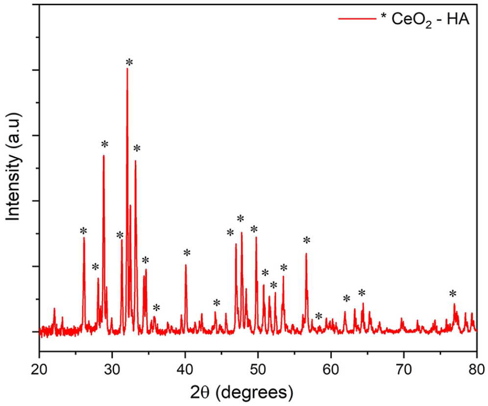
XRD Pattern of CeO2HA sample indicating distinct peaks.
The morphologies of the synthesized scaffold were examined using SEM, depicted in Figure 2. The microstructure of the scaffolds was studied by observing its morphology at a resolution of 300 nm. The presence of microporous morphologies inside the scaffold plays a crucial role in promoting cellular proliferation and vascularization. 27 The presence of these microstructures facilitates the interchange of gases, nutrients, and wastes. 26
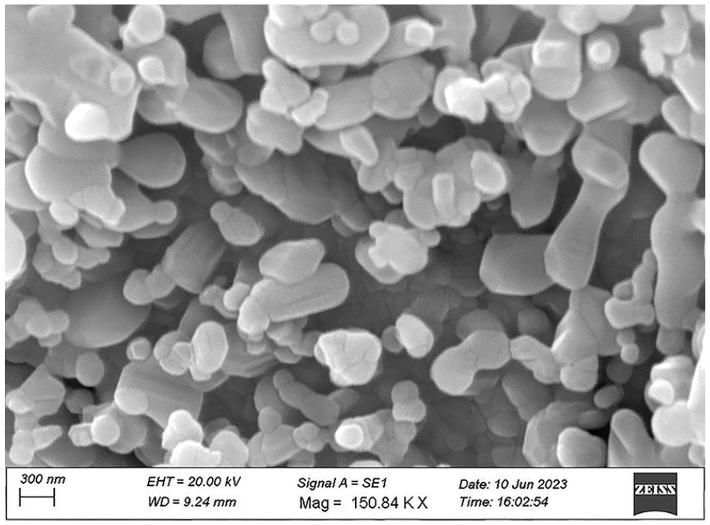
SEM analysis of CeO2-HA scaffold.
The swelling parameters of the scaffolds have been examined in PBS buffer and aqueous medium under typical in vitro (pH 7.4 at 37°C) conditions. The results shown in Figure 3 demonstrate that the swelling rate of the scaffolds varied significantly in the presence of CeO2. Scaffolds swell less in PBS media and more in aqueous media due to distinct physicochemical properties and interactions between various available functions and the media. 17 The phenomenon of scaffold swelling is an essential characteristic that plays a crucial role in promoting osteogenesis via its facilitation of cell adhesion and proliferation. During the process of swelling, the scaffolds experience an increase in both porosity and pore size, which in turn promotes the migration of cells, hence aiding cell development and enabling other cellular processes.
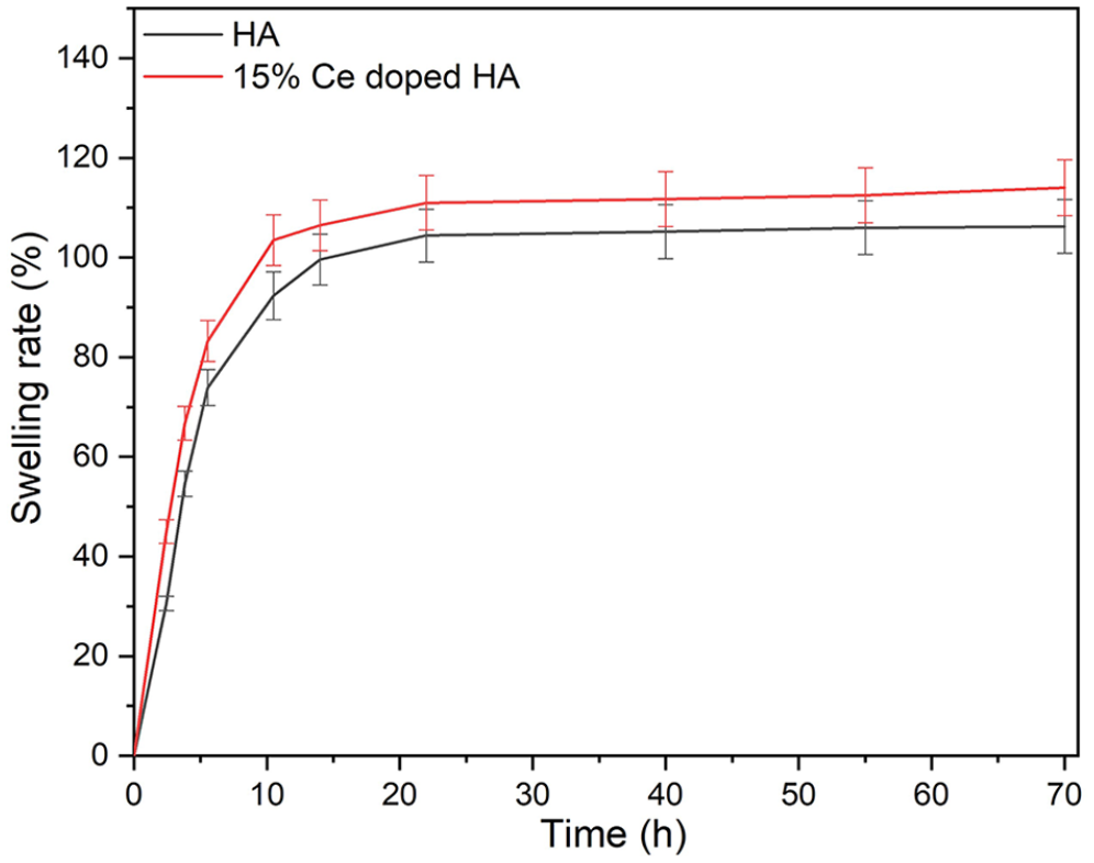
Swelling ratio of 15% CeO2-HA and pure HA.
Scaffold implantation for bone repair is an ongoing procedure. Cell adhesion is a critical process that develops as a result of cell and material surface contact and results in the production of new tissue, which may then be used to repair or regenerate bone tissues. The facilitation of cell adhesion to the scaffold matrix is a crucial process for promoting bone formation. According to an investigation by Leung et al., 27 the toxicity of cerium oxide nanoparticles varied, and this was attributable to how the nanoparticles interacted with cells as well as variations in their composition. It is necessary to undertake longer-term research to clarify the toxicity of CeO2 NPs. Measurements of optical density were employed in our investigation to evaluate bacterial activity. The data were gathered throughout a time span of 10 h, extending up to 30 h. The observation of bacterial growth was conducted on two samples: the control sample of HA and a sample of CeO2-HA. As shown in Figure 4, there was a notable decrease in the overall number of bacterial cells during the first 10 h period. Subsequently, there was an exponential fall in the bacterial count observed between the 20th and 30th hour in the presence of CeO2-HA. These features interact with the cellular membrane to promote their reproduction, attachment, and movement. The antibacterial properties of CeO2 against S. aureus have been reported in several investigations, exhibiting a significant and robust antibacterial impact. According to a study by Arumugam et al., 28 the two Gram-positive bacteria that were evaluated had a higher resistance to the toxicity of manufactured CeO2 NPs than did the five Gram negative bacteria.
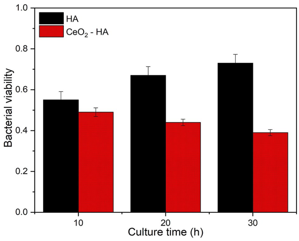
Bacterial viability of S. aureus on CeO2-HA and pure HA.
The cytotoxicity impact of the synthesized nanoparticle was evaluated using the MTT assay in vitro. In a 96-well plate with 100 µL of DMEM, MG63 cells were seeded for 24 h at a density of 104 cells per well. The measurement of optical density is a commonly used technique for evaluating bacterial growth in a culture. Consequently, the absorbance values of bacterial suspensions may be quantified using a photometer.29,30 Acc-ording to the data shown in Figure 5, it can be concluded that the introduction of CeO2 into HA does not have an adverse effect on the development of osteoblast cells, as compared to the use of pure HA. To validate this phenomenon, MTT assay has been used, in which major variations were seen in osteogenic cell development between day 7 and day 14, as compared to the first day, which could be due to the early cellular reaction. CeO2-HA has excellent biocompatibility, making it a highly suitable material for coating applications. The use of CeO2-HA as a coating material confers non-toxic properties to the coated surface. According to a research conducted by Ren et al., 30 the application of CeO2 NPs to human periodontal ligament stem cells (hPDLSCs) resulted in enhanced cell proliferation. The presence of CeO2 NPs inside the cells is responsible for safeguarding the physiological function of stem cells via the elimination of excessive reactive oxygen species (ROS) generated as a result of oxidative damage. The use of CeO2 nanoparticles can help to decrease the harm caused by ROS in cell components such as DNA, lipids, and proteins. Moreover, these nanoparticles have demonstrated improved biocompatibility, making them highly promising for use in tissue engineering within both the dental and medical fields. 31
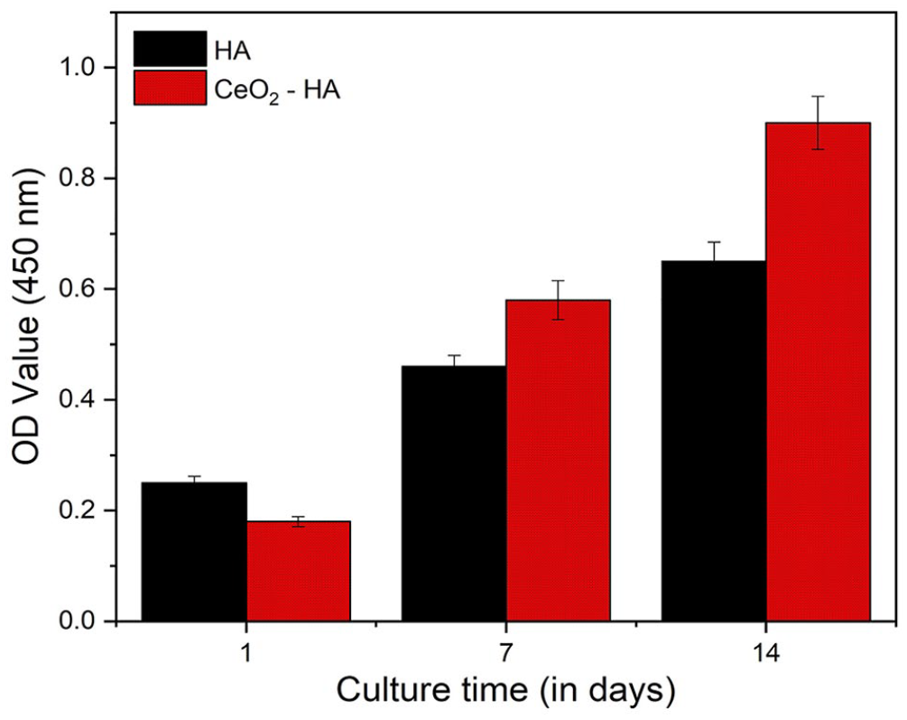
MTT assay of cells cultured on CeO2-HA and pure HA.
The MG-63 human osteoblast-like cell line (NSSC Pune, India) generated from human osteosarcoma has been utilized in this work, and the scaffolds have supported cell growth against it. According to the relative expression of genes associated with osteogenesis, such as osteocalcin (COL-1), Runt-transcription factor-2 (RUNX2), and osteocalcin (OCN), surfaces enriched in CeO2-HA which greatly boosted osteogenic expression. The evaluation of gene expression levels of COL-1, RUNX2, and OCN in MG63 cells cultured on CeO2HA and HA samples was conducted on the 1st, 7th, and 14th day. The gene expression levels of CeO2-HA were found to be significantly greater than those of HA (p < 0.05).
The upregulation of osteoblast-lineage cell proliferation and differentiation is governed by the reciprocal regulation of RUNX2. As observed on days 1 and 7, Figure 6 shows that there was little change in the proliferation rate. However, on day 14, a significant change in cell growth was observed. Which induces the proliferation of mesenchymal cells and their commitment into osteoblast-lineage cells. 30
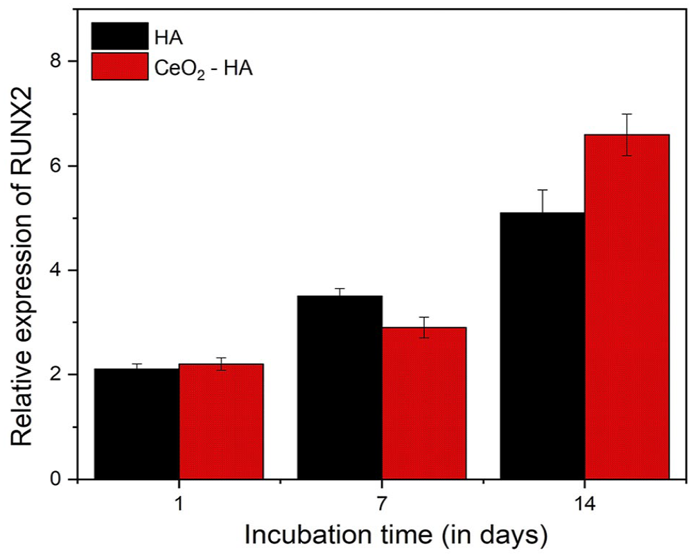
Osteogenic gene expression of RUNX2 on CeO2-HA and pure HA.
The extracellular protein molecules COL-I and OCN play a crucial role in the formation of bone tissue, making them of utmost importance in the composition of the bone matrix. OCN is seen to be expressed throughout the later stages of osteogenic differentiation. The gene expression levels of COL-1 (Figure 7), and OCN (Figure 8) for MG63 cells on HA and CeO2-HA increased from 1 to 14 days. 115% CeO2-HA revealed higher gene expression levels as compared to HA (p < 0.05). As a result, the examination of OCN and COL-1 may provide insights into the underlying mechanisms of osteoblastic development. Hence, we conducted an assessment of the biological efficacy of CeO2-HA on the COL-1 and OCN activities in MG-63 cells. The results of the mRNA analysis demonstrated that the addition of 15% CeO2HA to MG-63 cells resulted in increased expression levels of COL-I and OCN.32
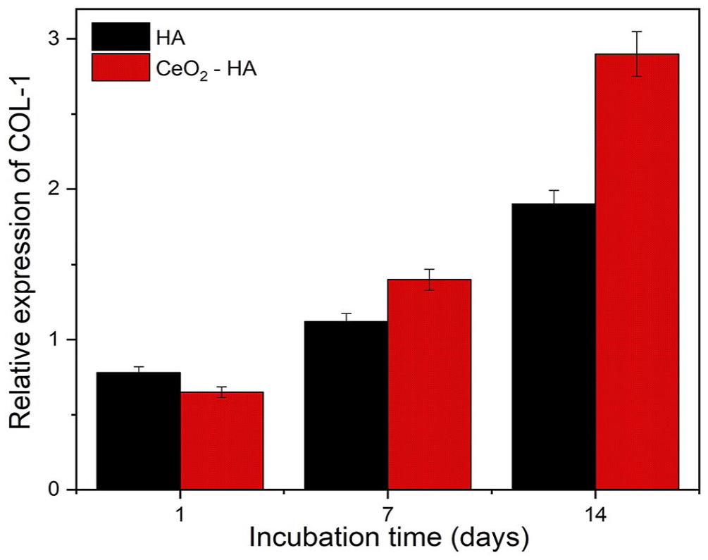
Osteogenic gene expression of COL-1 on CeO2-HA and pure HA.
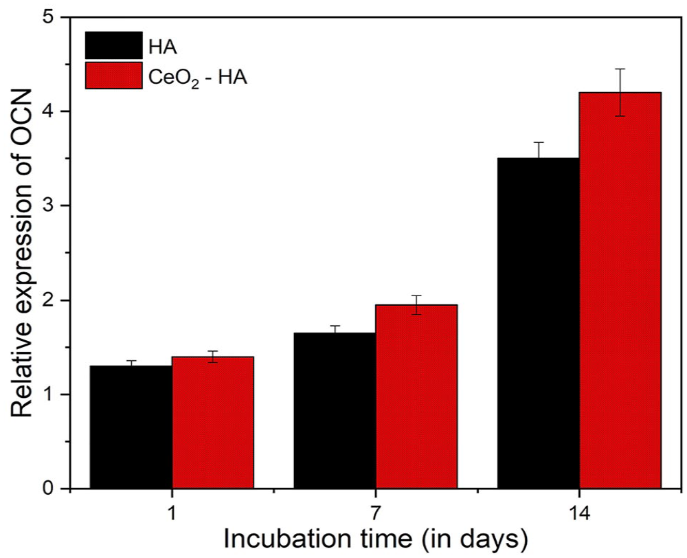
Osteogenic gene expression of OCN on CeO2-HA and pure HA.
Conclusion
The sole purpose of this investigation was to create a porous scaffold by synthesizing CeO2-HA nanocomposites through ballmilling technology. Since HA nanoparticles are biocompatible and may provide a novel approach to treat ailments, they have been incorporated into biomaterials and tissue engineering. XRD examination verified the purity of the CeO2-HA phase, meanwhile, SEM results determined the morphology of the synthesized scaffold.
In addition to its antioxidative property against S. au-reus, as confirmed by a bacterial viability test (Figure 4), CeO2 exhibits pro-oxidative behavior, meaning that it can induce oxidative stress and thus manifest toxicity toward various bacterial cells. 16 In vitro studies have shown that the presence of CeO2-HA NPs influenced the expression of osteogenesis-related genes such COL-1, OCN, and RUNX2. Due to its antioxidant properties, which may protect cells from damage caused by infections, CeO2 is gaining attention for use in biomedical sectors.
Footnotes
Declaration of conflicting interests
The author(s) declared no potential conflicts of interest with respect to the research, authorship, and/or publication of this article.
Funding
The author(s) received no financial support for the research, authorship, and/or publication of this article.