
Abstract
The challenges in achieving optimal outcomes for wound healing have persisted for decades, prompting ongoing exploration of interventions and management strategies. This study focuses on assessing the potential benefits of implementing a nano-gelatin scaffold for wound healing. Using a rat skin defect model, full-thickness incisional wounds were created on each side of the thoracic-lumbar regions after anesthesia. The wounds were left un-sutured, with one side covered by a gelatin nano-fibrous membrane and the other left uncovered. Wound size changes were measured on days 1, 4, 7, and 14, and on day 14, rats were sacrificed for tissue sample excision, examined with hematoxylin and eosin, and Masson’s trichrome stain. Statistical comparisons were performed. The gelatin nanofibers exhibited a smooth surface with a fiber diameter of 260 ± 40 nm and porous structures with proper interconnectivity. Throughout the 14-day experimental period, significant differences in the percentage of wound closure were observed between the groups. Histological scores were higher in the experiment group, indicating less inflammation but dense and well-aligned collagen fiber formation. A preliminary clinical trial on diabetic ulcers also demonstrated promising results. This study highlights the potential of the nano-collagen fibrous membrane to reduce inflammatory infiltration and enhance fibroblast differentiation into myofibroblasts during the early stages of cutaneous wound healing. The nano-fibrous collagen membrane emerges as a promising candidate for promoting wound healing, with considerable potential for future therapeutic applications.
Impact statement
For decades, wound healing has presented persistent challenges, with the optimal interventions and management strategies for achieving improved outcomes remaining elusive. This study has identified that the implementation of a nano-gelatin scaffold holds significant promise for enhancing wound healing. Specifically, the nano-collagen fibrous membrane demonstrated the capacity to diminish inflammatory cell infiltration and facilitate the differentiation of fibroblasts in the early stages of cutaneous wound healing. The implications are noteworthy, suggesting that the nano-fibrous collagen membrane has the potential to foster wound healing and could offer substantial prospects for future therapeutic advancements.
Introduction
Wound healing is a complex process influenced by various factors, with acute wounds, chronic wounds, and complicated wounds representing distinct categories necessitating specific durations and management approaches. Despite their differences, these wounds share common repair phases: coagulation and hemostasis, inflammation, and proliferation. 1 Managing full-thickness or deep partial-thickness skin defects, especially in extensive cases, remains a reconstructive challenge in contemporary clinical practice. 2 Non-healing wounds pose complications, ranging from local infections to systemic issues, often requiring amputation. Current surgical treatments, such as skin grafts or substitutes, have limitations, 3 underscoring the need for innovative therapies in chronic wound management. 4
Standard wound management techniques include debridement and antibiotic treatment. 5 Surgical debridement, involving the removal of infected tissues, accelerates healing by reducing pathogens and biofilm.5 –7 Similarly, antibiotics aim to decrease bacterial burden, facilitating wound healing.6,7 However, these methods have drawbacks, with sharp debridement risking increased inflammation and antibiotic treatment potentially disrupting the body’s microbiome and pH.5,8,9 Delivery limitations, such as inadequate penetration for topical antibiotics, further complicate their effectiveness. 5
To enhance the efficacy of debridement and antibiotic treatment, prioritizing wound healing promotion through appropriate wound dressings is crucial. 10 Proper dressings foster tissue renewal, reduce inflammation and scar formation, and enhance the release of growth factors, ultimately promoting wound closure.9 –11 Various regenerative modalities, including epidermal substitutes and dermal regenerative substitutes, have been explored to improve outcomes in reconstructive procedures. 12
Traditional epidermal coverage alone, such as skin grafts, may not provide stable cover for restoring the structure and function of normal skin in cases involving dermal and epidermal defects. Excessive wound contraction and scar formation present functional and aesthetic challenges. Successful incorporation of a skin substitute requires dynamic cellular/extracellular matrix interaction, with design principles considering collagenous forms, inherent resistance, porosity, and hydration to promote intrinsic regeneration. 13
Micro-needling has been reported to accelerate skin regeneration by creating microinjuries that evoke various inflammatory, proliferative, and remodeling reactions. 14 Additionally, nanotechnology has emerged as a revolutionary technique in wound dressing production. Nanofibers and nanoparticles, applied to wounds, enhance surface area coverage, absorption capacity, therapeutic agent delivery, and provide a conducive environment for cell growth. 15 Gelatin and collagen, known for promoting cell migration and providing tissue structural support, hold promise as nanomaterials for advanced wound dressings.1,5,15,16 To optimize the effectiveness of debridement and antibiotic treatment, prioritizing wound healing promotion through suitable dressings is crucial. Nanotechnology has emerged as a revolutionary technique in wound-dressing production forwound care. In this study, we aim to elucidate whether implementing a nano-gelatin scaffold will yield beneficial effects on wound healing.
Materials and methods
Fabrication of electro-spun gelatin membrane
The electro-spun nano-fibrous gelatin membrane was fabricated following previously reported methods by our research team. 17 In brief, a homogeneous solution containing 10 g of gelatin (G1890, Type A, porcine, Sigma-Aldrich, USA) in 100 mL dd-H2O was prepared at 45°C–50°C. The solution was loaded into a 10 mL syringe (with an inner diameter 1.5 mm needle), and the internal temperature was maintained between 50°C using a heater (AREX-6, VELP Scientifica, USA). High-voltage power supply (SC-PME50, Cosmi Global Co., Ltd., Taiwan) set at 20 kV was employed to generate electro-spun fiber mats, collected on an aluminum foil-covered collector positioned 5 cm away from the needle tip. The resulting nano-fibrous membrane used in this study had a thickness of 160 ± 20 μm. For analysis, the membranes were fixed with a 2.5% glutaraldehyde solution for 2 h, dehydrated using a series of ethanol concentrations (50% to 100%), sputter-coated with gold, and examined with a scanning electron microscope (S4500; Hitachi, Tokyo, Japan).
The composite fabrics analyzed in this study were ground and mixed thoroughly with potassium bromide, and then analyzed by using Fourier-transform infrared spectroscopy (FTIR) with the IR spectra analyzed set at a range of 450–4000 cm−1.
Surgery and wounding model
Rats undergoing incisional wounds were anesthetized with ketamine (50 mg/kg) and diazepam (50 mg/kg). The back fur was shaved, and skin cleaned with alcohol and povidone-iodine. Using a No. 15 blade and scalpel, two full-thickness incisional wounds (20 mm × 25 mm) were made in the thoracic-lumbar regions of each rat (Figure 1 ). A total of 12 wounds in six rats were included, with six assigned to the sham group and six to the experimental group. The wounds were left un-sutured, and one side of each rat was covered with the gelatin nano-fibrous membrane while the other side remained uncovered. Wounds were allowed to heal by secondary intention, with surgical day considered as day 0. Wound size changes were measured on days 1, 4, 7, and 14.
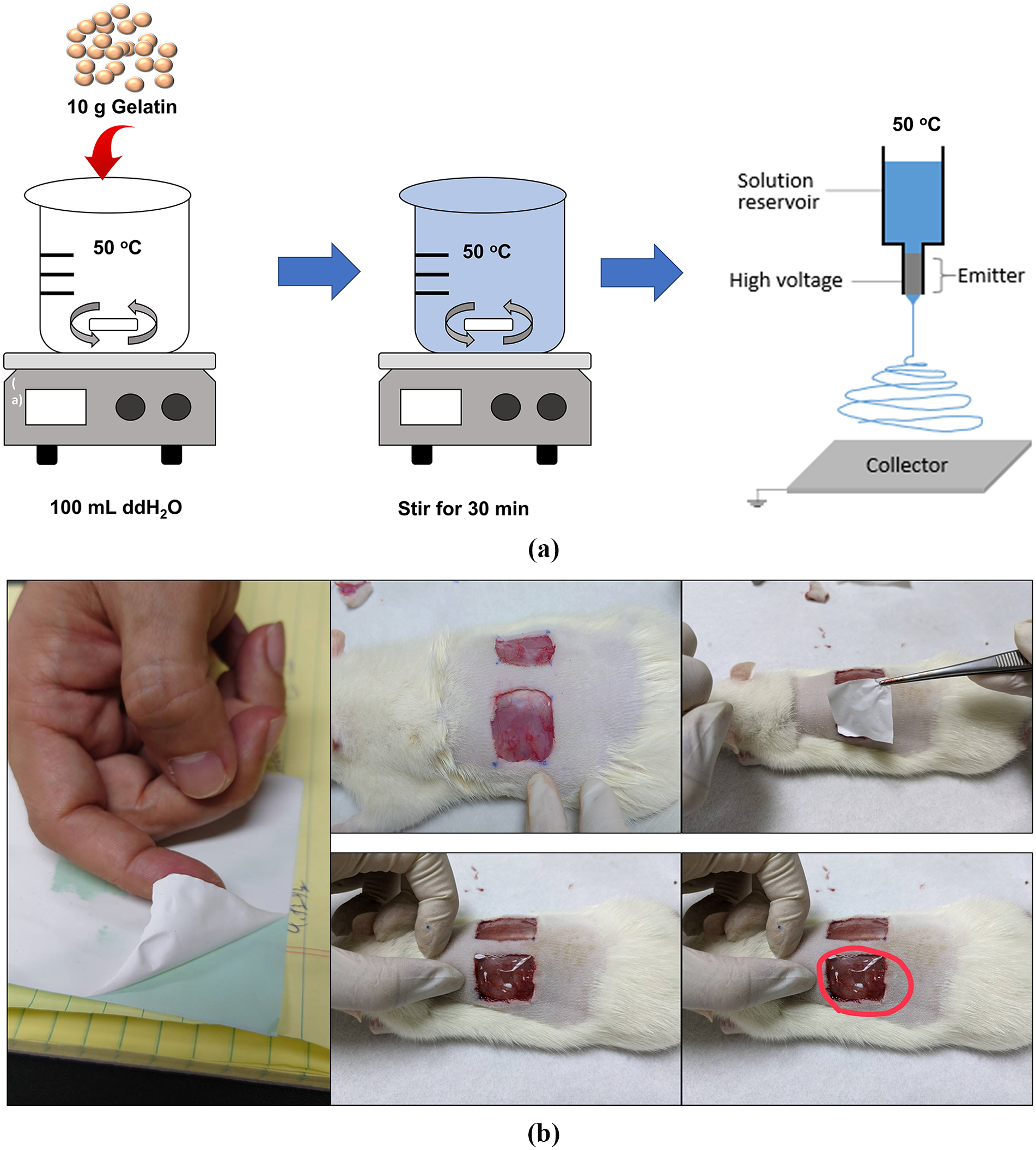
The preparation of gelatin nanofibrous scaffold and surgery of skin wounding model: (a) the preparation of gelatin nanofibrous scaffold and (b) surgery and wounding model.
Histopathological evaluation
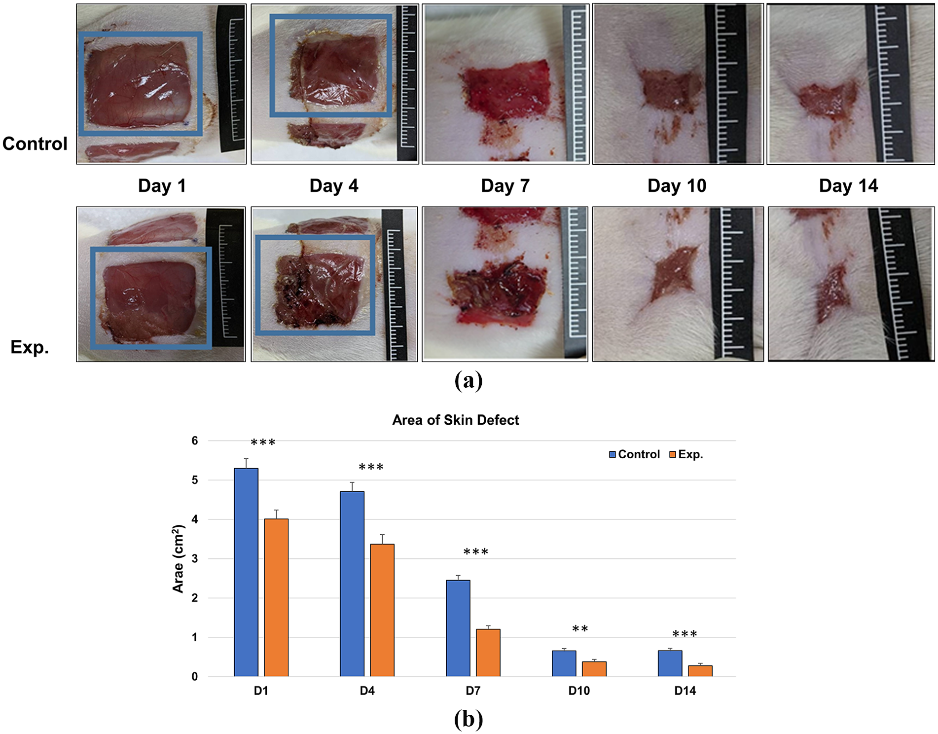
On day 14, rats were sacrificed after measuring the wound defects. Tissue samples from the wound bed and normal adjacent skin were excised, fixed in 10% neutral buffered formalin, and subjected to histopathology examination. Trimmed specimens were embedded, and sagittal sections were cut. Staining was performed with hematoxylin and eosin, as well as Masson’s trichrome stain. Ten random zones from each sample were selected for histomorphometric analyses. Semi-quantitative scoring of histopathological lesions was conducted using light microscopy (DM2700M, Leica) at ×400 magnification. Scoring included fibroblasts, neutrophils, macrophages, blood vessels, and re-epithelization, following a modified procedure reported by Schwartz et al. 8 and Gál et al. 18 (Table 1). Mean values were used for statistical comparison.
Explanation of scales used in the semi-quantitative evaluation of histological sections.
ST: surrounding tissue; DL: demarcation line; SCT: subcutaneous tissue; GT: granulation tissue.
Statistical analysis
Pathologic lesion grades in the test article group were recorded and compared to the sham group. Microsoft Excel (2010) for Windows® was used for statistical analysis. Unpaired Student’s t-test was employed for comparing parametric data collected from histopathological grading.
Results
Microstructure of gelatin nano-fibrous membrane
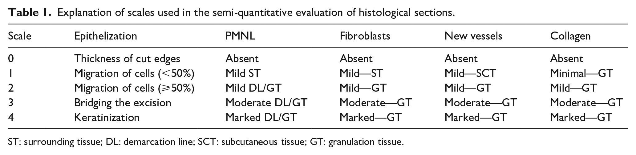
Microstructure of Gelatin Nano-Fibrous Membrane: The morphology of the gelatin nano-fibrous membrane was examined using scanning electron microscopy (SEM), and the surface characteristics and fiber diameter were analyzed. Gelatin nanofibers exhibited a smooth surface with a diameter of 260 ± 40 nm, and porous structures with proper interconnectivity were observed (Figure 2(a)).
Microstructure and FTIR pattern of gelatin nano-fibrous membrane: (a)Microstructure of gelatin nano-fibrous membrane. The gelatin nanofibers exhibited a smooth surface with a diameter of 260 ± 40 nm, and porous structures with proper interconnectivity were observed. (b) The FTIR pattern showed the functional group of nano-fibrous membrane, the peak of amide, phosphate, hydroxide, and the carbonate group all suggest that nano-fibrous membrane was similar to the natural collagen.
Characterization of nano-gelatin fibers (NGF)
The FTIR pattern showed the functional group of nano-fibrous membrane in Figure 2(b), a broad band appearing between 3010 and 3650 cm−1 (peaked at 3290 cm−1) indicates O–H stretching. The transmittance bands near 1635, 1536, and 1451 cm−1 in the FTIR spectrum are due to C–N stretching in the amides I, II, and III groups; a carbonyl carbon bond peak appeared at 1062 cm−1. In the FTIR spectrum, the peak of amide, and the carbonate group suggests that the characteristic of gelatin nano-fibrous membrane was quite similar to the natural collagen (Figure 2(b)).
Macroscopic analysis for wound closure
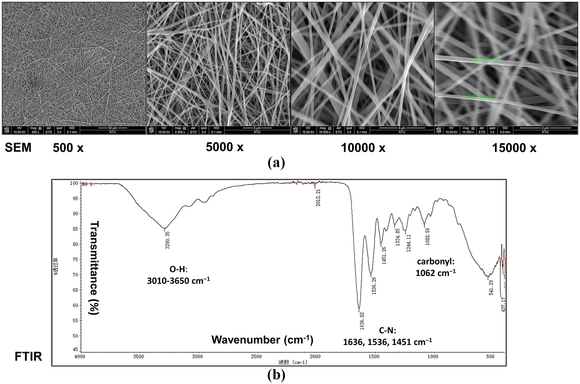
Representative photographs of wounds from each experimental group at different time points are depicted in Figure 3(a). Notably, significant differences were observed between the nano-fibrous membrane-covered and control groups concerning the percentage of wound closure throughout the 14-day experimental period (Figure 3(b)).
Grossly size changes of wounds and quantitative evaluation of the wound healing. (a) Grossly size changes of wounds. Photographs taken at different time points over the 14-day experiment illustrate the changes in wound area on rats with and without the gelatin nano-fibrous membrane. (b) Quantitative evaluation of the wound healing.
Wound re-epithelialization, cellular content, and granulation tissue formation
At the conclusion of the study (day 14), the healing area, ranging from non-complete to complete, was examined in both control and experimental groups. Histopathological examination revealed less inflammatory cell reaction in the skin sections of the experiment group compared to the control group. The histopathological scores for skin wound healing in the nano-fibrous membrane (experiment) group were superior to those in the control group (Figure 4(a)). Masson’s trichrome stain on day 14 showed dense and well-aligned collagen fibers across the entire wound area in the experiment group, contrasting with sparse and dispersed collagen fibers in a thin layer under the wound in the control group (Figure 4(a)).
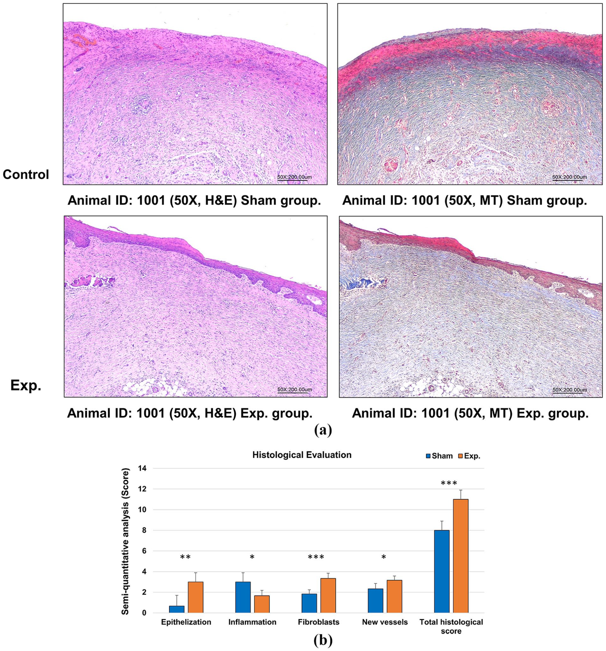
Representative sections and histological evaluation of skin wound healing: (a) representative sections in skin wound healing model and (b) histological evaluation of the skin in wound healing model.
In hematoxylin and eosin staining, significant differences were observed between the experiment and control groups in the mean scores for wound re-epithelialization, granulation tissue formation, and cellular contents during the 14-day experimental period (Figure 4(b)). On day 14 after surgery, semi-quantitative analysis revealed significantly increased scores for fibroblasts, new vessels, and epithelialization in the experiment group (p < 0.05), while the inflammation score significantly decreased. The total histological score remained significantly higher in the experiment group (p < 0.001). Detailed data on histopathologic findings and related statistical analysis are provided in Supplemental Appendices 1–3.
Clinical case presentation
In November 2022, a 72-year-old male with a history of type-2 diabetes mellitus exceeding 30 years presented. Poorly controlled blood sugar with elevated glycated hemoglobin (HbA1c >10%) led to a chronic, unhealed, and infected wound on his left leg (measuring up to 61 mm × 11 mm with surrounding erythematous changes) persisting for over 2 months. Following daily soft debridement with wet dressing for 15 min and subsequent coverage with a sterile gelatin nano-fibrous membrane (Sheng Chen International Biomedical Co., Ltd., Hsin-Chu, Taiwan, ROC), the wound size decreased to 34 mm × 4 mm on day 27, further reducing to 17 mm × 4 mm on day 44, and eventually achieving complete healing by day 55 (Figure 5). The condition remained stable for over 6 months at the last visit.
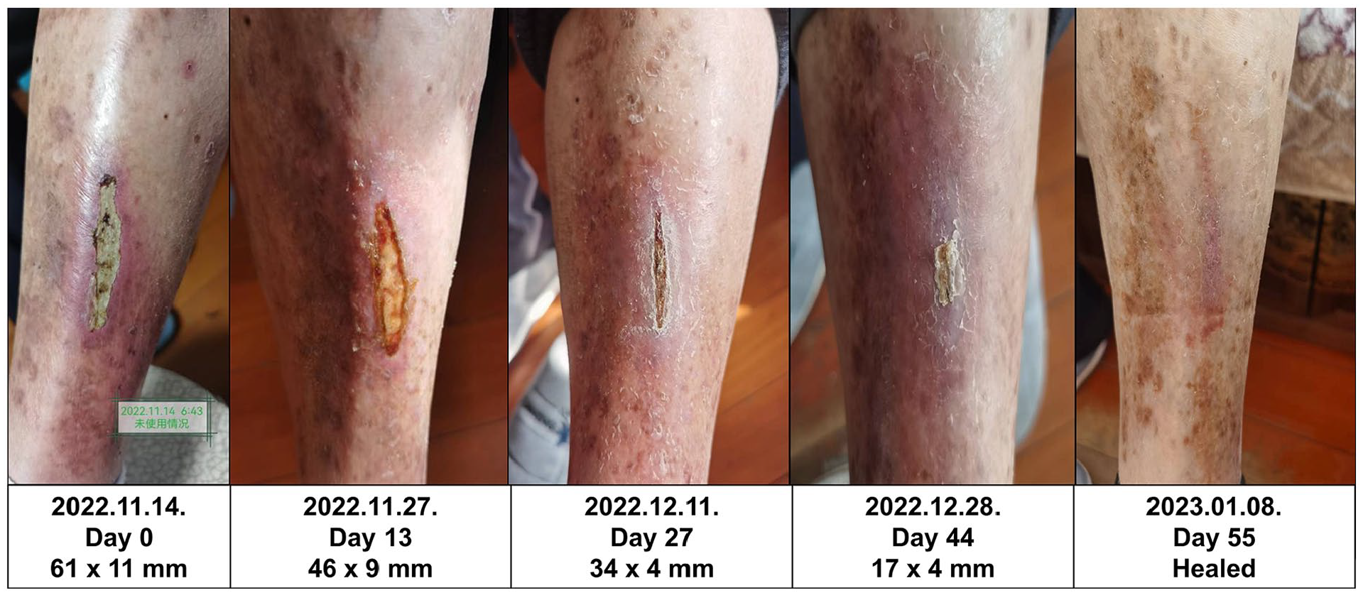
Clinical case presentation.
Discussion
Nanotechnology presents a transformative potential to enhance biomaterials with advantageous physical, chemical, and biological properties. The substantial specific surface area of nanomaterials facilitates the adsorption of bioactive molecules and cells, while porous nanofibrous structures can separate molecules and retain stem cells.19,20 Nanostructured collagen membranes have demonstrated potential for stimulating early-stage soft oral tissue regeneration and serving as a constituent for dermal bioequivalents. 21 In the biomedical field, electrospinning nanotechnology has been employed to produce polymeric nanofibers, incorporating drugs for wound healing, burns, and diabetic ulcers. Electro-spun nanofibers enable three-dimensional (3D) cell seeding and culturing in vitro, offering promise for treating chronic wounds and inspiring novel strategies for various organ systems, marking a new era of “regenerative and personalized medicine.” 3 This article presents a nanotherapeutic approach utilizing tissue-engineered nanofiber skin substitutes, including a case from a preliminary clinical trial in a chronic wound of a diabetic patient.
In our study, the experimental group demonstrated a superior wound repair rate throughout the 14-day period compared to the control group, as evidenced by macroscopic and histological analyses. Shrinkage of the skin defect appeared as early as the first day post-wounding, suggesting potential roles of attracted myofibroblasts in the treated wounds. 22
Neutrophils and activated monocytes releasing TNF-α were noted in the wound area, with implications for fibroblast mitogenic effects and macrophage-mediated delays in wound healing. 23 Insufficient vascularization is one of the major factors limiting the clinical success of tissue-engineered constructs. 24 In our study, the experiment group displayed better granulation tissue and new vessel formation, contributing to enhanced wound healing. Therefore, we suggested the possibility that nanofibrous collage membrane might help to promote wound healing after the rat skin defect model.
Skin, as the body’s natural physical barrier, plays a crucial protective role and requires appropriate wound dressings for effective treatment. 23 Traditional dressings may cause pain and scar formation during changes, 25 making nanofiber-based dressings an attractive alternative due to their high cell attachment rates and increased growth factor release. 16 In a preliminary clinical case trial, the nanofibrous membrane exhibited promising results, leading to remarkable wound closure in 55 days compared to the typical 12-week healing period for chronic wounds. 11
Decellularized extracellular matrices (ECM) and stem cells have been explored as wound graft materials in skin regenerative medicine.26,27 Skin breakdown enhances the synthesis and penetration of active ingredients, with activated platelet growth factors potentially playing a key role in stimulating fibroblasts to produce collagen and elastin. 14 The gradual release of growth factors from wounds can effectively promote the healing of skin defects by encouraging cell proliferation, facilitating collagen deposition, and supporting tissue revascularization. 28 The utilization of epidermal stem cells in the construction of tissue-engineered skin is also a viable approach for the reconstruction of skin defects. 29
In our study, the nanofiber scaffold, associated with chelating functional motifs in collagen/gelatin, may attract circulating stem cells and their growth factors to rebuild skin defects. The enhanced wound healing in the experimental group is characterized by increased cellular infiltration, granulation tissue formation, induced angiogenesis, and re-epithelization. The nano-fibrous collagen membrane demonstrated a significant reduction in inflammation, introducing a dual role as an anti-fibrogenic scaffold.
It is conceivable that the improved wound healing observed in the experimental group is characterized by anincrease in cellular infiltration, granulation tissue formation, induced angiogenesis, and re-epithelization post-surgery. Notably, the nano-fibrous collagen membrane used in this study significantly mitigated inflammation in the rat experimental group, corroborated by similar findings in the preliminary clinical case presentation. This outcome presents novel evidence for the potential of an anti-fibrogenic scaffold, playing a dual role in alleviating fibrosis and expediting wound healing. 30 The nano-fibrous collagen membrane holds promise as a potential delivery system, showcasing extreme potential for future therapeutic applications.30,31 However, the underlying mechanism of this function requires further investigation in future studies. Prospective randomized controlled trials (RCTs) are essential to provide evidence of the clinical and cost-effectiveness of this technology for future clinical applications.
Conclusion
This study demonstrates that the nano-gelatin fibrous membrane has the potential to reduce inflammatory infiltration and likely enhance the differentiation of fibroblasts into myofibroblasts during the early stages of cutaneous wound healing in skin defects of rats. All data generated from this study consistently indicate superior wound healing effects in the experimental group compared to the sham group.
Supplemental Material
sj-docx-1-jao-10.1177_03913988241244661 – Supplemental material for Enhancing cutaneous wound healing: A study on the beneficial effects of nano-gelatin scaffold in rat models
Supplemental material, sj-docx-1-jao-10.1177_03913988241244661 for Enhancing cutaneous wound healing: A study on the beneficial effects of nano-gelatin scaffold in rat models by Jason Sun, Yi-Chung Lai, Yi-Wen Lin, Chih-Hsiang Fang and Jui-Sheng Sun in The International Journal of Artificial Organs
Footnotes
Acknowledgements
Histological Assessment for Skin Healing Model in Rats was performed at Master Laboratory Co., Ltd., Animal Laboratory, Zhudong Township, Hsinchu County 31053, Taiwan (R.O.C.), by Chien-Hao Chen, D.V.M., RLATG (Pathologist/Veterinarian).
Author contributions
Jason SUN: Data collection, data analysis, and manuscript drafting. Yi-Chung Lai: Active discussion of experimental design, data collection, and assembly. Yi-Wen Lin and Chih-Hsiang Fang: Performing the animal study. Jui-Sheng Sun: Study supervision, assistance with study conceptualization and design, analysis and interpretation of data, and critical revision of the article for important intellectual content. All authors have read and approved the final submitted manuscript.
Availability of data and materials
The datasets used and/or analyzed in the current study are available from the corresponding author upon reasonable request.
Declaration of conflicting interests
The author(s) declared no potential conflicts of interest with respect to the research, authorship, and/or publication of this article.
Funding
The author(s) disclosed receipt of the following financial support for the research, authorship, and/or publication of this article: This work was supported by the National Science and Technology Council, Taipei, Taiwan, ROC [NSTC 112-2314-B-039 -049 -MY2], the En Chu Kong Hospital, New Taipei City, 237, Taiwan, ROC, and the National Taiwan University Hospital, Taipei, Taiwan, ROC.
Ethics approval and consent to participate
Authorization for the study’s execution was obtained from the Research Ethics Committee, National Taiwan University Hospital, Taipei, Taiwan, ROC.
Consent for publication
Not applicable.
Commercial associations
ach author certifies that they have no commercial associations (e.g. consultancies, stock ownership, equity interest, and patent/licensing arrangements) that might pose a conflict of interest in connection with the submitted article.
Location of work
All work was conducted at National Taiwan University, National Taiwan University Hospital (Taipei City, Taiwan).
Supplemental material
Supplemental material for this article is available online.
References
Supplementary Material
Please find the following supplemental material available below.
For Open Access articles published under a Creative Commons License, all supplemental material carries the same license as the article it is associated with.
For non-Open Access articles published, all supplemental material carries a non-exclusive license, and permission requests for re-use of supplemental material or any part of supplemental material shall be sent directly to the copyright owner as specified in the copyright notice associated with the article.