






Abstract
Cyclophosphamide (CPA) is an anticancer drug used in the treatment of a variety of neoplastic lesions. On the other hand, treatment with CPA was accompanied by different toxic effects on different body organs. The present work was conducted to study the effect of fenugreek seed extract on histomorphometrical and ultrastructural changes induced by CPA in testes of albino mice. Twenty animals were given CPA (7.0 mg/kg body weight) three times/week orally for 8 weeks and were killed after 4 and 8 weeks. Testis of CPA-treated mice showed many histological alterations including appearance of irregular seminiferous tubules, reduction in the number of all spermatogenic cells, degeneration of Leydig cells and appearance of intertubular hemorrhage. Concerning the ultrastructural changes, abnormalities in spermatogonia (A and B), spermatocytes, round and elongated spermatids were observed. Degenerated Sertoli cells and degenerated interstitial tissue with abnormal Leydig cells were also seen. Moreover, administration of CPA to animals significantly increased malondialdehyde (MDA, lipid peroxidation marker) and decreased superoxide dismutase (SOD) and catalase (CAT). These changes were time-dependent. Treating animals with CPA and fenugreek seed extract (0.4 g/kg body weight) led to an improvement in the histological and ultrastructural pictures of the testis together with reduction in the level of serum MDA and increase in the activities of serum SOD and CAT. In conclusion, the results of the present work indicated that fenugreek had ameliorative effect against testis damage induced by CPA and this may be mediated by its potent antioxidant activities.
Introduction
Alkylating agents are commonly used in the treatment of cancer. Cyclophosphamide (CPA) is extensively used as an anticancer drug as well as an immunosuppressive agent. It is used in the treatment of different types of cancers (Evans et al., 1980). Davidson et al. (1990) reported that CPA has been used in the chemotherapy of lymphocytic leukemia, multiple myloma and ovarian carcinoma. There is a body of evidence that cyclophosphamide can induce antitumor effects not only by directly affecting dividing cancer cells but also by indirectly augmenting T-cell concomitant antitumor immune responses (Turk et al., 2004). CPA has been used extensively in various inflammatory disorders, including multiple sclerosis (Kieseier and Jeffery, 2010). Oral CPA therapy for 12 months, in a case with unilateral active serpiginous choroiditis, was effective for improving vision and preventing recurrences (Sahin, 2010). Calabreis and Parks (1980) mentioned that CPA is a highly reactive chemical that transfers alkyl group to important cell constituents by combining with nucleophilic groups such as amino, sulfhydryl, carboxyl, hydroxyl and phosphate. These agents alkylate DNA and more specifically, the purine base, guanine in which the 7 nitrogen is highly nucleophilic. On the other hand, treatment with CPA was accompanied by different side effects (Styler et al., 1996). CPA has significant detrimental effects on male reproduction. CPA exposure caused oligospermia and azoospermia in both animal models (Elangovan et al., 2006) and humans (Garolla et al., 2006). CPA was found by many authors to induce testicular atrophy, histopathological alterations and suppression of spermatogenesis (Ceribaşi et al., 2010; Matsui et al., 1995; Wetzels, 2004).
Medicinal plants and herbs play an important role in pharmacological preparations and medicine for hundreds of years. Along with traditional medicine, they are used in the treatment of various illnesses. Fenugreek (Trigonella foenum graecum) is an annual herb and belongs to the family leguminosae. The seeds of fenugreek are commonly used in India, Egypt and in other oriental countries as a spice in food preparations due to their strong flavor and aroma (Kaviarasan et al., 2007). The seeds are also used as herbal medicine in many parts of the world for carminative, tonic and aphrodisiac effects (Xue et al., 2007). Fenugreek seeds are known to exhibit hypoglycemic, hypolipidaemic, antifertilitic, antiandrogenic, antinociceptive and wound healing properties and are a good source of dietary fibers (Muralidhara et al., 1999). Fenugreek seeds reversed the diabetic state and protect cellular structures, in vivo and in vitro, from oxidative damage (Kaviarasan et al., 2007; Sricharoen et al., 2008; Thakran et al., 2004). The present work was conducted to study the possible ameliorative effect of aqueous extract of fenugreek seeds on histomorphometrical, ultrastructural and biochemical changes induced in testis of mice by the anticancer drug, CPA.
Materials and methods
Experimental animals
Eighty healthy adult male albino mice (Mus musculus) approximately 3 months old weighing 20 ± 5 g were used in the present study. Animals were kept in the laboratory under constant condition of temperature (24 ± 2°C) for at least 2 weeks before and throughout the experimental work. They were maintained on a standard rodent diet composed of 20% casein, 15% corn oil, 55% corn starch, 5% salt mixture and 5% vitaminized starch (Egyptian Company of Oils and Soap, Kafr-Elzayat, Egypt). Water was available ad libitum. All the experiments were done in compliance with the guide for the care and use of laboratory animals (National Research Council, 1985).
Materials used
Cyclophosphamide
CPA (Cytoxan®, CAS No. 50-18-0) is an anti-neoplastic compound obtained from Eimc Company, Cairo, Egypt. CPA was dissolved in distilled water and given to experimental mice orally at a dose level of 7.0 mg/kg body weight (equivalent to the therapeutic dose for humans; Paget and Barnes, 1964).
Fenugreek seeds
Dried and fresh batches of fenugreek seeds were purchased from local market, Shebin El-kom, Egypt. Seeds were washed with distilled water to get rid of extraneous matter, air-dried and ground into a fine powder in a mixer. The powder was mixed with distilled water (1 g of seed powder per 100 ml) in a vortex cyclomixer for l0 min and then centrifuged at 10,000 rpm and the supernatant was collected. The supernatant was used as the aqueous extract for feeding the animals and was freshly prepared. In this study, each animal was given orally 1 ml of the final aqueous extract containing 0.4g/kg body weight fenugreek seeds (Thirunavukkarasu et al., 2003). Chemical profile of fenugreek seed extract has been described by various researchers and the main identified constituents include polyphenols (Gupta and Nair, 1999; Wang et al., 2010).
Treatments
Animals were divided into four groups, 20 animals in each group. The first group served as a control, while animals of the second group were given fenugreek at a dose level of 0.4 g/kg body weight orally three times/week for 8 weeks. Animals of the third group were given CPA at a dose level of 7.0 mg/kg body weight orally three times/week for 8 weeks. Animals of the fourth group were given CPA (7.0 mg/kg body weight) and fenugreek (0.4 g/kg body weight) orally at the same time for 8 weeks. Half of the animals from each group were killed by cervical decapitation after 4 weeks and the rest of animals were killed after 8 weeks.
Histological and ultrastructural studies
Animals were dissected and their testes were removed. The left testis was used for histological study, while the right one was used for ultrastructural examination. For histological preparations, the testis was fixed in alcoholic Bouin’s fluid, dehydrated, cleared and embedded in paraffin wax. Paraffin sections of 5 micron thickness were prepared and stained with Ehrlich’s haematoxylin and eosin. The diameter and germinal epithelial height of seminiferous tubules were measured from the spermatogenic cells on the inner surface of the basement membrane through the most advanced cell types lining the lumen of the tubules.
For electron microscopy, small pieces (1 mm thick) of testis were fixed in 2.5% glutaraldehyde, buffered at pH 7.2 with 0.1 M sodium cacodylate and post fixed in 1% osmium tetroxide. After dehydration in gradual series of ethanol, the tissue was embedded in Epon 812. Ultrathin sections were cut and mounted on copper grids. The sections were stained with uranyl acetate and lead citrate (Dickersin, 2000). The grids were then examined and photographed using Jeol transmission electron microscope 100 CX, unit of Electron Microscope, Alexandria University, Egypt.
Biochemical assays
For enzymes determination, blood samples were obtained from the inferior vena cava and then centrifuged. Sera were stored at –20°C until assayed for the biochemical parameters. The extent of lipid peroxidation was estimated as the concentration of thiobarbituric acid reactive products (malondialdehyde, MDA) according to Ohkawa et al. (1979). The activity of superoxide dismutase (SOD) was measured using the methods of Rest and Spitznagel (1977). Catalase (CAT) activity was determined from the rate of decomposition of H2O2 (Aebi et al., 1974).
Statistical analysis
The results were expressed as mean ± SD of different groups. The differences between the mean values were evaluated by analysis of variance (ANOVA) followed by Student’s t test using Minitab 12 computer program (Minitab Inc., State College, PA, USA). Values of p < 0.05 were considered statistically significant.
Results
Biochemical results
Malondialdehyde
Data in Figure (1a) revealed significant (p < 0.05) increase in the level of MDA in sera of animals treated with CPA for 4 or 8 weeks compared with the control group, the mean values were 30 ± 2.2 and 40.13 ± 1.7 n mol/ml, respectively. Animals treated with CPA and fenugreek showed decrease in MDA levels in sera compared with CPA group; after 8 weeks the mean value was 20.06 ± 1.8. No significant change was recorded between control and fenugreek-treated group.

Effect of different treatments on (a) malondialdehyde (MDA; nmol/ml) level, (b) superoxide dismutase (SOD; µmol/ml) and (c) catalase (CAT; µmol/ml) activities.
The antioxidant enzymes (SOD and CAT)
Treating mice with CPA for 4 or 8 weeks showed decrease in the activity of SOD and CAT in sera compared with control group. The mean values of CAT and SOD were 0.30 ± 0.01 and 21.83 ± 1.58µmol/ml, respectively, in CPA group after 8 weeks. Animals given CPA and fenugreek showed significant increase in the activity of SOD and CAT compared with CPA group. The mean values of CAT and SOD in CPA and fenugreek group after 8 weeks were 1.06 ± 0.15 and 61.3 ± 5.7, respectively. No significant change was obtained in SOD and CAT activity in sera of mice treated with fenugreek for 4 or 8 weeks (Figures 1 b and c).
Morphometric results
Figures (2 a and b) showed the morphometric changes in the diameter of seminiferous tubules and their epithelial heights. Treating mice with CPA for 4 or 8 weeks showed significant decrease (p < 0.05) in the diameter of the tubules and their epithelial heights. On the other hand, animals treated with CPA and fenugreek for 4 or 8 weeks showed highly significant increase (p < 0.001) in the diameter of seminiferous tubules and their epithelial heights in comparison with CPA group. No significant changes were recorded in the diameter of the tubules or the epithelial height in fenugreek-treated mice compared with control animals.
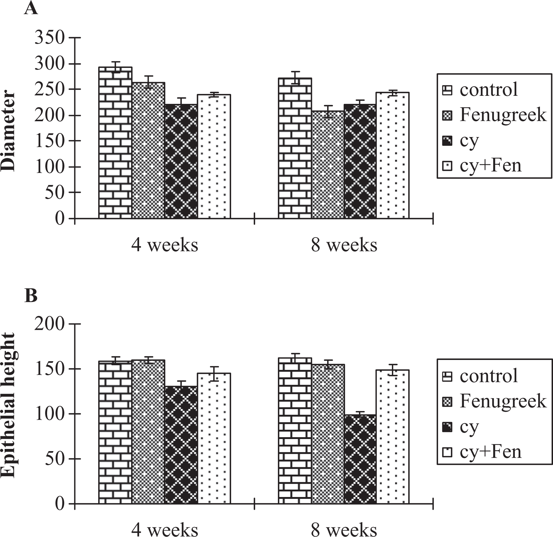
Effect of different treatments on (a) diameter and (b) the epithelial height of seminiferous tubules (mean ± SD).
Histological observations
Figure (3a) showed testis of control mouse. No histological alterations were observed in animals treated with fenugreek three times/week for 4 or 8 weeks. Testis of animals administered with CPA for 4 weeks exhibited a distinct histological difference when compared with control. In testes of these animals, large numbers of seminiferous tubules appeared irregular and sometimes were with thick basement membranes (Figure 3b). Intertubular hemorrhage and cytoplasmic vacuolization of the spermatogonia were observed. Most of the nuclei of the spermatocytes appeared with undetectable chromatin materials. These histopathological alterations were more obvious in animals treated with CPA for 8 weeks. In these specimens, the seminiferous tubules were more affected; there was decrease in the diameter of the tubules and enlargement of their lumens. In the same specimens, hemorrhage was seen in the degenerated intertubular connective tissue. The seminiferous tubules were degenerated with irregular boundaries and some spermatogenic cells were exfoliated in the lumen of the tubules. Some spermatogenic cells appeared vacuolated with darkly stained nuclei. In addition, the sperm bundles were less abundant. Sloughing off the germinal epithelium at some points was also seen (Figure 3c). Examination of testes of animals treated for 4 weeks with CPA and fenugreek revealed less prominent histopathological changes when compared with the group treated with CPA for the same period. The seminiferous tubules were compact with each other. The spermatogenic layers appeared somewhat normal. In the same specimen, a number of mitotic figures were seen. Advanced degree of improvement was seen in testes of animals treated for 8 weeks with CPA and fenugreek. Most of the seminiferous tubules restored its normal structure but few abnormalities were still seen (Figure 3d).
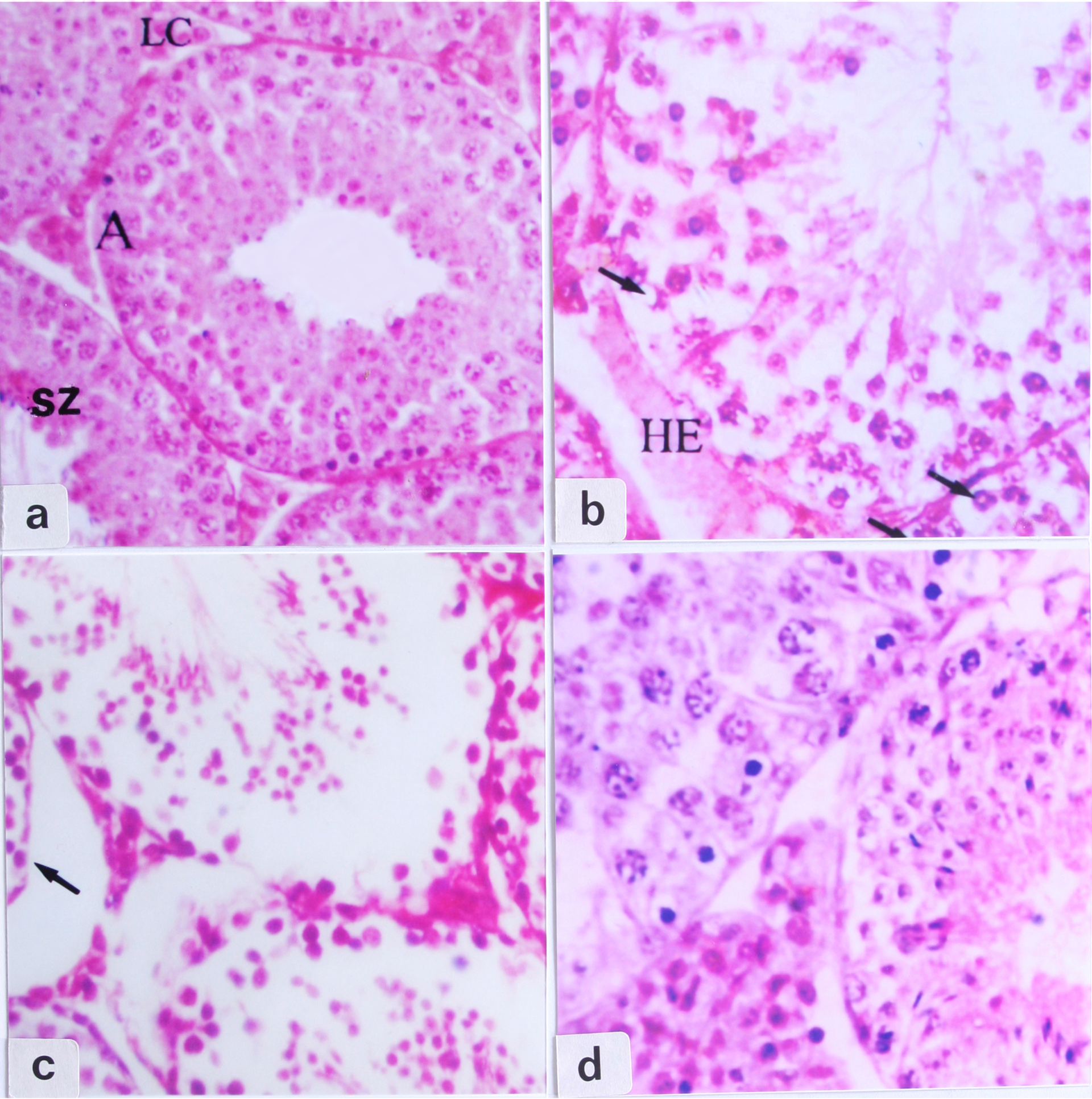
Sections of testes of (a) a control mouse showing spermatogonia A (A), spermatozoa (SZ) and Leydig cell (LC), (b) CPA-treated mouse for 4 weeks showing rupture of the spermatogenic layers, intertubular hemorrhage (HE), cytoplasmic vacuolization of the spermatogonia (arrows), (c) an animal treated with CPA for 8 weeks showing degeneration and sloughing off of the germinal epithelium (arrow) and (d) an animal treated with CPA and fenugreek for 8 weeks showing improvement of seminiferous tubules (haematoxylin and eosin (H&E), ×400).
Ultrastructural observations
Control group
In normal testis, there are two types of spermatogonia: A and B. Type A spermatogonia appeared oval shaped with flattened nucleus oriented parallel to the basal lamina, with a fine, light and non-condensed chromatin granulation (Figure 4a). Type B spermatogonia were round in shape and their nuclei characterized by heavily stained chromatin patches (Figure 4b). Sertoli cell extends from the basal lamina to the lumen of the seminiferous tubule. It appeared with a large pale oval nucleus with infolded nuclear membrane, a relatively homogeneous chromatin material and one or two nucleoli. The cytoplasm contains abundant smooth endoplasmic reticulum, ovoid Golgi apparatus and spherical or cylinder-shaped mitochondria. The spermatocytes are differentiated into two types; primary and secondary spermatocytes. Primary spermatocyte appeared with large spherical nucleus with fine granular chromatin and clusters of Golgi apparatus accumulated at one pole of the cell. The secondary spermatocytes are rarely seen among the germinal cells. They are smaller in size than the late primary spermatocytes and their nuclei are spherical with centrally located chromatin clumps. Round spermatids in Golgi phase and different stages of acrosomal cap formation were seen (Figure 4c). The elongated spermatids with dark nuclei (condensed chromatins) and normal tail formation were observed. Figure (4d) illustrated Leydig cell contained large nucleus with a thin rim of heterochromatin. The cytoplasm of Leydig cell is eosinophilic rich with small lipid droplets, mitochondria of a moderate size and abundant smooth endoplasmic reticulum, which account for their eosinophilia. Oral treatment with fenugreek for 4 or 8 weeks did not show any alternation in testis structure when compared with control animals.

Electron micrographs of portions of seminiferous tubules of control mice showing (a) normal basal lamina (BL) and spermatogonia A (A; ×7500), (b) spermatogonia B (B), Sertoli cell (SC) and primary spermatocyte (PS; ×5000), (c) round spermatids (RS; ×5000) and (d) Leydig cell (LC), with fat droplets (FD; ×7500).
Treated groups
When animals were treated with CPA for 4 weeks, different ultrastructural changes were seen in their testes. Basal lamina appeared degenerated, while spermatogonia appeared with irregular nuclei (Figure 5a). Irregular round spermatids with abnormal darkly stained nuclei were observed (Figure 5b). In addition, abnormal, fragmented and elongated spermatids were illustrated in Figure (5c). Within the interstitial tissue, degenerated Leydig cells with irregular nuclei were observed (Fig. 5d).

Electron micrographs of portions of seminiferous tubules of animals treated with CPA for 4 weeks showing (a) degenerated basal lamina (BL) and degenerated spermatogonium A (A; ×4000), (b) abnormal round spermatids (RS) with abnormal nucleus (N; ×5000), (c) fragmented elongated spermatids (arrows; ×7500) and (d) abnormal Leydig cell (LC) with irregular nucleus (N; ×7500).
Seminiferous tubules obtained from animals treated with CPA for 8 weeks were severely affected. Figure (6a) illustrated degenerated basal lamina, abnormal Sertoli cell and type A spermatogonium with irregular cell membrane and degenerated nucleus. The spermatocytes appeared with degenerated cytoplasmic organelles. The germinal epithelium showed many spaces between the spermatogenic cells. Abnormal round spermatids with abnormal nuclei were seen in Figure (6b). Figure (6c) showed abnormal round spermatid with degenerated acrosomal cap and malformed elongated spermatids. Furthermore, interstitial tissue appeared degenerated and contained apoptotic Leydig cells (Figure 6d).

Electron micrographs of portions of seminiferous tubules of mice treated with CPA for 8 weeks showing (a) degenerated basal lamina (BL), degenerated spermatogonium A (A), and abnormal Sertoli cell (SC; ×7000), (b) abnormal round spermatids (RS) with abnormal nuclei (×4000), (c) abnormal round spermatid (RS) and abnormal elongated spermatids (arrows; ×7500) and (d) apoptotic Leydig cell (LC; ×5000).
Testes of animals treated with CPA and fenugreek for 4 weeks showed some improvement. Most spermatogenic cells appeared in the normal location within the seminiferous tubules, while some of them still showed different abnormalities. Figure (7a) showed normal basal lamina with myoid cell, and normal type B spermatogonia with normal nucleus.

Electron micrographs of portions of seminiferous tubules of mice treated with CPA and fenugreek showing (a) basal lamina (BL) containing a thin myoid cell (MC), and type B spermatogonia (B; ×7500), (b) normal Sertoli cell (SC) and normal spermatogonia B (B; ×5000), (c) normal round spermatid with acrosomal cap (RS) and elongated spermatid during tail formation stage (arrow; ×5000) and (d) normal Leydig cell (LC; ×5000).
Advanced degree of improvement was observed after treatment for 8 weeks with CPA and fenugreek. Fugure (7b) showed normal Sertoli cell and normal type B spermatogonia resting on normal basal lamina. Figure (7c) showed normal round spermatid with normal acrosomal cap and elongated spermatids during the tail formation stage. In the same specimen, Leydig cells appeared mostly normal (Figure 7d).
Discussion
Treating mice with CPA revealed several histomorphological alterations such as reduction in the diameter of the seminiferous tubules, degeneration of the interstitial tissue, loss and degeneration of the spermatogenic cells. These alterations were more prominent in animals treated for 8 weeks. These results are in agreement with many investigators who reported that a period of 3–6 weeks of CPA treatment leads to testicular toxicity. Gamal El-Din et al. (1993) reported decrease in the number of both primary spermatocytes and spermatids in both rats and mice treated with daily therapeutic dose of CPA for 1 week. Elangovan et al. (2006) reported that administration of CPA once a week for 5 weeks caused oligospermia, azoospermia and testicular damage in mice. DNA damage in post-meiotic germ cells was induced in male Sprage Dawley rats given CPA for 6 weeks (Sawyer and Brown, 2000). Tripathi and Jena (2008) reported that treating mice with CPA once per week for 5 weeks caused germ cell toxicity, and astaxanthin had protective effect against this toxicity. The particular sensitivity of the reproductive tissues to CPA is due to the high proliferating activity (Jarrelle et al., 1991). Matsui et al. (1995) studied testicular toxicity of CPA in rats by quantitative morphometery of spermatogenic cycle stage and indicated that the toxic effect of CPA on spermatogensis was detected from Day 7 after single administration.
Concerning the ultrastructural changes within the testis in the present study, degenerated lamina propria, degenerated spermatogenic stages and abnormal Sertoli cells were detected. In addition, interstitial tissue contained degenerated Leydig cells with apoptotic nuclei, degenerated organelles and large number of vacuoles. El-Seedy et al. (2005) indicated that marked increase in sperm abnormality induced by CPA in mice proved the ability of this drug to interfere with different processes of spermatogenic cells. The same authors also detected extensive mitochondrial damage presented as extensive vacuolization and loss of cristae in sperms of these animals. They concluded that these abnormalities may be resulted directly from DNA damage or at specific level of differentiation of spermatozoa during development.
It was reported that the toxic effect of CPA is attributed to its metabolites phosphamide mustard and acrolin (Plowchalk and Mattison, 1991). Metabolism of CPA takes place in the liver and undergoes metabolic activation by cytochrome p 450 isoenzyme 2B. The major circulating metabolite of CPA, 4-hydroxy cyclophosphamide, is in equilibrium with aldophosphamide, which is spontaneously broken down to produce phosphoramide mustard and acrolein (Zhang et al., 2005). Metabolites of CPA (phosphamide mustard and acrolein) were found to reduce steroidogensis (Ataya et al., 1988; Hoorweg-Nijman et al., 1992).
Examination of sera of animals treated with CPA in the present study revealed a significant reduction in the activities of SOD and CAT and increase in lipid peroxidation. Similarly, Kehrer and Biswal (2000) indicated that CPA-induced depletion of glutathione peroxidase which is primarily mediated by interaction of its reactive metabolite acrolein with glutathione peroxidase. The same authors also indicated that acrolein not only interacts with glutathione peroxidase but also with cysteine which is one of the constituent amino acids of glutathione peroxidase. Reduction in glutathione, CAT and SOD also recorded in various tissues as a result of CPA treatment (Haque et al., 2003; Lopez and Luderer, 2004). Türk et al. (2010) reported that CPA induced testicular lipid peroxidation and apoptosis in male rats.
Lipid peroxidation has been reported to increase in different tissues including testis of CPA-exposed animals (Adams and Klaidrnan, 1993; Rezvanfar et al., 2008; Uchida, 1999). It has been suggested that this take place by binding to nucleophilic amino acids leading to the depletion of glutathione peroxidase (Kehrer and Biswal, 2000).
In the present study, when aqueous extract of fenugreek seeds was administered to the animals together with CPA, it improved the histopathological, ultrastructural and biochemical changes induced by CPA in the testis. Similarly, Khalil (2004) indicated that when diabetic rats were treated with aqueous extract of fenugreek seeds, marked recovery of testis and well-developed spermatogenic activity and Leydig cells were seen. Hamden et al. (2010a) reported that oral treatment of diabetic rats with fenugreek steroids improved the histological appearance of testis and epididymis with significant decrease in sperm shape abnormalities. Thakran et al. (2004) indicated that administration of Trigonella seed powder for 3 weeks to diabetic rats prevented the ultrastructural alterations observed in hepatocytes. Hamden et al. (2010b) reported fenugreek oil restored almost a normal architecture of kidney in diabetic rats, which may be attributed to its immunomodulatory activity along with its antioxidant potential.
The biochemical results in the present study indicated that treatment with CPA and fenugreek improved the elevated antioxidant enzymes and decreased lipid peroxidation. Hamden et al. (2010b) reported that the increased level of lipid peroxidation in diabetic rats decreased significantly after treatment with fenugreek oil. The authors attributed these results to its antioxidant potential. Gupta and Nair (1999) attributed the antioxidant effect of fenugreek to its richness in flavonoids. Choudhary et al. (2001) suggested that fenugreek seeds could modulate the activity of SOD, CAT and glutathione-S-transferase. The authors added that fenugreek seeds seem to have a dual effect on the tissue as is evident from the enhanced antioxidant status at lower doses and pro-oxidant action at higher doses. Wang et al. (2010) isolated 10 different flavonoids namely, 5,7,3’-trihydroxy-5’-methoxylisoflavone, biochanin A, formononetin, irilone, tricin, daidzein, calycosin, orientin-2’’-O-p-trans-coumarate, vitexin-2’’-O-p-trans-coumarate and tricin-7-O-beta-D-glucopyranoside from fenugreek seeds. Belguith-Hadriche et al. (2010) confirmed the concept that the antioxidant activity of fenugreek could be attributed to the presence of flavonoids which act as scavengers of reactive oxygen species. In conclusion, CPA was found to impair mice testicular structure by inflicting oxidative stress. Fenugreek seed extract effectively ameliorated the toxicity induced by CPA in testes of mice through enhancement of the activities of the antioxidant enzymes (CAT and SOD) and reduction in lipid peroxidation. These findings raise the possibility that fenugreek seed extract may be used as an adjuvant therapy, potentially protecting the testes from oxidative action related to CPA and might help ultimately to prevent the devastating adverse effect of CPA in clinical practice.
Footnotes
This research received no specific grant from any funding agency in the public, commercial or not-for-profit sectors.