
Abstract
Occupational exposure in a copper smelting industry may produce various adverse health effects including cancer. Despite a number of well-documented studies reporting an increased risk of cancer among copper smelter workers, the data on genotoxic effects in this industry are scarce. In view of the above, an assessment of DNA damage in peripheral blood leukocytes by Comet assay from copper smelter workers was undertaken. Additionally, the proton-induced X-ray emission (PIXE) analysis was assessed to determine the metal content of samples. The study was conducted with all workers from a copper smelter (males; n = 11), and a control group (n = 11) was recruited. The results of our study showed a significant increase (p < 0.001; Mann-Whitney test) in DNA damage in peripheral blood lymphocytes of smelter workers, compared to the controls (p < 0.001; Mann-Whitney test). No correlation between DNA damage or metal concentration and age mean or time of exposure was found under study. Our findings indicate that copper smelter workers have increased levels of DNA damage in somatic cells, suggesting a potential health risk for the workers. PIXE results show the presence of copper, iron, and other metals.
Introduction
Humans have been in contact with metals almost since the beginning of our existence. In fact, one cannot even think on human evolution without considering the great role played by metals in mankind’s development. Metals are involved in a wide variety of biological processes and hence are found in all living organisms. Some metals are essential for human nutrition; others are found as contaminants in foodstuffs. Copper is very common in the environment. It occurs naturally in rock, soil, water, sediment, and air, as well as in plants and animals (ATSDR, 2007).
In humans, copper is a respiratory irritant. Workers exposed to copper dust report a number of symptoms that are suggestive of respiratory irritation, including coughing, sneezing, thoracic pain, runny nose, and others, according to available epidemiologic data, which associated mainly with exposure to arsenic (ATSDR, 2007). Other risks reported are in relation to nervous system (Blom et al., 1985; Lilis et al., 1985) and renal function (Lilis et al., 1984).
There are limited data for humans and no data for animals on the carcinogenicity of inhaled copper (IARC, 1998; Lewijska et al., 2007). Similarly, data do not exist to indicate that copper can cause birth defects in humans (IARC, 1998). Despite a number of studies reporting an increased risk of cancer among copper smelters workers, the data on genotoxic effects in this industry are scarce (Lewijska et al., 2007). The few studies indicate that such effects can occur, but almost all has not been adequately documented. The findings of the study conducted among Swedish workers in Rönnskär copper smelter indicated an increase in the frequency of chromosome aberrations in the leukocytes of the exposed workers (Beckman et al., 1977; Nordenson et al., 1978). Another study with individuals from three copper smelters in southwestern Poland showed a significant increase in micronucleus frequency in peripheral blood leukocytes and in buccal epithelial cells (Lewijska et al., 2007).
Taking into account the epidemiologic evidence of increased cancer risk among copper smelters workers and the scarcity of data on the genotoxic effects in these populations, the present study was undertaken to investigate the latter end point in more detail. To this end, DNA damage was assessed in peripheral blood leukocytes from workers employed in copper smelters in southern Brazil.
Materials and methods
Study population and sample collection
The study involved 22 male subjects divided into 2 groups. The first group consisted of 11 smelters employed in smelting plants located at Cachoeirinha, Brazil. The smelters had varying duration exposure (0.2–25 years), and they were in the age group of 30–57 years. The group of copper smelter workers was formed by all individuals employed in the same smelting plant. The second group, comprising 11 subjects (26–66 years), was the control group. Healthy male individuals living in Cachoeirinha, who did not report any exposure to known genotoxins formed the control group.
All the individuals examined in the study were required to answer a Portuguese version of a questionnaire from the International Commission for Protection against Environmental Mutagens and Carcinogens (Carrano and Natarajan, 1988) and participate in a face-to-face questionnaire which included standard demographic data (age, gender, etc.) as well as questions relating to medical issues (exposure to metals, vaccinations, medication, etc.), life style (smoking, coffee, alcohol, diet, etc.), and their occupation (number of hours worked per day, time exposed to metals, use of protective measures, etc). In all the groups, individuals who smoked more than 5 cigarettes per day for at least 1 year were considered smokers.
Blood samples were obtained from individuals on the same day during the morning shift. All blood samples were collected using venipuncture and heparinized vacutainers and processed as quickly as possible to avoid the damage associated with storage, the blood cell samples being transported to the laboratories at or below 8°C and processed within 8 h of collection in order to minimize the loss of DNA and damage due to the DNA repair processes.
This study was approved by the Brazilian National Ethical Committee on Research (Comissão Nacional de Ética em Pesquisa—CONEP) and informed written consent was obtained from each individual prior to the start of the study.
Comet assay
The alkaline Comet assay was performed as described by Singh et al. (1988), with the modifications suggested by Tice et al. (2000) and Heuser et al. (2007). Blood cells (5 μl) were embedded in 95 μl of 0.75% low melting point agarose and when the agarose had solidified on the slides (two slides per whole blood sample), they were placed in lysis buffer (2.5 M NaCl, 100 mM EDTA, and 10 mM Tris; pH 10.0–10.5) containing freshly added 1% (v/v) Triton X-100 and 10% (v/v) dimethyl sulfoxide (DMSO) for a minimum of 1 h and a maximum of 2 weeks. After treatment with lysis buffer, the slides were incubated in freshly made alkaline buffer (300 mM NaOH and 1 mM EDTA; pH > 13) for 20 min and the DNA was electrophoresed for 20 min at 25 V (0.90 V/cm) and 300 mA after which the buffer was neutralized with 0.4 M Tris (pH 7.5) and the DNA stained. For silver staining, the slides were fixed for 10 min in trichloroacetic acid 15% (w/v), zinc sulfate 5% (w/v), glycerol 5% (v/v), washed 3 times in distilled water and dried for 2 h at 37°C. The dry slides were rehydrated for 5 min in distilled water and then stained (sodium carbonate 5% w/v, ammonium nitrate 0.1% w/v, silver nitrate 0.1% w/v, tungstosilicic acid 0.25%, formaldehyde 0.15% w/v, freshly prepared in the dark) and shaken constantly for 35 min. The stained slides were washed twice with distilled water and submerged in the stop solution (acetic acid 1%) and washed again (Villela et al., 2007). To demonstrate the electrophoresis conditions and efficiency, negative and positive controls from human blood, collected in the laboratory, were used for each electrophoresis treatment. For a positive control, slides were exposed to UV radiation (254 nm). The results of each electrophoresis was considered only if the negative and positive controls demonstrated negative and positive results, respectively.
Images of 100 randomly selected cells (50 cells from each of 2 replicate slides) were analyzed for each person. Comet image were visually allocated to one of the five classes depending on tail size (class 0 = no tails, class 4 = longest tails) to give a single-DNA damage score for each subject and hence for each group studied, the group damage index (DI) ranging from 0 (no tails on any cells, i.e. 100 cells × 0) to 400 (all cells with maximally long tails, i.e. 100 cells × 4; Collins et al., 1997; Silva et al., 2000). The damage frequency (DF [%], i.e. the proportion of cells with altered migration) was calculated based on the number of cells with tails versus the number of cells without tails. All the slides were scored blindly.
Analysis by PIXE
The metal content of the blood samples was analyzed through the particle-induced X-ray emission (PIXE) technique (Johansson et al., 1995). Briefly, blood samples were dried at 60°C for 6 h, ground into a fine powder, homogenized, and pressed into approximately 3-mm thick pellets, and were subsequently placed in the target holder inside the reaction chamber. During the experiments, the pressure inside the reaction chamber was about 10−5 mbar. The experiments were carried out at the Ion Implantation Laboratory of the Physics Institute of the Federal University of Rio Grande do Sul (IF-UFRGS). A 3 MV Tandetron accelerator provided a 2.0 MeV proton beam with an average current of 5 nA at the target. The X-rays produced in the samples were detected by a Si(Li) detector with an energy resolution of about 160 eV at 5.9 keV. The spectra were analyzed with the GUPIX software package, developed at the University of Guelph (Ontario, Canada), and the data are expressed in parts per million ([ppm] Campbell et al., 2000; Maxwell et al., 1995).
Statistical analysis
The normality of the variables was evaluated using the Kolmogorov-Smirnov test. Student’s t test was used to compare the basic characteristic of study populations and data normally distributed. The statistical analysis of differences in Comet assay for negative control and smelters were carried out using the nonparametric Mann–Whitney U test. Correlations between different variables were determined by Spearman rank correlation test as appropriate. The critical level for rejection of the null hypothesis was considered to be a p value of 5%. All analyses were performed with the SPSS/PC statistical software package.
Results
The main demographic characteristics of the unexposed control and copper smelters workers are presented in Table 1. No significant differences in average age were detected between the groups (t test). The studied exposed and control groups generally had similar age and number of smokers. Five volunteers were considered smokers (smoking five or more cigarettes per day) in both studied groups. No statistical differences were found in smoking habit between the groups. With regard to the use of protective measures, 100% of copper smelters used silicone gloves against dermal contact with solvents. All smelting plants had workplace ventilation.
Evaluation of genetic damage in Brazilian copper smelter workers: main demographic characteristics of studied subjects
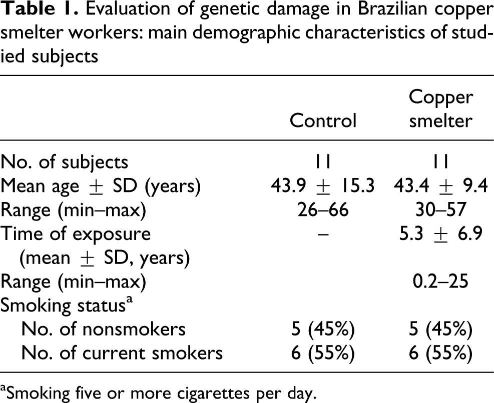
aSmoking five or more cigarettes per day.
The Comet assay data for unexposed control and copper smelters workers groups are presented in Table 2. Data are shown only in DI because only class 1 was observed for every individual, and DI value is the same as that of DF. No differences were observed between smokers and nonsmokers or less than and more than 40 years old or less than and more than 2 years of exposition to copper. The Comet assay analysis of the values (mean ± SD), considering all individuals as the same group, indicated a significant increase in DI for the copper smelters workers group in relation to the control shown in Figure 1 (p ≤ 0.001; Mann–Whitney U test).
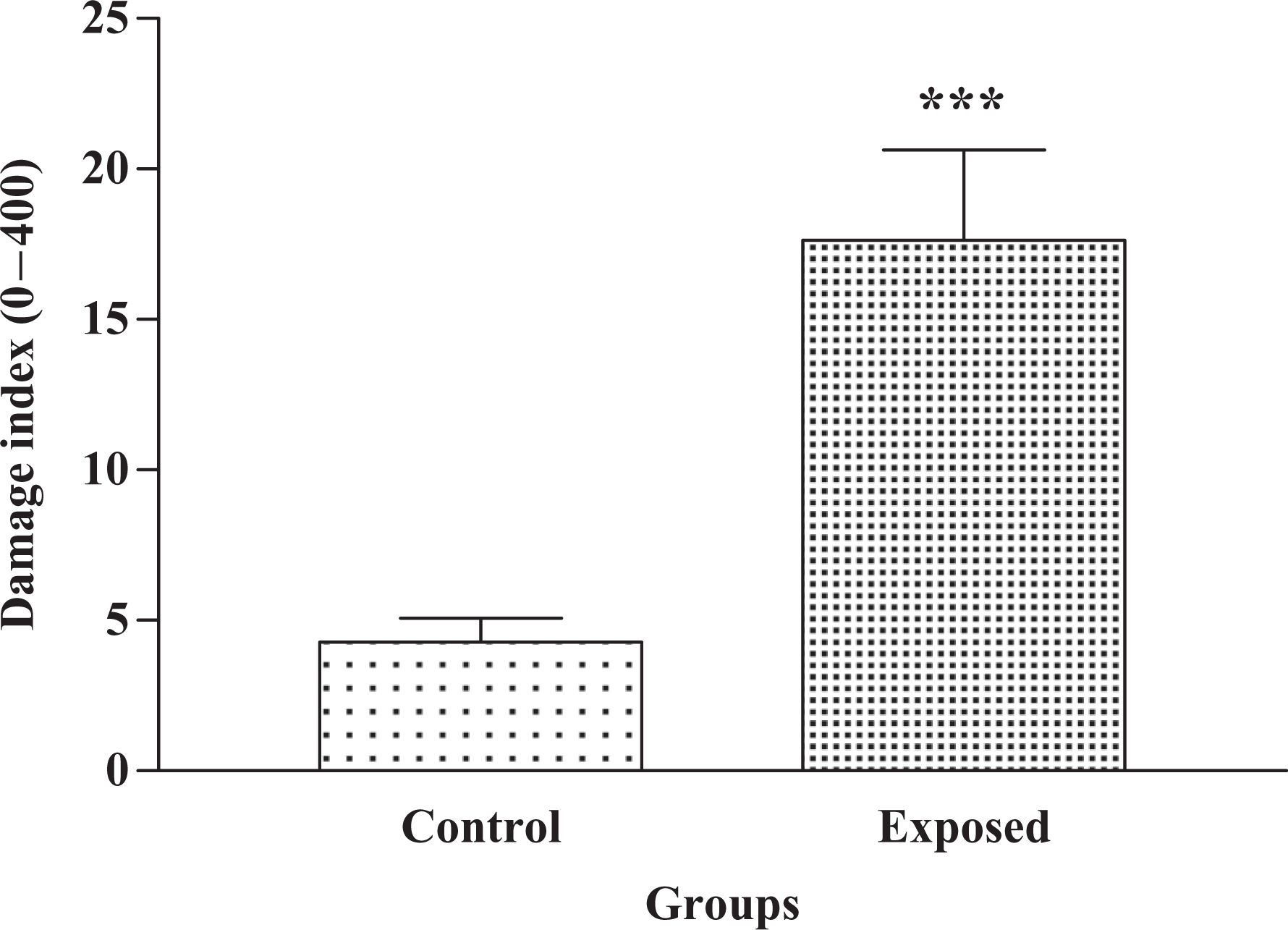
Mean and standard error of the control (n = 11) and exposed group (copper smelter workers; n = 11). ***p < 0.001 in relation to control individuals (Mann-Whitney U test).
Mean values of damage index (mean ± SD) obtained by Comet assay from unexposed control individuals and copper smelters workers
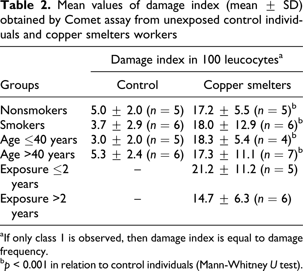
aIf only class 1 is observed, then damage index is equal to damage frequency.
b p < 0.001 in relation to control individuals (Mann-Whitney U test).
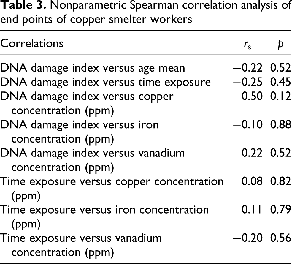
With respect to the significant results observed by Comet assay, we used the Spearman rank test to investigate the possible correlations between age and DNA DI, time exposure and DNA DI, and time exposure and metal concentration. There were no significant correlations between all results (Table 3).
Nonparametric Spearman correlation analysis of end points of copper smelter workers
The inorganic elements detection by PIXE analyses content in the peripheral blood samples of unexposed control individuals and copper smelters workers are shown in Table 4. Most of the inorganic elements related to human activities were present in the samples, and the concentrations (mean ± SD) of copper, iron, zinc, sodium, magnesium, aluminum, phosphorus, sulfur, chlorine, potassium, calcium, vanadium, cobalt, bromide, and rubidium were obtained, showing some differences between copper smelter workers in relation to the unexposed control. The limit of the level of copper in whole blood in nonexposed individuals is 1–2.17 ppm (Moreno et al., 1999; ATSDR, 2007). Arsenic could not be detected in blood samples (detection limit was about 27 ppm).
Inorganic element detection by PIXE analyses in the blood samples of unexposed individuals and copper smelter workers
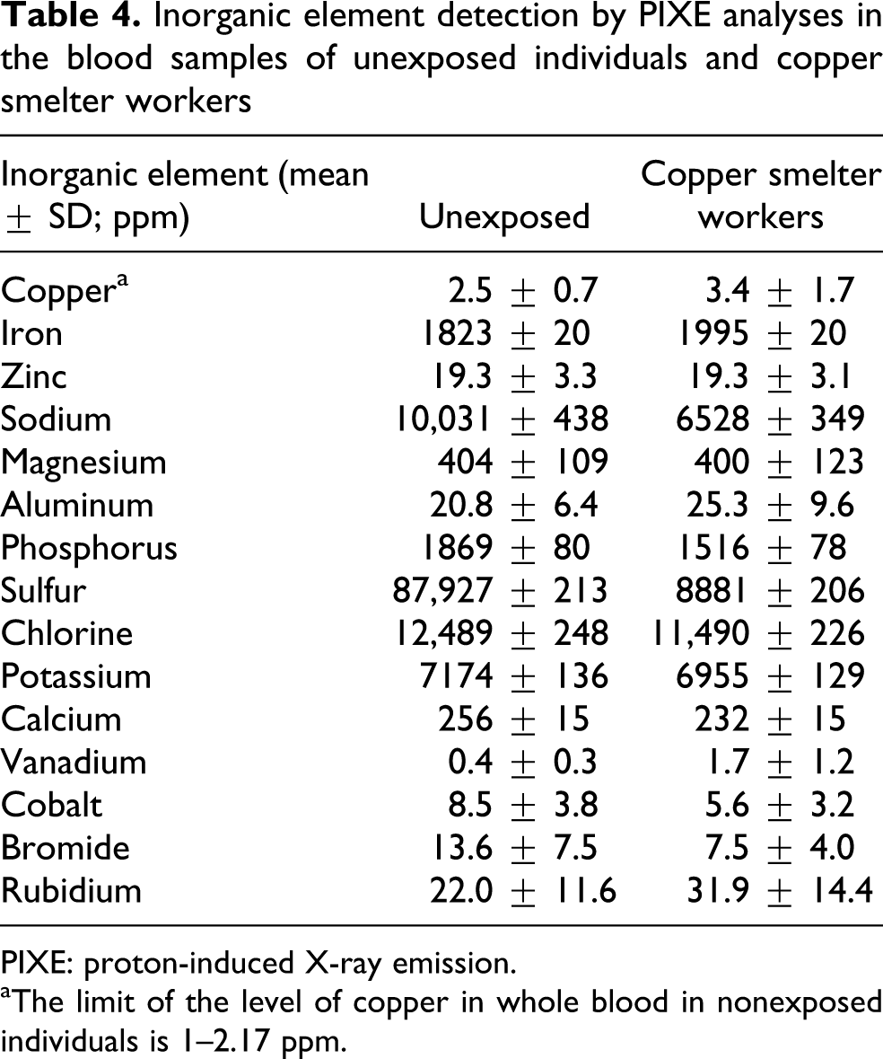
PIXE: proton-induced X-ray emission.
aThe limit of the level of copper in whole blood in nonexposed individuals is 1–2.17 ppm.
Discussion
The present results revealed a significant increase in comet assay DNA damage in peripheral blood of the workers occupationally exposed in a Brazilian copper smelter. Beckman et al. (1977), Nordenson et al. (1978), Palus et al. (2005), Paiva et al. (2006) and Lewijska et al. (2007) were the only authors that associated chromosomal alterations in the blood samples of copper smelter workers. However, all of them indicated arsenic as the major health hazard, none of them found results associating copper in their results. In our results from PIXE no arsenic was detected in blood samples of workers; PIXE for the speciation of arsenic compound can be discussed, and the improvement in the technique with decrease in the limit of detection of the analyses should be made. In the present study, results of biomonitoring measurements were also limited to PIXE analyze; however, it is obvious that a role of other agents in inducing the observed genotoxic events, like polycyclic aromatic hydrocarbons (PAHs), cannot be excluded. No associations were found between age or exposure time and Comet assay parameters or between age or exposure time and metals detected in this study.
PIXE has been successfully employed to detect trace elements in plants and organisms because of its multielemental character, high sensitivity, simplicity, and high sample throughput (Mireles et al., 2004). The PIXE technique demonstrated small increases in concentrations of copper, iron, magnesium, and aluminum in copper smelter workers in relation to unexposed control.
Copper levels can be measured in tissues, body fluids, and excreta. Whole blood, serum, and urine copper levels have been established in healthy individuals. It has been demonstrated that copper levels in the body increase with increased exposure after acute poisoning. Elevated tissue copper levels should be a sufficient indicator of exposure and the possibility of intoxication (ATSDR, 2007). Copper is relatively easy to absorb. About 70% of dietary copper (about 1 mg) is absorbed daily by the human adult, and larger amounts are recycled from digestive tract secretions (perhaps an additional 3 mg Cu per day). In most mammals, copper is also easy to excrete. It has become clear that when excess copper is administered, or alternatively when there is a lack in the diet, the body adjusts its net excretion (Dunn et al., 1991; Linder, 1991; Turnlund et al., 1989, 1998a, b). Generally, also, excess copper is not ‘stored.’ As a result, the total copper content in the body tends to be constant. It has thus been generally accepted that copper homeostasis is mainly controlled by regulation of excretion (Linder et al., 2003). This fact can explain our lack of association between age or exposure time and copper concentration or Comet parameters in this study.
Iron and copper, these two metal ions, influenced each other’s transport. Both the presence of iron and copper leads to the generation of reactive oxygen species (ROS) that can attack biomolecules directly, with the consequent enhancement in membrane lipid peroxidation, DNA damage, and protein oxidation (Videla et al., 2003). The ROS generated by this process could attack DNA, leading to base damage and DNA strand excision. The true strand breaks and/or the strand breaks formed as an intermediate step in excision repair of altered bases could explain the increased DNA damage in the comet assay. Our results showed only classes 0 and 1, which showed a possible exclusion of double strand break of DNA. These results are in accord with those of other authors who associated metal exposition and single strand break (Hengstler et al., 2002; Villela et al., 2007).
Our data are in agreement with those of Bolognesi et al. (1999), Guecheva et al. (2001), and Villela et al. (2007), which reported Cu-induced DNA damage detected by the alkaline elution technique. Shubber et al. (1998) studied women using the copper-containing intrauterine contraceptive device (IUCD), they observed a combination of high copper plasma level, chromosomal aberrations, and the increased frequency of sister chromatid exchange with a positive correlation between the long-term use of the IUCDs and DNA damage.
Vanadium, the other metal detected, is a transition metal widely distributed in the environment and in biological systems, it is a major trace element in fossil fuels and also found associated with copper smelter emissions. Research on biological influence of vanadium has gained major importance because it is well known that it exerts potent toxic, mutagenic, and genotoxic effects on a wide variety of biological systems, including that recently compounds as the vanadium pentoxide has been classified by the International Agency for Research on Cancer (IARC, 2006) as a possible carcinogenic agent for humans. Information about the clastogenic effects of vanadium compounds is limited and controversial, and data about its mutagenic and genotoxic potential in bacterial, yeast, and plants are inconclusive. On the other hand, results obtained in mammalian cells, both in vivo and in vitro, indicate that vanadium compounds produce mutagenic and genotoxic responses, however, the most evident action exhibited by this metal compounds is their ability to disrupt microtubule function, and consistent cytotoxic and cytostatic effects. By these reasons, some authors classify vanadium as a weak mutagen (Rodriguez-Mercado and Altamiro-Lozano, 2006).
To conclude, our results showed that occupational exposure in copper smelters may pose some risk of increased DNA DI in peripheral blood leukocytes of the workers, mainly related with the metals copper, iron, and vanadium. Although these metals were present in blood samples of workers, no clear association could be made between these metals and DNA damage (without correlation). Finally, further knowledge of DNA damage mechanisms in peripheral blood workers occupationally exposed in copper smelters is necessary in order to support repair studies by comet assay. However, the best remedy for occupational exposure is prevention. Workers in many occupational settings are exposed to certain genotoxic agents. These workers may not be aware that they have been exposed to genotoxic agents nor do they know the type and amount of agent to which they have been exposed. Therefore, there is a need to educate those who work with metals about the potential hazard of occupational exposure and the importance of using protective measures.
Footnotes
Acknowledgments
The authors express their gratitude to all the individuals who volunteered to participate in this study. The authors also thank the technicians of the Laboratório de Implantação Iônica, Instituto de Física, UFRGS for the PIXE analysis, and the Laboratório de Genética Toxicológica, ULBRA, that supported this research.
Conflict of interest
The authors declared no conflicts of interest.
Funding
This research received no specific grant from any funding agency in the public, commercial, or not-for-profit sectors.