
Abstract
Effect of standardized Bacopa monniera (BM; family: Scrophulariaceae) extract (100 and 300 mg/kg) against sodium fluoride (NaF; 100 and 200 ppm)-induced behavioural, biochemical, and neuropathological alterations in mice was evaluated. Akinesia, rotarod (motor coordination), forced swim test (depression), open field test (anxiety), transfer latency (memory), cholinesterase (ChE), and oxidative stress (superoxide dismutase, catalase, glutathione peroxidase, and lipid peroxidation) were determined in mice treated with NaF for 30 days alone and in combination with BM. NaF induced motor incoordination, depression, and memory impairment, and these were prevented by coadministration of BM in mice. However, NaF did not alter the weight gain, feed/water consumption, and anxiety profile. Suppression of ChE levels and increased oxidative stress were observed in mice treated with NaF. Coadministration of BM significantly improved the memory, ChE levels, and antioxidant enzymes but failed to alter the fluoride levels in NaF-treated mice. Histopathological studies revealed that BM protected the neuropathological alterations induced by NaF.
Introduction
Fluorosis, a state of chronic poisoning from long-term exposure to excessive quantities of fluorine, is a serious health problem in countries where the fluoride content of water is more than 1 ppm (Kaushik et al., 2001). Due to its relatively large electronegativity, fluoride combines with many elements and forms ionized fluorides that are capable of accumulating in the body causing tissue and organ damage (Eraslan et al., 2007). In addition to the well-known effects of fluorosis on the skeleton and teeth, the neurotoxicity of fluoride was also confirmed in humans and animals (Zhang et al., 2007). Studies on animals demonstrated that chronic exposure to fluoride produces memory impairment, neuropathological changes, increased oxidative stress, and affects neurotransmitters in certain regions of brain (Niu et al., 2009). Even though, the fluoride toxicity is well characterized, only few studies have been so far carried out to reverse the effects of fluoride using agents such as calcium carbonate (Ekambaram and Paul, 2001, 2002), black-berry fruit juice (Hassan and Abdel-Aziz, 2010), and vitamins and minerals (Chinoy and Memon, 2001). However, no previous study exists to our knowledge on the alleviation of fluoride-induced behavioural changes, such as motor incoordination, depression, anxiety, and memory impairment in mice.
Bacopa monniera (BM) (L.) Wettst (Brahmi, family: Scrophulariaceae), a traditional ayurvedic medicinal plant, is extensively used for centuries for the treatment of epilepsy (Mathew et al., 2010), insomnia, anxiety (Sandhya et al., 2012), and also as a memory enhancer (Stough et al., 2008). Besides, BM has been reported for its antidepressant, adaptogenic, and antioxidant properties in experimental animals. Several clinical studies have confirmed the beneficial actions of BM and its pharmacological actions are mainly attributed to the bacosides present in the alcoholic extract of the plant (Russo and Borrelli, 2005). Based on these merits, the present study aimed to evaluate the standardized extract of BM against fluoride-induced behavioural, biochemical, and neuropathological alterations in female mice.
Materials and methods
Chemicals
Standardized BM extract was a generous gift from M/s Natural Remedies (Bangalore, Karnataka, India). It was derived from an alcoholic extract of the BM herb (herb to extract ratio, 20:1). The extract was standardized to contain total bacosides 40.0–50.0%, along with a number of chemical constituents, namely bacoside A3 (>2.7%), bacopaside II (>3.6%), Jujubogenin isomer of bacopa saponin C (>4.5%), bacopa saponin C (>3.0%), bacopaside I (>4.5%), apigenin (0.1–0.5%), and luteolin (0.1–0.8%). Sodium fluoride (NaF) was obtained from Himedia (Mumbai, Maharashtra, India), and all the other chemicals and reagents used were of AR grade obtained from SRL Ranbaxy (Mumbai, Maharashtra, India).
Animals
Swiss albino female mice (25–27 g) were procured from the central animal facilities of JSS College of Pharmacy (Ootacamund, Tamil Nadu, India). The mice were housed in colony cages at the temperature of 25 ± 2°C and relative humidity of 40–65%, with a 12:12-h light:dark cycle. The mice had free access to standard pellet chow and drinking water. The study was approved by Institutional Animal Ethics Committee and the work was carried out as per Committee for the Purpose of Control and Supervision of Experiments on Animals (CPCSEA) guidelines, New Delhi.
Preselection of mice
Before grouping, mice were trained for 2 days in order to remain for 180 s on a rod rotating at constant speed of 25 r/min (Jones and Roberts, 1968). Three trials per day for 2 days were enough for the mice to learn this task. On the day of the test, only the mice that were able to stay balanced on rotating rod for 180 s were selected for the study.
Drug preparation and administration
The dose of NaF to be treated was fixed based on the previously reported study (Chioca et al., 2008). The mice were exposed to NaF (100 and 200 ppm) for 30 days by dissolving it in drinking water and allowed to have free access to water. Dose for the BM extract (100 and 300 mg/kg) was fixed based on a previous study (Rao et al., 2000). The BM extract was administered orally as a fine suspension in 0.3% (w/v) carboxy methyl cellulose (CMC) daily to NaF (100 or 200 ppm)-exposed mice. All the behavioural tests were carried out 1 hour after administration of BM extract.
Experimental design
Mice were divided into seven groups of seven female mice each. Group I served as control and mice were given access to nonfluoridated water, and in addition 0.3% (w/v) CMC was orally administered daily. Groups II and III received NaF 100 and 200 ppm, respectively, through drinking water and in addition 0.3% (w/v) CMC was administered orally for both the groups. Groups IV and V received NaF 100 ppm, and BM was coadministered orally at the doses of 100 and 300 mg/kg, respectively. Groups VI and VII received NaF 200 ppm, and BM was coadministered orally at the doses of 100 and 300 mg/kg, respectively.
Body weight, water, and feed intake
Weekly body weight of the mice was measured during the study period. In order to calculate the daily water/feed intake, a measured amount of water/feed was kept in the cages and the left over was measured 24 h later.
Akinesia
Akinesia was measured by observing the latency to move all the four limbs and the test was terminated if the latency exceeded 120 s. Each mouse was initially acclimatized for 5 min on a wooden elevated (30 cm) platform (40 × 40 cm2). Using a stopwatch, the time taken by the mice to move all the four limbs was recorded (Luthra et al., 2009). Three trials were conducted in each mouse at 5 min intervals. The experiment was performed on the 30th day of the study period.
Rotarod test
On 30th day of the study period, mice were subjected to rotarod test to assess the motor coordination of mice based on the ability of mice to stay on the rotating rod (25 r/min) with a cut-off time of 180 s as described previously.
Forced swim test
Mice were subjected to swim test in a water beaker (20 × 15 × 15 cm3). The temperature of the water was maintained at 27 ± 2°C. The mice were left to swim in the water for 10 min before being removed, dried with paper towels, and returned to their home cage. The procedure was repeated again in 24 h for a 5-min test session and the duration of immobility was observed. Mice were judged to be immobile whenever it remained floating passively in the water in a slightly hunched but upright position, its nose just above the surface (Porsolt et al., 1978). The training session was given on day 29 and test session was conducted on day 30 of the study period.
Open field test
The open-field area was made of acrylic transparent walls and black floor (30 × 30 × 15 cm3) divided into nine squares of equal area. The experiment was carried out in a sound attenuated room under dimly lighted illumination. The open field instrument was cleaned with 70% alcohol after each test session to prevent the next mouse from being influenced by the odours deposited in the urine and faeces of the previous mouse. Each mouse was placed in the central square and observed for 10 min. The following behaviour were recorded: ambulation: the number of grid lines it crossed with all the four paws; immobility period and time spent in centre; rearing: by counting the number of times the mice stood on its hind limbs; grooming: number of times the mice made these responses, namely grooming of the face, licking/cleaning, and scratching various parts of the body; defaecation: the number of faecal boli excreted during the test period (Sonavane et al., 2002). The experiment was performed on 30th day of the study period.
Transfer latency using EPM
The elevated plus maze (EPM) test was used to evaluate learning/memory in NaF-treated mice and was performed as described previously by Biala and Kruk (2008). The experimental apparatus is shaped like a ‘plus’ sign and consists of a central platform (5 × 5 cm2), two open arms (30 × 5 cm), and two equal-sized closed (30 × 5 × 15 cm3) arms opposite to each other. The maze is made of dark plexiglass, elevated to a height of 50 cm above the floor, and illuminated by dim light. The test consisted in placing mice at the end of one open arm facing away from the central platform. The time taken by each mouse to move from the open arm to either of the enclosed arms transfer latency (TL) was recorded. If the mice did not enter the enclosed arm within 90 s, they were forced to enter into the enclosed arm and permitted to explore this maze for an additional 60 s; in these cases, TL was recorded as 90 s. Each group was subjected twice to the TL procedure (acquisition and retention trials, interval between trials was 24 h). Entry into one arm was recorded when a mice placed all four paws and past the line dividing the central square from the open arms. The test arena was wiped with a damp cloth after each trial. This test was carried out on days 29 and 30 of the study.
Blood, brain tissue collection, and homogenate preparation
On 30th day of the study, the blood was collected by retro-orbital sinus puncture under light ether anaesthesia in heparinized tubes to estimate the total cholinesterase (ChE) levels in plasma. Then the mice were killed by decapitation, and the brain tissues were immediately removed, washed using chilled saline solution, dried, weighed, and homogenized in potassium phosphate buffer (0.01 M, pH 7.4) containing 10% potassium chloride (KCl) in homogenizer. The homogenate was centrifuged at 2000 r/min for 10 min, and the resultant supernatant was kept for antioxidant levels, total protein, and sodium–potassium adenosine triphosphatase (Na+–K+-ATPase) estimations. Six mice from each group were used for the estimation of fluoride in brain as well as biochemical estimations, and one mouse brain from each group was processed for histopathology. For morphological observation by light microscopy, brain tissue was fixed in 10% buffered neutral formaldehyde solution and embedded in paraffin. Paraffin sections of thickness 5 μm were prepared for microscopic study. Then tissues were dipped in water and then transferred to haematoxylin and eosin stain for 10 min at 60°C. The stained sections were washed in running water to remove excess stain and then upgraded for dehydration through different grades of alcohol. Slides were cleared with xylene and mounted with DPX to make them permanent. The slides were observed at 100×.
Total ChE level
Total ChE level in the plasma was measured by the method indicated by Ellman et al. (1961) using Labkit Diagnostic Kit (UK). The ChE level was expressed in units per litre.
Superoxide dismutase
Superoxide dismutase (SOD) was assayed by taking 0.05 mL of brain homogenate followed by the addition of 0.3 mL of sodium pyrophosphate buffer (0.025 M, pH 8.3), 0.025 mL of phenazine methosulphate (186 μM), and 0.075 mL of nitroblue tetrazolium (300 μM in buffer of pH 8.3). Reaction was started by the addition of 0.075 mL of nicotinamide adenine dinucleotide (780 μM in buffer of pH 8.3). After incubation at 30°C for 90 s, the reaction was stopped by the addition of 0.25 mL glacial acetic acid. Then the reaction mixture was stirred vigorously and shaken with 2.0 mL of n-butanol. The mixture was allowed to stand for 10 min and centrifuged. n-Butanol of 1.5 mL alone served as blank. The colour intensity of the chromogen was read at 560 nm using ultraviolet (UV) spectrophotometer (Kakkar et al., 1984). The SOD activity was expressed in units per milligram protein.
Catalase
Catalase (CAT) measurement was carried out based on the ability of CAT to inhibit the oxidation of hydrogen peroxide (H2O2). Potassium phosphate buffer of 2.25 mL (65 mM, pH 7.8) and 100 μL of the brain homogenate or sucrose (0.32 M) were incubated at 25°C for 30 min. H2O2 (7.5 mM, 650 μL) were added to initiate the reaction. The change in absorbance at 240 nm was measured for 2–3 min. dy/dx for every minute for each assay was calculated, and the result was expressed in units per milligram protein (Beers and Sizer, 1952).
Glutathione peroxidase
Glutathione peroxidase (GPx) was measured by taking 50 μL of brain homogenate, 50 μL of ethylenediaminetetraacetic acid (EDTA, 5.0 mM), 50 μL of sodium azide (10 mM), 50 μL of nicotinamide adenine dinucleotide phosphate (0.2 mM), 50 μL of reduced glutathione (1 mM) in 0.6 mL of sodium phosphate buffer, pH 7.2, and 50 μL of Triton X-100 (0.01%, v/v). Finally, glutathione reductase of about 1 unit was added. The reaction mixture consisted of all the above reagents in a final volume of 2.8 mL. The reaction started by the addition of H2O2 (2.6 mM) and the absorbance was read at 340 nm against a blank. The decrease in absorbance was read for about 5 min (Paglia and Valentine, 1967). GPx activity was expressed in units per milligram protein.
Lipid peroxidation
Malondialdehyde and other thiobarbituric acid reactive substances (TBARSs) are quantified by their reactivity with thiobarbituric acid (TBA) in acidic conditions. Homogenate of 0.2 mL was taken and 0.8 mL saline, 0.5 mL of butylated hydroxyl toluene, and 3.5 mL TBA reagent (0.8%) were added and incubated at 60°C in a boiling water bath. After cooling, the solution was centrifuged at 2000 r/min for 10 min. The reaction generated a pink-coloured chromophore, the absorbance of the supernatant was read using UV spectrophotometer at 535 nm against the blank. The amount of TBARS was expressed in nanomoles per gram protein (Ohkawa et al., 1979).
Na+–K+-ATPase activity assay
Na+–K+-ATPase was assayed by taking 250 μL of Tris-HCl (184 mM, pH 7.5) buffer followed by the addition of 50 μL of 600 mM NaCl, 50 μL of 50 mM KCl, along with 50 μL of 1 mM sodium EDTA, and 50 μL of 80 mM ATP. The reaction mixture was preincubated at 37°C for 10 min. Then 25 μL of 10% brain homogenate was added to the test alone and further incubated at 37°C for 1 h. The reaction was immediately arrested by the addition of 10% Tri-Chloro Acetic acid (TCA). The control reaction was correspondingly performed by adding 25 μL of 10% homogenate only after arresting the reaction. The precipitate was removed by centrifugation at 3500 r/min for 10 min (Sovoboda and Mossinger, 1981). From the 50 μL of the supernatant, the liberated inorganic phosphorus was measured by the method of Fiske and Subbarow (1925).
Total protein estimation
The protein content of brain homogenates was quantified by the method of Lowry et al. (1951) using bovine serum albumin as a standard.
Estimation of fluoride in brain
Fluoride levels in the mice brain of different groups were determined by the method of Birkel (1970). Brain homogenate was dried for 24 h at 105°C. About 200 mg of the dry sample was dissolved in an acid mixture (equal parts of 11.6 M perchloric acid and 14.3 M nitric acid) and neutralized with citrate buffer to a pH 5.5 with an alkaline mixture of 7.8 M sodium hydroxide and 1.0 M trisodium citrate. The process was carried out in a closed compartment. The sample thus obtained was used after appropriate dilutions for recording the fluoride content on a fluorimeter (Orion R 94-09, St. Louis, Missouri). The values were calculated from a standard curve. The amount of fluoride was expressed in microgram of fluoride per gram of dry tissue.
Statistical analysis
The collected data were subjected to one-way analysis of variance (ANOVA) followed by Dunnett’s multiple comparison test. p-Values of <0.05 were considered as significant. The analysis was carried out using Graph pad prism software with version 4.0.
Results
Water/feed intake and body weight
Water/feed consumption and weekly body weight analysis (data not shown) did not show any statistically significant changes in all the groups of NaF and BM extract-treated groups when compared with nonfluoride-treated group.
Akinesia
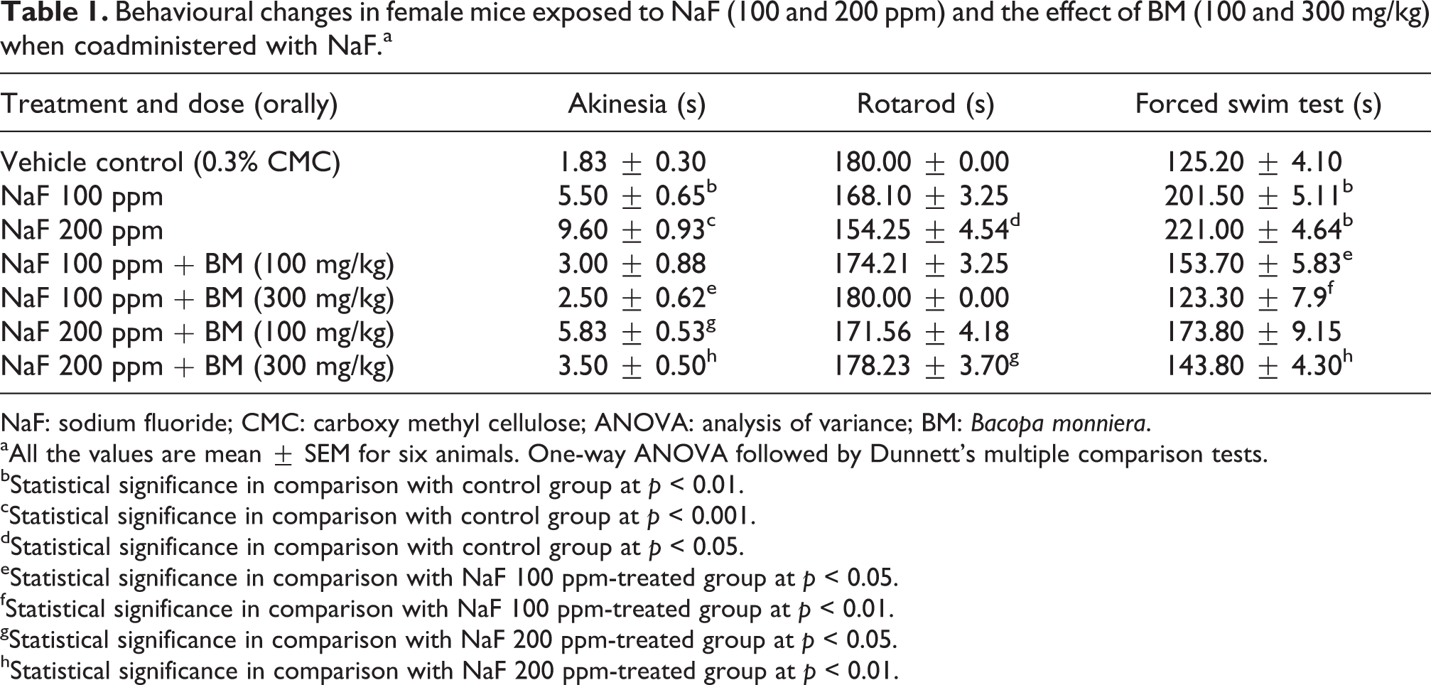
Mice that received NaF 100 and 200 ppm through drinking water exhibited akinesia in a significant manner when compared with nonfluoride-treated group. NaF 100 ppm-treated mice when coadministered with BM 300 mg/kg exhibited significant decrease in akinesia (p < 0.05) when compared with NaF 100 ppm-treated group. NaF 200 ppm-treated mice when administered with BM 100 and 300 mg/kg showed significant decrease in akinesia at p < 0.05 and p < 0.01, respectively, when compared with NaF 200 ppm-treated group (Table 1).
Behavioural changes in female mice exposed to NaF (100 and 200 ppm) and the effect of BM (100 and 300 mg/kg) when coadministered with NaF.a
NaF: sodium fluoride; CMC: carboxy methyl cellulose; ANOVA: analysis of variance; BM: Bacopa monniera.
aAll the values are mean ± SEM for six animals. One-way ANOVA followed by Dunnett’s multiple comparison tests.
bStatistical significance in comparison with control group at p < 0.01.
cStatistical significance in comparison with control group at p < 0.001.
dStatistical significance in comparison with control group at p < 0.05.
eStatistical significance in comparison with NaF 100 ppm-treated group at p < 0.05.
fStatistical significance in comparison with NaF 100 ppm-treated group at p < 0.01.
gStatistical significance in comparison with NaF 200 ppm-treated group at p < 0.05.
hStatistical significance in comparison with NaF 200 ppm-treated group at p < 0.01.
Rotarod test
NaF 200 ppm received mice exhibited significant shortening of rotarod endurance time (p < 0.05) when compared with nonfluoride-treated group. NaF 200 ppm-treated mice when coadministered with BM 300 mg/kg increased the rotarod performance significantly (p < 0.05) when compared with NaF 200 ppm-treated group (Table 1).
Forced swim test
Group of mice that received NaF 100 and 200 ppm showed significant increase in immobility time in forced swim test (p < 0.01). NaF 100 ppm received mice when coadministered with BM 100 and 300 mg/kg exhibited significant decrease in immobility period at p < 0.05 and p < 0.01, respectively, when compared with NaF 100 ppm-treated group. NaF 200 ppm-treated mice when coadministered with BM 300 mg/kg significantly (p < 0.01) decreased the immobility period when compared with NaF 200 ppm-treated group (Table 1).
Open field test
Number of lines crossed
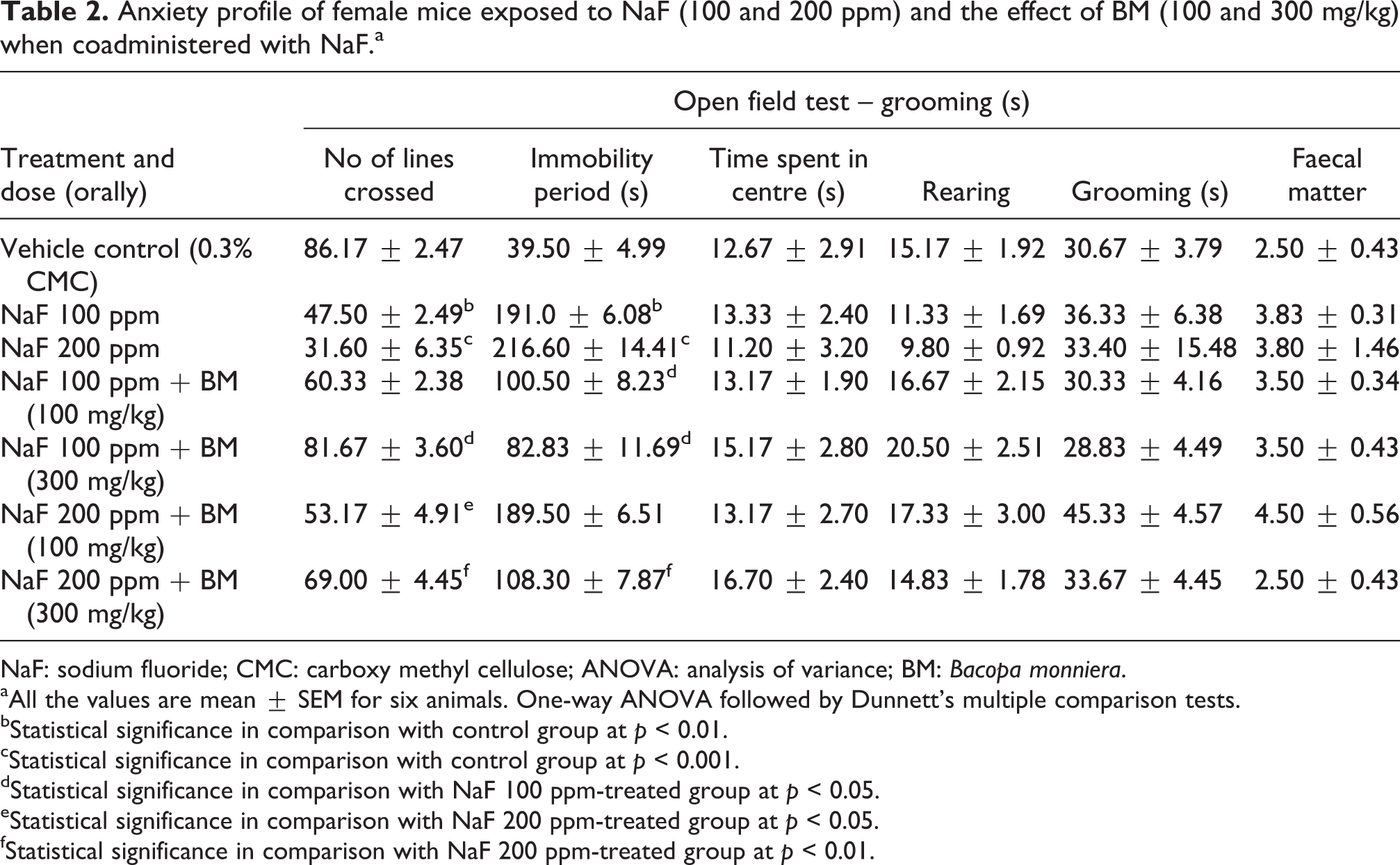
NaF 100 and 200 ppm-treated mice exhibited significant decrease in number of lines crossed when compared with nonfluoride-treated group of mice. NaF 100 ppm-treated mice when coadministered with BM 300 mg/kg exhibited significant (p < 0.05) increase in the number of lines crossed when compared with NaF 100 ppm-treated group. NaF 200 ppm-treated mice when coadministered with BM 100 and 300 mg/kg showed significant increase in the number of lines crossed at p < 0.05 and p < 0.01, respectively, when compared with NaF 200 ppm-treated group (Table 2).
Anxiety profile of female mice exposed to NaF (100 and 200 ppm) and the effect of BM (100 and 300 mg/kg) when coadministered with NaF.a
NaF: sodium fluoride; CMC: carboxy methyl cellulose; ANOVA: analysis of variance; BM: Bacopa monniera.
aAll the values are mean ± SEM for six animals. One-way ANOVA followed by Dunnett’s multiple comparison tests.
bStatistical significance in comparison with control group at p < 0.01.
cStatistical significance in comparison with control group at p < 0.001.
dStatistical significance in comparison with NaF 100 ppm-treated group at p < 0.05.
eStatistical significance in comparison with NaF 200 ppm-treated group at p < 0.05.
fStatistical significance in comparison with NaF 200 ppm-treated group at p < 0.01.
Open field test – immobility period
NaF 100 and 200 ppm-treated mice exhibited significant increase in immobility period in open field test when compared with nonfluoride-treated group of mice. BM 100 and 300 mg/kg when coadministered with NaF 100 ppm treated mice exhibited significant decrease in immobility period at p < 0.05 when compared with NaF 100 ppm-treated group. BM 300 mg/kg when coadministered with NaF 200 ppm significantly (p < 0.01) decreased the immobility period when compared with NaF 200 ppm-treated group. Other parameters such as time spent in centre, rearing, grooming, and number of faecal matter did not show any significant changes in NaF- and BM-coadministered groups (Table 2).
TL in EPM
Mice received NaF 100 and 200 ppm showed significant (p < 0.01) memory impairment when compared with nonfluoride-treated group. BM 100 and 300 mg/kg when coadministered with NaF 100 ppm significantly improved the memory retention at p < 0.05 and p < 0.01 when compared with NaF 100 ppm-treated group. NaF 200 ppm-treated mice when coadministered with BM 100 and 300 mg/kg significantly (p < 0.01) improved the memory retention capacity of mice when compared with NaF 200 ppm-treated group (Figure 1).
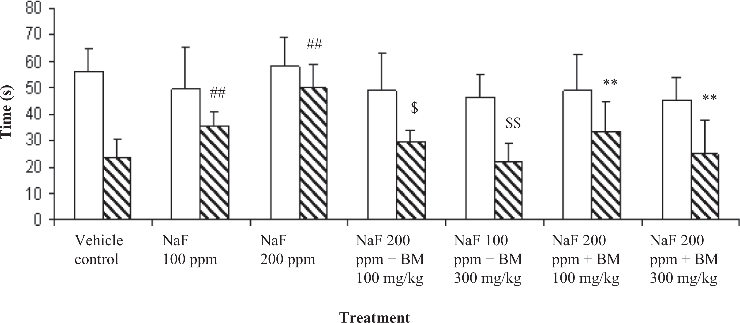
Independent effect of NaF (100 and 200 ppm) and the effect of BM (100 and 300 mg/kg) when coadministered with NaF in transfer latency in elevated plus maze test in female mice. NaF: sodium fluoride; BM: Bacopa monniera.
Total ChE level
NaF 100 and 200 ppm-treated mice showed significant decrease in total ChE level at p < 0.01 and p < 0.001, respectively, when compared with nonfluoride-treated group. BM 300 mg/kg when coadministered with NaF 100 ppm significantly elevated the total ChE level (p < 0.05) when compared with NaF 100 ppm-treated group. NaF 200 ppm-treated mice when coadministered with BM 100 and 300 mg/kg elevated the total ChE level significantly at p < 0.05 and p < 0.01, respectively, when compared with NaF 200 ppm-treated group (Figure 2).
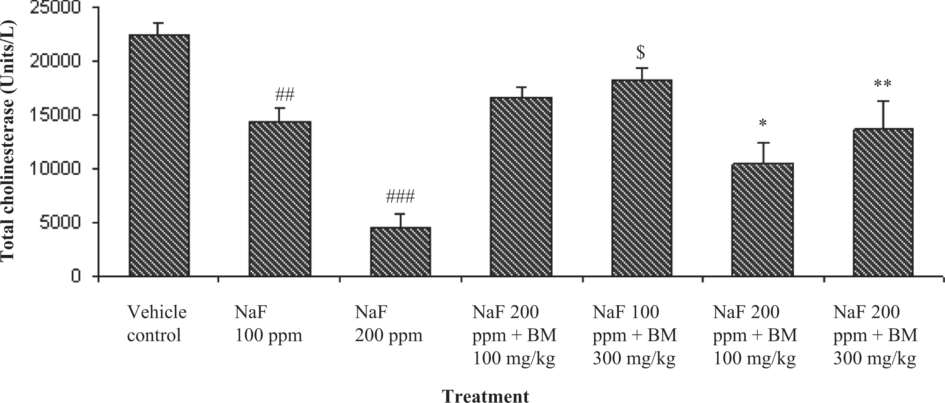
Independent effect of NaF (100 and 200 ppm) and the effect of BM (100 and 300 mg/kg) when coadministered with NaF in plasma total cholinesterase levels in female mice. NaF: sodium fluoride; BM: Bacopa monniera.
SOD, CAT, GPx activities, and LPO levels
NaF 100 and 200 ppm-treated mice exhibited dose-dependent significant decrease in the activities of SOD, CAT, and GPx as well as increase in levels of lipid peroxidation (LPO) when compared with nonfluoride-treated group of mice. BM 100 mg/kg when coadministered with NaF 100 ppm significantly increased GPx activity and decreased LPO levels when compared with NaF 100 ppm-treated mice. BM 300 mg/kg when coadministered with NaF 100 ppm significantly increased the SOD, CAT, and GPx activities as well as decreased LPO levels when compared with NaF 100 ppm-treated group. NaF 200 ppm-treated group when coadministered with BM 100 mg/kg significantly increased the SOD and GPx activities as well as decreased the levels of LPO when compared with NaF 200 ppm-treated group. BM 300 mg/kg when coadministered with NaF 200 ppm significantly increased SOD, CAT, and GPx activities as well as decreased the LPO levels when compared with NaF 200 ppm-treated group (Table 3).
Oxidative changes in female mice exposed to NaF (100 and 200 ppm) and the effect of BM (100 and 300 mg/kg) when coadministered with NaF.a
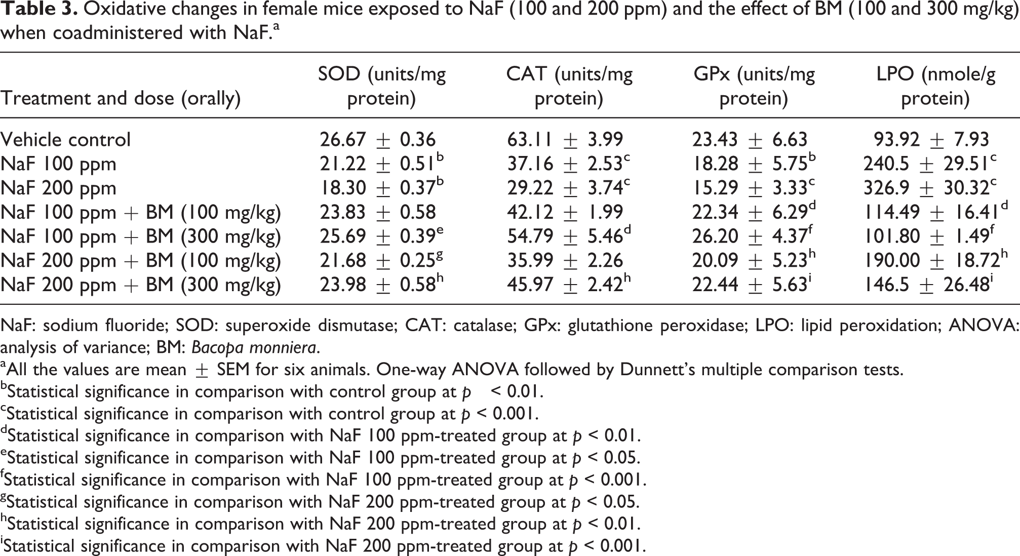
NaF: sodium fluoride; SOD: superoxide dismutase; CAT: catalase; GPx: glutathione peroxidase; LPO: lipid peroxidation; ANOVA: analysis of variance; BM: Bacopa monniera.
aAll the values are mean ± SEM for six animals. One-way ANOVA followed by Dunnett’s multiple comparison tests.
bStatistical significance in comparison with control group at p < 0.01.
cStatistical significance in comparison with control group at p < 0.001.
dStatistical significance in comparison with NaF 100 ppm-treated group at p < 0.01.
eStatistical significance in comparison with NaF 100 ppm-treated group at p < 0.05.
fStatistical significance in comparison with NaF 100 ppm-treated group at p < 0.001.
gStatistical significance in comparison with NaF 200 ppm-treated group at p < 0.05.
hStatistical significance in comparison with NaF 200 ppm-treated group at p < 0.01.
iStatistical significance in comparison with NaF 200 ppm-treated group at p < 0.001.
Na+–K+-ATPase levels
NaF 100 and 200 ppm-treated mice showed significant decrease in Na+–K+-ATPase levels at p < 0.05 and p < 0.01, respectively, when compared with nonfluoride-treated group. BM 300 mg/kg when coadministered with NaF 100 ppm significantly elevated the Na+–K+-ATPase levels (p < 0.01) when compared with NaF 100 ppm-treated group. NaF 200 ppm-treated mice when coadministered with BM 100 and 300 mg/kg elevated the Na+–K+-ATPase levels significantly at p < 0.05 and p < 0.001, respectively, when compared with NaF 200 ppm-treated group (Figure 3).
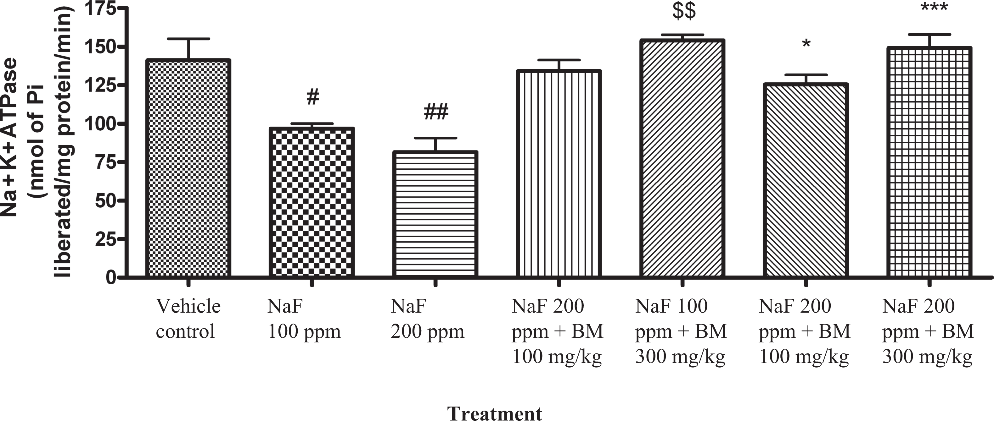
Independent effect of NaF (100 and 200 ppm) and the effect of BM (100 and 300 mg/kg) when coadministered with NaF in brain Na+–K+-ATPase levels in female mice. NaF: sodium fluoride; BM: Bacopa monniera; Na+–K+-ATPase: sodium–potassium adenosine triphosphatase.
Fluoride levels in brain
NaF 100 and 200 ppm treated mice showed significant increase in fluoride levels in mice brain at p < 0.01 and p < 0.001, respectively, when compared with nonfluoride-treated group. Treatment with BM at both the dose levels did not alter the fluoride levels of NaF 100 and 200 ppm-treated mice brain (Figure 4).
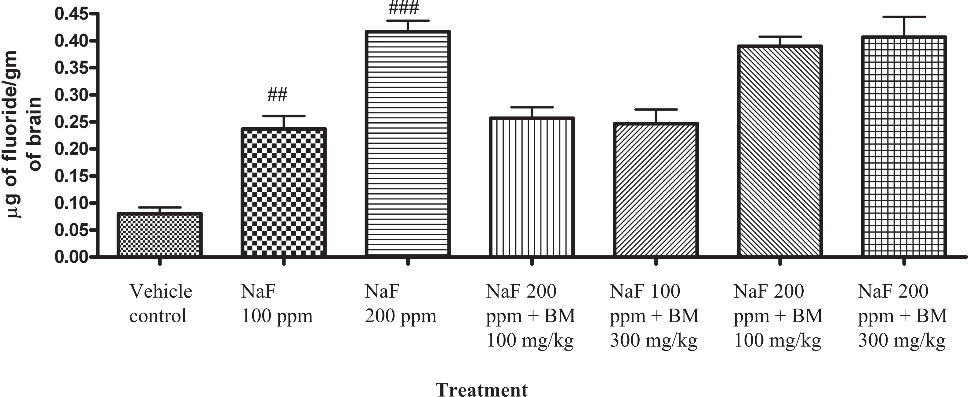
Fluoride content in mice brain.
Histopathology
Mice treated with NaF 100 ppm for 30 days exhibited oedema and pyknotic nerve cells, whereas mice treated with NaF 200 ppm exhibited oedema and severe microcytic degeneration of the nerve fibres in brain. BM 100 and 300 mg/kg protected the NaF 100 and 200 ppm-induced brain damage in mouse brain (Figure 5(a) to (g)).
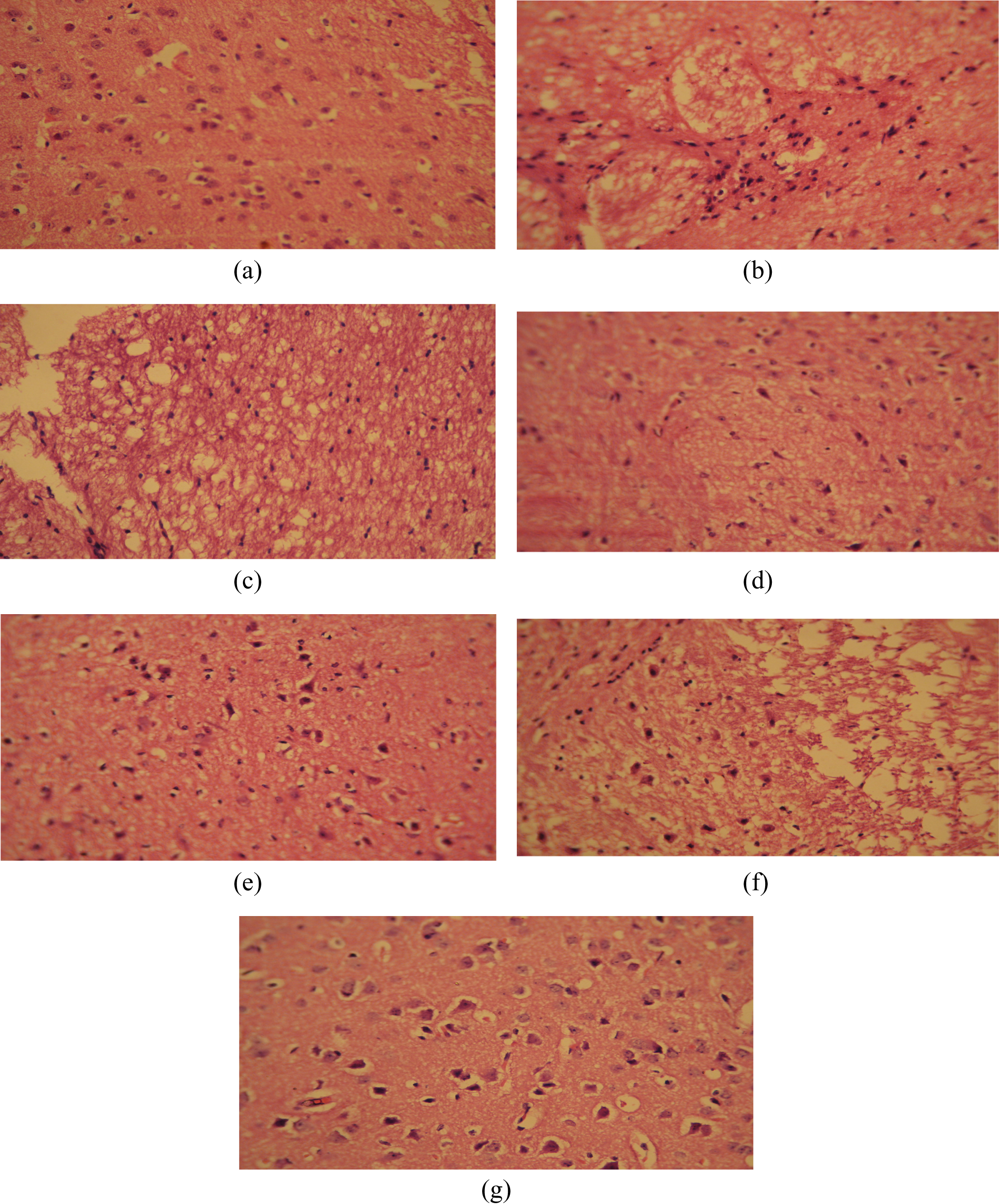
Microscopic study of lateral sections of mice brain (100×). Histological sections of brain were stained with haematoxylin and eosin. (a) Control mice brain showing intact neurons with normal glial and astrocytes surrounded by nerve fibres. (b) Mice exposed to NaF 100 ppm for 30 days clearly showing oedema with separated nerve bundles and closely packed pyknotic nerve cells. (c) Mice exposed to NaF 200 ppm for 30 days showing oedema, microcytic degeneration of the nerve fibres. (d) Mice administered with BM 100 mg/kg along with NaF 100 ppm showing irregular pyknotic nuclei and oedema of the brain with the absence of neural cell proliferation. (E) Mice administered with BM 300 mg/kg along with NaF 100 ppm showing only mild proliferation of cells with no oedema and normal astrocytes. (f) Mice administered with BM 100 mg/kg along with NaF 200 ppm showing oedema with pyknotic nuclei. (g) Mice administered with BM 300 mg/kg along with NaF 200 ppm showing normal astrocytes and no oedematous cells. NaF: sodium fluoride; BM: Bacopa monniera.
Discussion
The objective of the present study was to evaluate the effect of BM extract against fluoride-induced behavioural, biochemical, and neuropathological alterations in female mice. We selected female mice for the present study because female mice were more sensitive to fluoride exposure and behavioural alterations at adult stage than male mice (Mullenix et al., 1995).
In the present study, in the NaF (100 and 200 ppm)-treated mice for 30 days, we did not observe a decrease in body weight, feed, and water consumption; however, mice exhibited behavioural changes. In contrast to this, Ekambaram and Paul (2001, 2002) observed a decrease in body weight and showed certain behavioural changes in 500 ppm NaF-treated animals. These studies also found a reduction in feed and water consumption, which could lead to body weight reduction and could accounted for the behavioural changes observed. But in the present study, the behavioural changes can be considered as specific and direct effects of fluoride because NaF did not produce any alterations in the body weight of mice.
Mullenix et al. (1995) reported that exposure to high doses of fluoride for a prolonged period of time results in neurotoxic effects. In the present study, we observed motor incoordination and decrease in ChE levels in the NaF-treated mice. A disturbance in the activity of ChE in blood is likely to impair cholinergic transmission at the neuromuscular junction resulting in a suppression of motor coordination. The data presented here provide further support to the ChE-suppressing action of fluoride (Paul et al., 1998). Thus, an impaired neuromuscular system can be accounted for akinesia and decreased locomotion in the open field test in NaF-treated mice. Decreased rotarod performance in NaF-treated mice suggests that there is impairment in their ability to integrate sensory input with appropriate motor commands to balance their posture and adjust their limb movements on metallic rod is an indicative of cerebellar dysfunction (Nandhu et al., 2011). Our histopathological findings confirm that there is cerebellar damage in NaF-treated mice brain that may be due to the accumulation of fluoride in neurons resulting in strong morphological changes, clustering, and degeneration (Mullenix et al., 1995). In the present study, BM extract prevented the cerebellar damage and the inhibition of ChE by fluoride. The active constituents of BM extract, that is bacosides, are known to enhance nerve impulse transmission. The bacosides are also found to aid in repair of damaged neurons by enhancing kinase activity, neuronal synthesis, and restoration of synaptic activity, and ultimately nerve impulse transmission (Mathew et al., 2010).
The observed increased immobility period in the FST in NaF-treated mice may be due to the depression or fatigueness in NaF-treated mice, which cannot be definitely said with the present study. In fluorosis, membrane-bound ATPases (thiol dependent) are attacked by free radicals generated due to fluoride and inactivate Na+–K+-ATPase pumps (Cornelius et al., 2011), which leads to ATP depletion and thereby fatigueness (McMorrow et al., 2011). In the present study, BM showed significant antioxidant activity and hence by improving the thiol status, BM may have protected the inhibition of Na+–K+-ATPases by fluoride. Similar results have been reported by Anbarasi et al. (2005) in which bacosides prevented the membrane-bound ATPases from cigarette smoking-induced damage. Besides, BM is known to protect mitochondrial damage that could have favoured the efficiency of ATP production (Sumathy et al., 2002). Thus, the antidepressant activity of BM extract observed in the present study may be due to its antioxidant and energy-promoting properties. Earlier reports also indicate that BM possess significant antidepressant activity in forced swim and learned helplessness models of depression (Sairam et al., 2002), which is in agreement with our present study results.
Fluoride is known to cause memory impairment by decreasing the number of nicotinic acetylcholine receptors, increasing the glutamate levels and by oxidative stress in the brain (Chioca et al., 2008). On the other hand, cognition-facilitating effect of BM was well validated in different models and known to exert its memory-enhancing effects through cholinergic upregulation, γ-aminobutyric acidergic modulation, antioxidant effects, protein synthesis in brain, and by modulation of brain stress hormones (Saraf et al., 2010).
Hassan and Abdel-Aziz (2010) identified that oxidative stress is one of the important factors in the mechanism of neurotoxic effects of fluoride (Hassan and Abdel-Aziz, 2010). In the present study, NaF-induced oxidative stress in brain and BM extract significantly reduced the oxidative stress which is evident from decreased LPO and increased activities of antioxidant enzymes in the brain. This antioxidant capacity may also be responsible for the neuroprotective effects of BM extract against fluoride-induced neuropathological changes in mice brain (Jyoti et al., 2007).
An interesting finding in the present study is that BM protected the toxicity induced by NaF; however, the levels of fluoride accumulated in the brain were not altered in the BM-treated group. This indicates that BM extract counteracts the NaF-induced toxicity by its neuroprotective and antioxidant mechanism and not by decreasing the levels of fluoride. Coadministration of calcium, which can decrease the levels of fluoride, along with BM will be more beneficial in combating the NaF toxicity.
Conclusion
Our investigation concludes that adult female mice exposed to NaF (100 and 200 ppm) through drinking water displayed behavioural, impaired cholinergic neurotransmission, increased oxidative stress, and neuropathological alterations. BM extract (300 mg/kg) protected the mice from NaF-induced behavioural alterations, restored the cholinergic system, decreased the oxidative stress, and prevented neuropathological alterations in mice brain. Hence, BM extract can be used to mitigate the effects of fluoride in the geographical areas in which high levels of fluoride in drinking water is seen.
Footnotes
Acknowledgement
The authors would like to acknowledge the Managing Director, M/s Natural Remedies, Bangalore, for providing the gift sample of standardized Bacopa monniera extract. The authors also thank the management of JSS College of Pharmacy for providing the facilities to carry out this research work.
Conflict of interest
The authors declared no conflicts of interest.
Funding
This research received no specific grant from any funding agency in the public, commercial, or not-for-profit sectors.