
Abstract
Phthalate compounds are widely used industrial chemicals; when incorporated into polyvinyl chloride, they are not covalently bound and released into the surrounding media. Some of them have estrogenic potential in vitro but data on in vivo studies are scanty. For the 3-day uterotrophic assay, di-n-butyl phthalate (DBP;10 and 100 mg/kg), butyl benzyl phthalate (BBP; 20 and 200 mg/kg), and diethylstilbestrol (DES, 40 µg/kg, positive control) were administered orally to immature female rats for three consecutive days from postnatal day (PND) 21. For the 20-day pubertal onset assay, DBP (10 and 20 mg/kg), BBP (20 and 200 mg/kg), and DES (6 µg/kg) were administered orally from PND 21 daily for 20 days. In the uterotrophic assay, in groups treated with higher dose of DBP and BBP, the uterine wet weight significantly decreased in the higher dose, and there were minor variations in the ovary wet weight, while the wet weight of these organs increased significantly in DES-treated group. In the 20-day pubertal assay, the weight of uterus and ovary declined significantly and changes in vaginal weight were nonsignificant in DBP- and BBP-treated groups. However, in DES-treated group nonsignificant elevation in vagina weight was observed. All the DES-treated animals showed the vaginal opening (VO) on day 26.17 ± 0.16. However, VO was not observed in any of the animals in control, vehicle control, BBP-, and DBP-treated groups up to PND 42, except in one animal each in vehicle control and DBP (100 mg/kg)-treated groups. The data indicated that both DBP and BBP were unable to induce elevation in the uterine and ovarian weight. While DES treatment can accelerate the growth of uterus and ovary and alter the onset of puberty and estrous cyclicity in prepubertal rats. These suggest that these compounds may not have estrogenic potential in vivo.
Keywords
Introduction
Phthalates are widely distributed industrial chemicals and are used for a variety of purposes, such as plasticizers that impart flexibility and durability to polyvinyl chloride (PVC) products and various other uses such as in cosmetics, food packaging materials, medicine coatings, lubricants, medical devices and tubing, children toys and in various other house hold products, and so on. When incorporated into PVC, phthalates are not covalently bound and therefore released into the surrounding environment. The uses of plastics have increased many folds worldwide in all walks of life. Due to large-scale use of plastic products in day-to-day life, thousands of workers are engaged in the manufacture of plastic and plastic-containing products as well as recycling plastic industries are exposed to these chemicals. Furthermore, general population as well as vulnerable groups, that is, children and pregnant women, are also exposed to these chemicals through food, water, and other household products containing phthalates. This situation poses great concern as some of them reported to have estrogenic potential in vitro.
A large number of chemical compounds are produced worldwide for various uses and some of them are capable of disrupting the endocrine system of the living beings. There is a concern that certain chemicals may have the potential to disturb normal sexual differentiation and development in animals and humans also (McLachlan, 1993, McLachlan and Korach, 1995). Phthalate compounds have been reported to have estrogenic activities mostly in vitro, which mimic endogenous estrogen (Harris et al., 1997; Moore, 2000). A number of studies demonstrated an estrogenic activity of phthalates in various in vitro tests. It is known that the potential of in vivo effect of xenoestrogens is of great interest in relation to their effect on human health, especially reproduction. There are few systemic studies (in vivo and in vitro) in relation to estrogenic activity of phthalate compounds (Milligan et al., 1998). In vitro studies indicated that small number of phthalates are weakly estrogenic and data are not available (Wang et al., 1997; Zacharewski et al., 1998) or inadequate to establish whether these are also estrogenic in vivo. The data on estrogenic potential of di-n-butyl phthalate (DBP) and butyl benzyl phthalate (BBP) in vivo are scanty. The female pubertal onset assay uses vaginal opening (VO) as an indicator of pubertal onset and is an extensively used assay for the detection of estrogenic or antiestrogenic activity (Marty et al., 1999, O’Connor et al., 2000). Furthermore, the organization for economic cooperation and development (OECD, 1999) also proposes that the uterotrophic assay is a screening test to test the estrogenic properties. Hence, estrogenic potential of DBP and BBP was investigated in vivo by employing both these tests.
Materials and methods
Chemicals
The test compounds such as DBP (CAS # 84-74-2) and BBP (CAS # 85-68-7), diethylstilbestrol (DES), and corn oil were procured from Greyhound Chromatography and Allied Chemicals (Merseyside, UK), Sigma Chemicals (Saint Louis, MO, USA), and MP Biomedical (Illkirch, France), respectively. Stock and working solutions of test substances were prepared in corn oil and stored in brown glass containers at low temperature.
Animals
The immature female rats (20 days old, weighing 23.8 ± 2.4 g) were selected from the animal house facility of the institute, and they were divided into different groups with minimum of six animals in each group. Animals were maintained on standard rat feed (Amrut feed, Pune, India) and water ad libitum. The animals were maintained at a room temperature of 25 ± 2°C, relative humidity of 60 ± 5%, and light–dark cycle. This study is a part of major project entitled “Studies on phthalate compounds on male reproduction of rats” approved by Institutional Animal Ethical Committee of National Institute of Occupational Health (NIOH), Ahmedabad, Gujarat, India.
3-Day uterotrophic assay
For 3-day uterotropic assay, the method of Kanno et al. (2001, 2003) was adopted. Immature female rats (20 days old) were treated orally with two different doses of DBP (10 and 100 mg/kg) and BBP (20 and 200 mg/kg), that is, 100 and 1000 times of reference dose (RfD) in corn oil, RfD—an estimate, with uncertainty spanning perhaps an order of magnitude, of a daily oral exposure to the human population, including sensitive subgroups that are likely to be without an appreciable risk of deleterious effects during a lifetime (US EPA, IRIS, 1990, 1993), and a single dose of DES (40 μg/kg) once daily for three consecutive days. Control and vehicle control (corn oil) were also maintained in similar fashion. Body weight was recorded before, during treatment, and prior to being killed, and clinical signs and symptoms were also observed daily (Table 1).
Body weight (mean ± SE) changes in immature female rats (21 days old) after DBP and BBP exposure for 20 days.
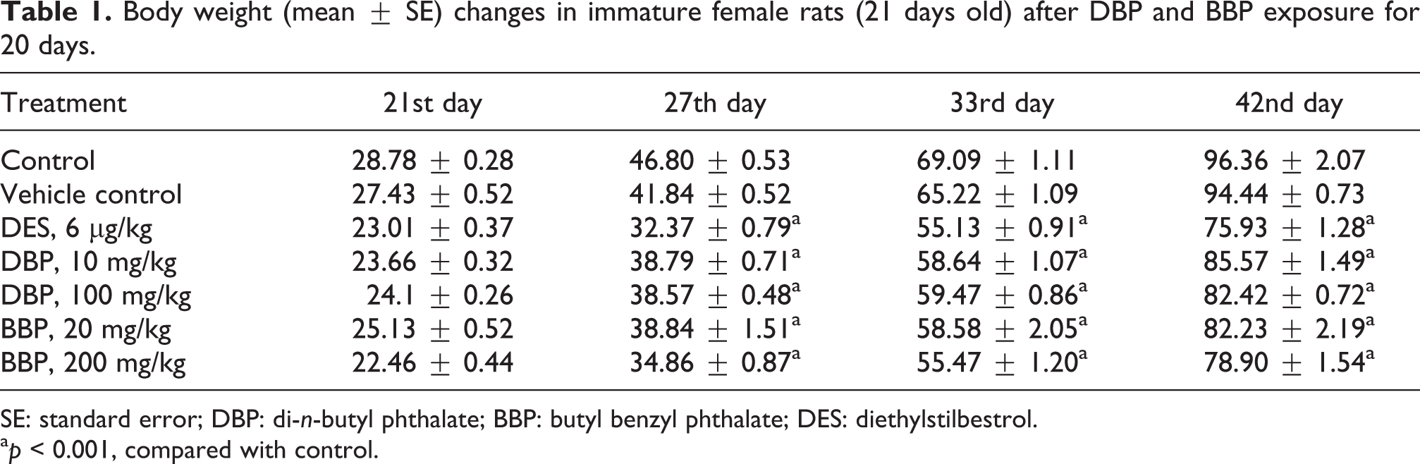
SE: standard error; DBP: di-n-butyl phthalate; BBP: butyl benzyl phthalate; DES: diethylstilbestrol.
a p < 0.001, compared with control.
Uterine and ovary weights
On day 4, animals were killed using carbon dioxide inhalation. The uterus was carefully dissected out and trimmed free of fat to avoid loss of the luminal contents. The body of the uterus was cut just above its junction with the cervix and at the junction of the uterine horns with the ovaries. The uterus and ovary were weighed with their luminal contents (wet weight) to the nearest of 0.1 mg and recorded using electronic balance.
20-Day pubertal female assay
For 20-day pubertal female assay (Kim et al., 2002) immature female rats were treated orally from postnatal day (PND) 21 with DBP (10 and 100 mg/kg), BBP (20 and 200 mg/kg) in corn oil, and DES (6 μg/kg) daily for 20 days. Simultaneously control and vehicle control groups were also maintained. The animals were examined daily and body weight of each rat was recorded before treatment, during treatment, and before necropsy.
VO and estrous cyclicity
Each animal was examined daily for VO from the beginning of PND 21. The appearance of small pinhole, a vaginal thread, and a complete VO was recorded on the day it was observed. The concern of female pubertal assay is with VO day, age at first estrous, and onset of estrous cyclicity (Kim et al., 2002). On the day of VO, the body weight was taken and the age was also noted. Vaginal lavage was collected from the day of VO by pushing surgical dropper into the vagina with normal saline (0.85%). The lavage fluid was smeared and viewed immediately under a microscope for the presence of leukocyte, nucleated epithelial cells, or cornified epithelial cells. Estrous cycle, that is, proestrous, estrous, metaestrous, and diestrous, was evaluated and the stages of estrous cycle were determined (Everett, 1989). Animals were killed on PND 42. The body of the uterus was cut just above its junction with the cervix and at the junction of the uterine horns with the ovaries. The vagina was removed from the uterus at the level of the uterine cervix and weight of ovaries; vagina and uterus were recorded using digital balance. The uterus was cleaned from fat bodies and weighed with luminal fluid. The data are expressed as mean ± standard error and significance between the two groups was analyzed using Student’s t test and depicted in Figures 1 to 4.
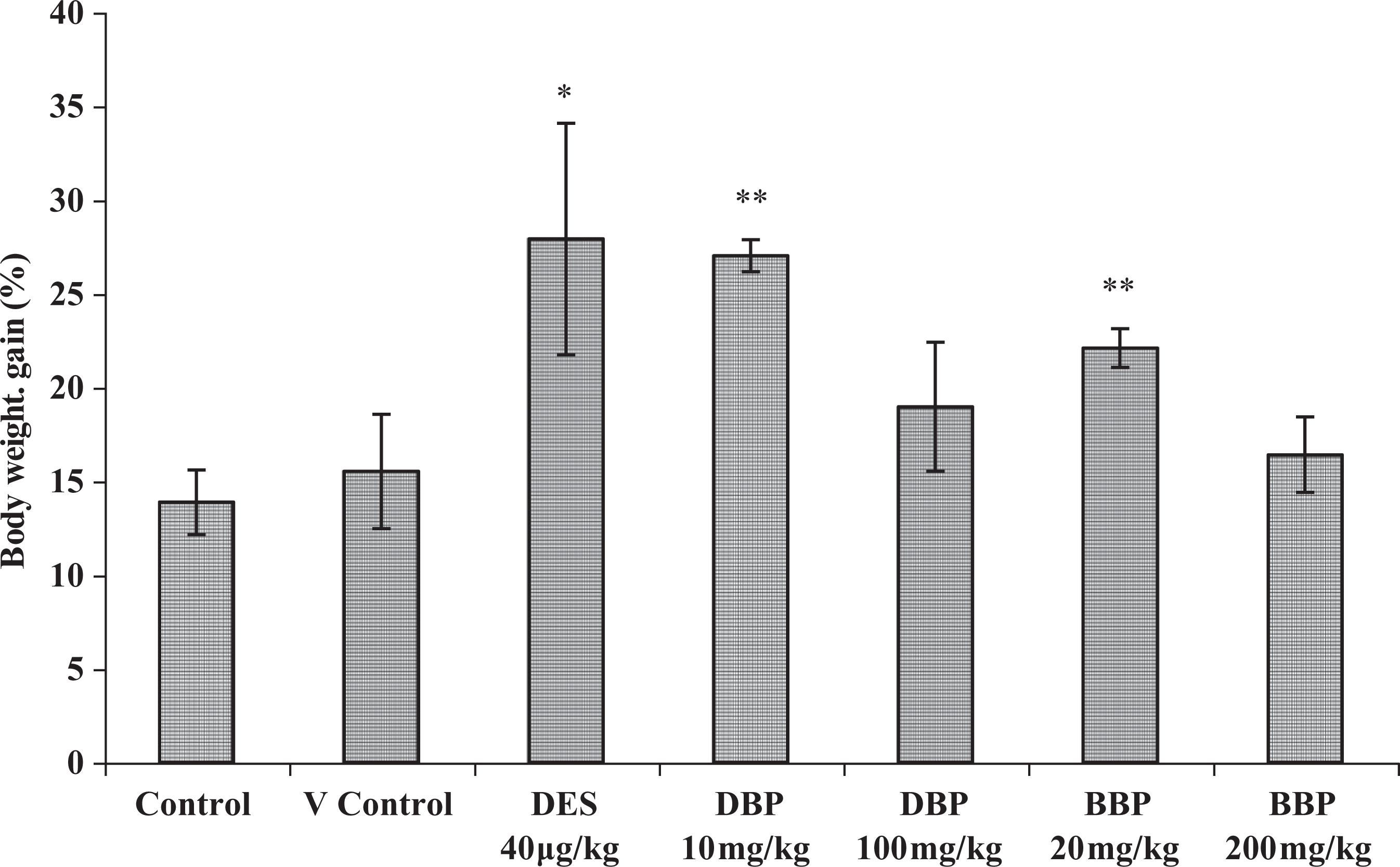
Body weight gain (%) (mean ± SE) in immature female rats after DBP and BBP exposure for 3 days. *p < 0.05, **p < 0.001, compared with control. SE: standard error; DBP: di-n-butyl phthalate; BBP: butyl benzyl phthalate.
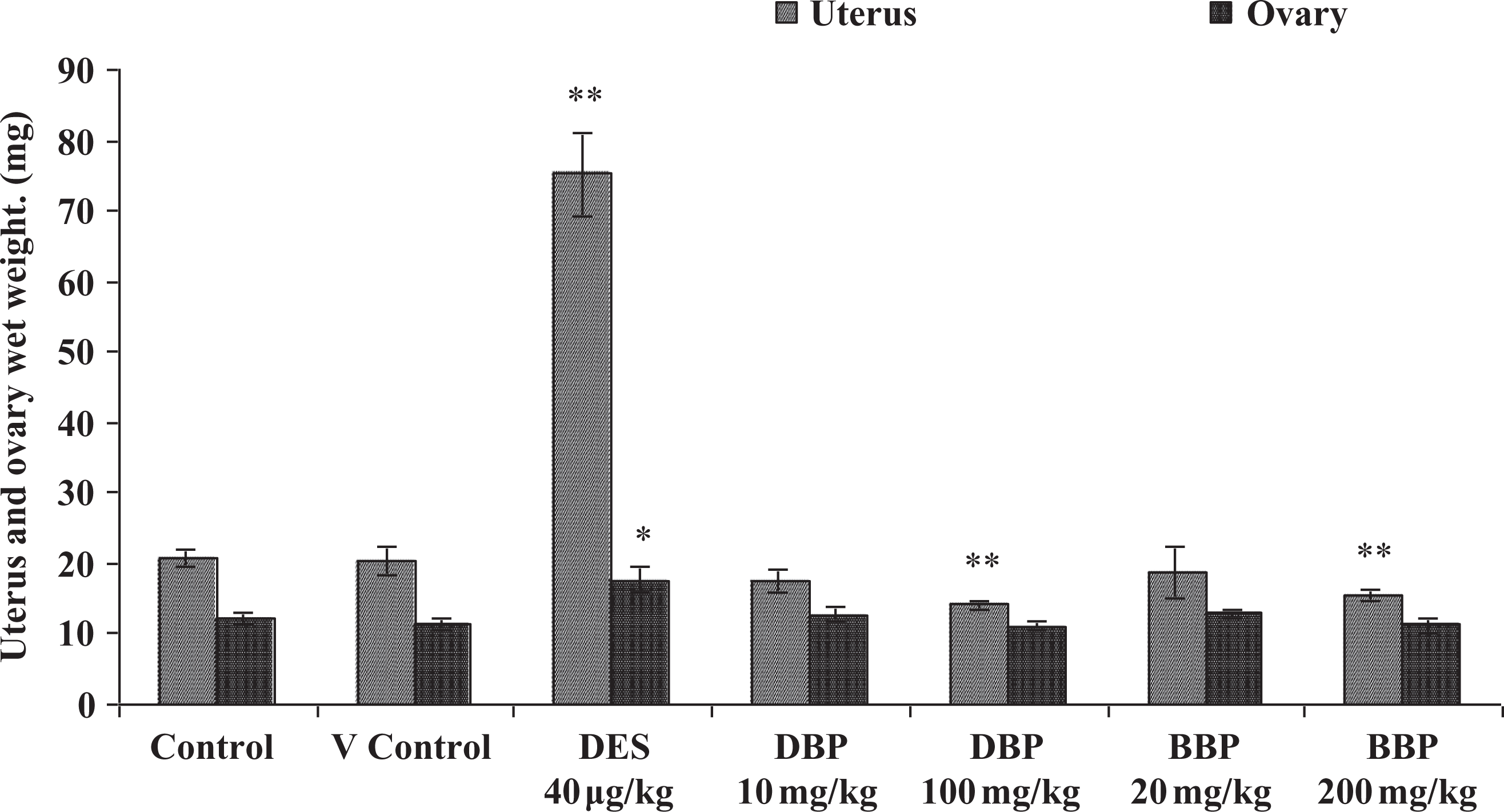
Uterus and ovary weight changes (mean ± SE) in immature female rats after DBP and BBP exposure for 3 days. *p < 0.05, **p < 0.001, compared with control. SE: standard error; DBP: di-n-butyl phthalate; BBP: butyl benzyl phthalate.
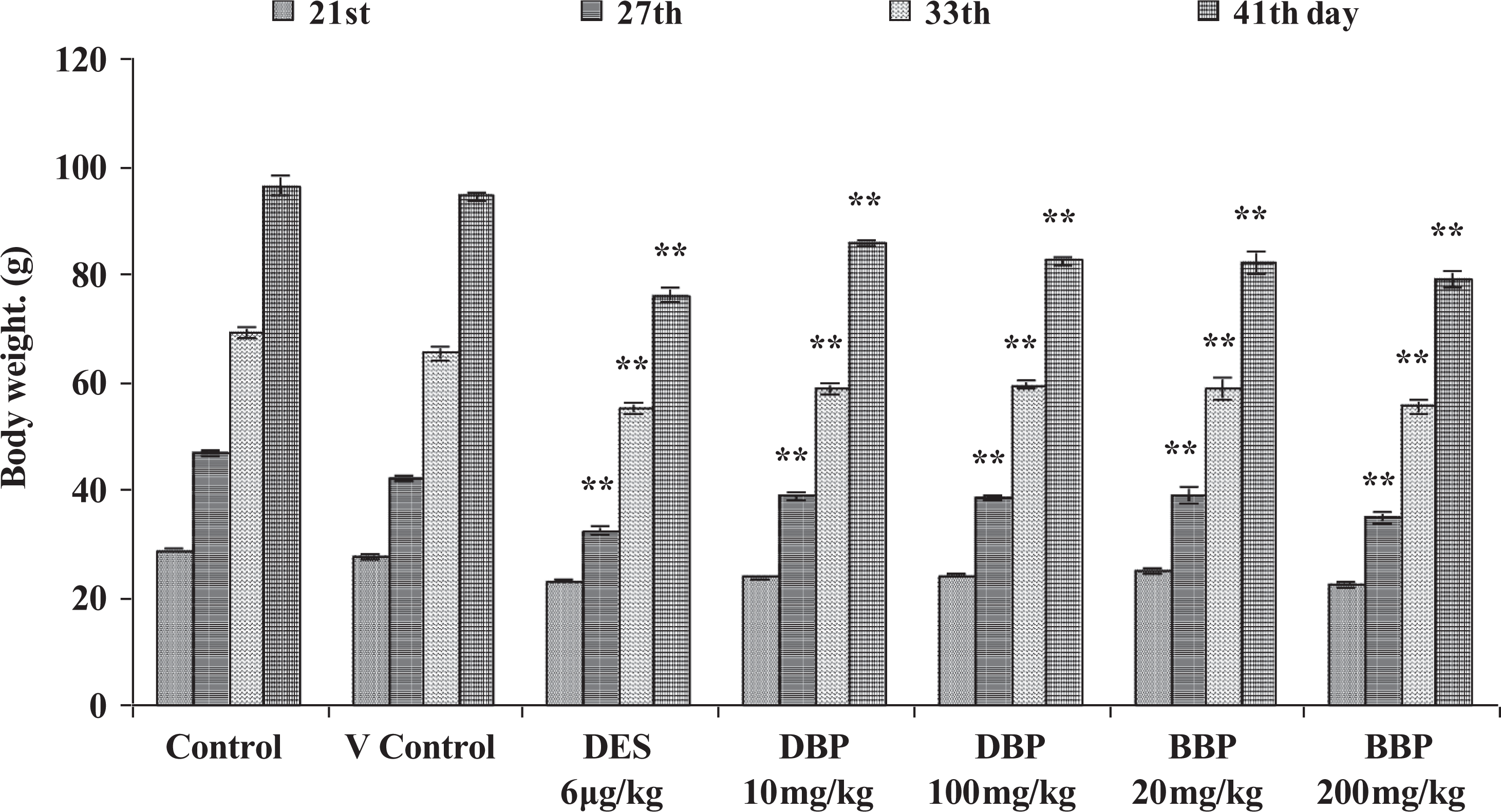
Body weight (mean ± SE) changes in immature female rats after DBP and BBP exposure for 20 days. (**p < 0.001) compared with control. SE: standard error; DBP: di-n-butyl phthalate; BBP: butyl benzyl phthalate.
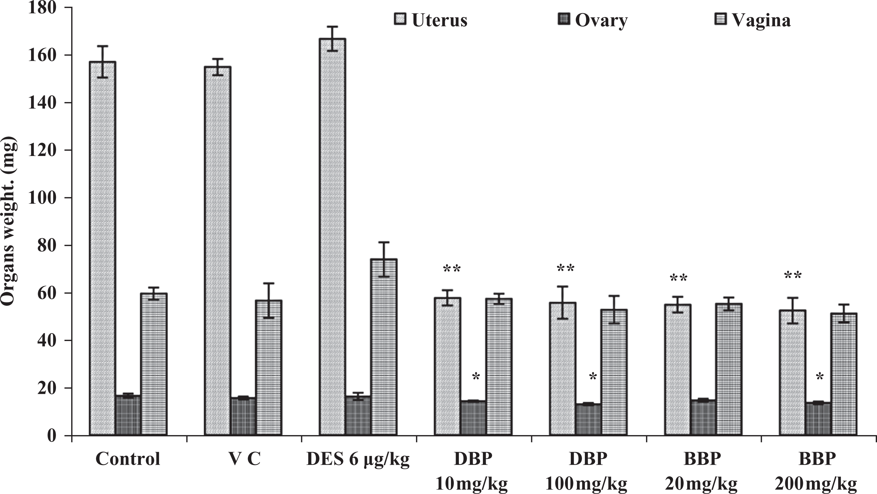
Organs weight (mean ± SE) changes in immature female rats after DBP and BBP exposure for 20 days. (*p < 0.05, **p < 0.001) compared with control. SE: standard error; DBP: di-n-butyl phthalate; BBP: butyl benzyl phthalate.
Results
3-Day uterotrophic assay
Clinical signs, body weight, and gross morphology
No abnormal clinical signs and symptoms were observed in any of the treated and nontreated animals during the study period. The data of body weight gain, uterine-, and ovary wet weight are depicted in Figures 1 and 2. The body weight increased significantly in the lower dose of both DBP and BBP (10 and 20 mg/kg) as well as in DES-treated groups, but elevation in body weight was nonsignificant in groups treated with higher dose of DBP and BBP with respect to control (Figure 1).
Uterine and ovary weight
Uterine wet weight increased by three- to fourfold following the treatment with DES. In contrast to this, the uterine wet weight decreased significantly in groups used in the study with higher dose of DBP (100 mg/kg) and BBP (200 mg/kg) with respect to control. A significant elevation in ovary wet weight was also observed in DES-treated group. But the alteration in the ovary wet weight was nonsignificant in both DBP- and BBP-treated groups (Figure 2). These data suggest that DES treatment induces significant elevation of uterine and ovary organ weight gain, while uterine weight gain was significantly lower in groups treated with high dose of DBP and BBP and ovary weight was also slightly lower in groups treated with higher dose of DBP and BBP.
20-Day pubertal female assay
Body weight
All the animals from DBP-, BBP-, and DES-treated and nontreated groups gained body weight during the study period, that is, PND 21 to 42 from their initial body weights. However, a statistically significant decline in body weight gain was observed at PND 27, 33, and 42 in both DBP- and BBP-treated groups at both dose level and also in DES-treated group as compared to control (Figure 3). However, the weight gain was lowest in DES-treated group as compared to BBP- and DBP-treated groups as well as nontreated groups.
Organ weight
The uterus and ovary weight decreased significantly in all the DBP- and BBP-treated groups except a nonsignificant decline in ovary weight in the group treated with lower dose of BBP (20 mg/kg). However, marginal elevation in uterus weight was observed in DES-treated group, which was statistically nonsignificant. The weight of vagina was more or less similar in both DBP- and BBP-treated group (Figure 4). However, the vaginal weight marginally increased in DES-treated group as compared to control.
Vaginal opening
The VO was checked in all the animals from PND 21 to PND 42. All the DES (6 µg/kg)-treated animals showed the VO on day 26.17 ± 0.16 and the mean body weight of these animals was 32.37 ± 0.78 g at the time of VO. VO was not observed in any of the animals in control and BBP-treated groups, except one animal each in vehicle control and group treated with higher dose of DBP (100 mg/kg) till PND 42.
Estrous cycle
The estrous cycle of individual animals was observed from the day after VO until the end of the study and the number of days in each stage of the estrous cycle and cycle length was recorded. The number of days in estrous cycle increased in DES-treated animals. Estrous cycle was not at regular intervals and was affected in all the DES-treated animals.
Discussion
The estrogenic potential of DBP and BBP in rats was assessed using 3-day uterotrophic and 20-day pubertal female assay with the end points, that is, alteration in uterus and ovary wet weight and VO in prepubertal female rats as an indicator of pubertal onset. The 3-day uterotrophic assay has been reported as a sensitive testing method to detect environmental agents with weak/high estrogenic activity (Kanno et al., 2001, 2003), and 20-day pubertal female assay detects alterations in thyroid hormone status, hypothalamic–pituitary–gonadal function, and inhibition of steroidogenesis (including aromatase), estrogens, and antiestrogens (Laws et al., 2000, Marty et al., 1999). In the uterotrophic assay, in groups treated with higher dose of DBP and BBP, the uterine wet weight decreased significantly and there were minor variations in the ovary wet weight, while the weight of these organs increased significantly in DES-treated group. In the 20-day pubertal assay, the weight of uterus and ovary declined significantly in DBP- and BBP-treated groups. Uterotrophic assay (uterine wet weight) in immature rat has been used as a sensitive parameter for evaluating estrogenic activity (Branham et al., 1988, 1993). Furthermore, it has been suggested that uterine weight change as an end point of estrogenic activity because wet weight was almost totally uninfluenced by the body weight changes (O’Connor et al., 1996, Odum et al., 1997). Earlier, a number of investigators reported that uterine wet weight increase in rodents is an established measure of the estrogenicity of a compound. Furthermore, uterus has also been reported to respond to the treatment of the animal with progesterone, testosterone, and other agents that are not characteristically estrogenic (Clark et al., 1980; Korach and McLachlan, 1995; Lindzey and Korach, 1997).
Furthermore, data revealed that DES can accelerate the growth of uterus and ovary and alter the onset of puberty and estrous cyclicity in prepubertal female rats. The result of the study indicates that both the compound may not have estrogenic potential in vivo and are unable to accelerate the growth of uterus and ovary. These corroborate with earlier studies on DBP and BBP as both these esters reported to fail to induce increase in uterine wet weight (Brady et al., 1998; Coldham et al., 1997; De George et al., 1997; Milligan et al., 1998).
Furthermore, VO was not observed in any of the DBP- and BBP-treated and nontreated groups except one animal each in DBP and vehicle control group, whereas all the DES-treated animals showed the VO on day 26.17 ± 0.16. Earlier, Kim et al. (2002) reported that DES could significantly advance the VO in all the DES-treated animals to 8.4 days earlier than control. Several studies have demonstrated that the onset of puberty in the female rat is associated with VO and the first estrus (Ramirez and Sawyer, 1965, Ramaley, 1981). In addition, it has been reported that phthalates weakly bind to receptor sites (Zacharewski et al., 1998) and may not possess the potential to promote the uterine growth in BBP-exposed immature female rats (Monsanto, 1996).
The present study suggests that both DBP and BBP may not have estrogenic potential in vivo. It has been reported that the absence of in vivo responses for DBP, BBP, and DEHP suggests that metabolic events may inactivate the estrogenic activity of the parent phthalate ester (Zacharewski et al., 1998). This is supported by earlier studies indicating that BBP is efficiently metabolized to the corresponding monophthalates (i.e. monobutyl phthalate and monobenzyl phthalate) and the monophthalate glucuronides in the male Fischer rat model (Eigenberg et al., 1986, Takahashi and Tanaka (1989)). Earlier studies conducted in a recombinant yeast strain have shown that monobutyl, monobenzyl, monoethylhexyl, and monooctyl phthalate failed to induce an estrogen receptor (ER)-mediated response (Harris et al., 1997). Furthermore, the adverse reproductive and developmental effects were observed in rodents exposed to DBP and BBP during gestation and lactation may not be due to the in vivo estrogenic activities of these phthalates suggested by Sharpe et al. (1995) and Wine et al. (1997).
The present study suggests both the compounds may not have estrogenic potential in vivo. However, both these compounds reported to have the estrogenic potential in vitro. Earlier, Zacharewski et al. (1998) reported that only selected phthalate esters (i.e. DBP, BBP, and DHP) exhibit weak ER-mediated activity in some in vitro assays at high concentrations but none of the selected eight phthalate esters elicited in vivo estrogenic responses from uterotrophic and vaginal cornification assays. The metabolites of these compounds in vivo may lose their estrogenic potential in vivo. Earlier, Moore (2000) reviewed the estrogenic potential of the phthalate esters and reported that the monoesters (the primary metabolites of the diesters in vivo) are inactive in similar in vitro tests. Furthermore, the diesters have not shown any estrogenic activity in numerous and diverse studies in vivo at doses eliciting systemic toxicity.
In conclusion, both the tested phthalate esters (DBP and BBP) did not show the estrogenic potential in vivo as both the esters were unable to accelerate the uterine wet weight and VO, whereas statistically significant responses were observed in DES-treated animals. The data further suggest that 3-day uterotrophic assay and 20-day pubertal female onset assay can be used as sensitive methods to detect environmental agents possessing estrogenic activity in vivo.
Footnotes
Acknowledgments
The authors are thankful to the Director, NIOH, and the staffs of the department and animal house facility for their support during the study.
Funding
This work was financially supported by Indian Council of Medical Research (ICMR), New Delhi, in the form of a research grant (to SK).