
Abstract
Silver (Ag) and gold nanoparticles (Au NPs) have wide applications. They are increasingly being used in the medical devices, biosensors, cancer cell imaging, and cosmetics. Increased applications of these NPs in the technological advances have also led to the risk of exposure to these particles. This study investigated the toxic effects of Ag and Au NPs (1 μM and 2 μM, oral) on mouse erythrocytes and tissues after 14 consecutive days’ exposure. Our results demonstrate significant increase in reactive oxygen species (ROS) and depletion of antioxidant enzyme status in erythrocytes and tissues. Hepatic and renal toxicity was evident from liver and kidney function tests. Inflammatory markers, interleukin-6 and nitric oxide synthase increased in plasma on administration following exposure to these NPs at both the doses. A more pronounced increase was noted in kidney metallothionein (MT) compared to liver MT on exposure to these NPs. Toxic potential of these NPs was further confirmed by increased 8-hydroxy-2′-deoxyguanosine levels in urine, a biomarker of DNA damage. Among the two NPs, Ag NP was more toxic at 2 μM dose compared to lower dose of 1 μM. The study suggests oxidative stress as the major mechanism responsible for the toxic manifestations induced by Ag and Au NPs.
Introduction
Engineered nanoparticles (NPs), besides providing wide range of industrial and biomedical applications, impelled the necessity to investigate their toxic effects on humans and the environment. Their unique physical properties, such as, small size, allow their transport across cell membranes and interaction with cellular organelles, leading to possible toxicity (Korani et al., 2011). Inert elements like gold (Au) can also become highly active at nano-dimensions, leading to the generation of harmful free radicals. Enhanced generation of reactive oxygen species (ROS) may disturb pro and antioxidant balance of the body, resulting in oxidative stress (Prabhakar et al., 2012). Thus a correlation between exposure to NPs and oxidative stress is possible. This study was planned to investigate (i) systemic toxicity following 14 days’ continuous exposure to silver (Ag) and Au NPs and (ii) oxidative stress plays role in eliciting these toxic effects.
Ag and Au NPs exhibit promising applications. Ag has been widely used as an antimicrobial agent. Coating of milk bottles to inhibit bacterial growth, administration of Ag nitrate solution to newborn babies in order to prevent neonatal conjunctivitis, and use of sulfadiazine creams on the skin of denuded patients to prevent bacterial growth are few examples (Samberg et al., 2010). Recently nano-sized Ag particles are in demand, particularly in the medicine and technology due to their highly effective nature. Ag impregnated catheters, wound dressings, and other wide range of medical devices are few common examples. They are also being used in washing machines to get clean and sterile clothes and are an ingredient for electro conduit slurry, air purifiers, inks of inkjets, etc. (Quadros and Marr, 2010). Besides having such a wide range of medical applications, safety profile of nano Ag is not well established and requires investigations. Au NPs, on the other hand, are being widely used in cancer therapy and in the treatment of rheumatoid arthritis (Alkilany and Murphy, 2010). Desirable properties such as strong and size-tunable surface plasmon resonance, fluorescence, and easy surface functionalization have made them a perfect choice for applications, such as, biosensors, cancer cell imaging, photothermal therapy, drug delivery, and radiotherapy (Zhang et al., 2010). Due to different modifications in the structure, such as, surface functional attachments, there have been reports indicating the toxic potential of these NPs (Abdelhalim and Jarrar, 2011).
After oral exposure, NPs tend to distribute in the kidneys, liver, spleen, lungs, brain, and gastrointestinal (GI) tract (El-Ansary and Al-Daihan, 2009). They become systemically available as soon as absorption is completed, leading to conditions, such as, argyria and sometimes detrimental consequences, such as, hepatic injury (Johnston et al., 2010). Potential toxic effects, such as, cell activation, generation of ROS and inflammation are being reported as NPs bind themselves to tissues, ultimately leading to cell death. At relatively higher concentrations, such as 2.7 μg/mL, they are necrotic to cell such as liver cells, leading to membrane rupture (Panyala et al., 2008; Piao et al., 2011). Ag NPs also interact with thiol-containing enzymes, leading to the alterations in the mitochondrial function (Johnston et al., 2010). It has recently been reported that Au NPs of smaller dimensions (5–15 nm) compared to micro-sized particles had wider organ distribution, with liver and spleen among the major targeted organs (Genter et al., 2012; Zhang et al., 2010). Au NPs induce oxidative stress in the body by producing nitric oxide (NO) (Jia et al., 2009). Au NPs on contact with fresh blood serum, catalyzes the generation of NO, which reacts rapidly with free radicals, leading to oxidation of DNA, proteins, and lipid molecules (Jia et al., 2009). These reactions trigger cellular responses ranging from subtle modulations of cell signaling to overwhelming oxidative injury, committing cells to necrosis or apoptosis (Stamler, 2004; Yang et al., 2006; De-silva et al., 2004). We thus investigated the plasma concentration of NO and inflammatory marker, interleukin-6 (IL-6), as both can be linked directly to the potential toxicity of nanomaterials.
Materials and methods
Chemicals
Au (gold colloidal solution, 20 nm in size, Sigma Aldrich catalogue # G1652-25 ML) and Ag NPs (Ag dispersion NP, 20 nm in size, Sigma Aldrich catalogue # 730793-25 ML) were procured from Sigma Aldrich (St. Louis, Missouri, USA) and all other laboratory chemicals were of analytical grade and were purchased from Merck (Germany), BDH chemicals (Mumbai, India), or Sigma (USA). Ultra pure water prepared by Millipore (New Delhi, India) was used throughout the experiment to avoid metal contamination and for the preparation of reagents/buffers used for various biochemical assays. Prior to dosing, the suspension was sonicated for 30 min in order to avoid formation of NP aggregates.
Animals
Male Swiss albino mice, weighing approximately 25–30 g, were obtained from the animal house facility of the establishment. All animals received humane care in compliance with the guidelines of the Committee for the Purpose of Control and Supervision of Experiments on Animals (CPCSEA). The Animal Ethical Committee of the institute also approved the protocols for the experiments. The animals were housed in stainless steel cages in an air-conditioned room with temperature maintained at 25 ± 2°C with 12 h alternating day and night cycles and relative humidity of 60%. Mice were given standard chow diet (Ashirwad Feeds, Chandigarh, India; metal content of diet, in ppm dry weight: Cu 10.0, Zn 45.0, Mn 55.0, Co 5.0, Fe 75.0) and water ad libitum throughout the experiment.
Experimental design
Study was conducted in order to compare the toxicity of NPs at two different doses of 2 μM and 1 μM. The animals were divided into seven groups of eight mice each and treated as below for 14 days through oral gavage. Group I – Control animals (received drinking water). Group II – Cadmium as cadmium chloride; CdCl2, 1 μM/kg (0.1 mg/kg/2 mL/ day), orally. Group III – Cadmium as cadmium chloride; CdCl2, 2 μM/kg (0.2 mg/kg/2 mL/day), orally Group IV – Ag NP, 1 μM/kg (0.1 mg/kg/2 mL/day), orally. Group V – Ag NP, 2 μM/kg (0.2 mg/kg/2 mL/day), orally. Group VI – Au NP, 1 μM/kg (0.2 mg/kg/2 mL/day), orally. Group VII – Au NP, 2 μM/kg, (0.4 mg/kg/2 mL/day), orally.
Animals exposed to bulk cadmium chloride were used as positive control for determining metallothionein (MT) induction in tissues. Selection of doses for both Ag and Au NPs was based on previous studies (Sardari et al., 2012; Zhang et al., 2010) The animals were dosed orally, once daily for 14 days and dosing was carried out at the same time between 11:00 h and 12:00 h. It is necessary to maintain regular dosing time to avoid any possible variations in results. After 14 days, blood was collected from orbital plexus of mice in heparinised tubes and used for various biochemical assays before anesthesia as induction of anesthesia combined with orbital puncture can cause more distress. Animals were anesthetized and euthanized by decapitation. Brain, liver, kidney, and spleen were removed, rinsed in cold saline, blotted, weighed, and used for various biochemical assays.
Separation of red blood cells (RBCs) and plasma
RBCs were isolated from blood following the method of Steck and Kant (1974). Briefly, blood was centrifuged at 1500 × g for 10 min at 4°C. Plasma and buffy coat were removed by aspiration. The RBCs were washed three times in phosphate buffer saline (0.1 M). The packed cell volume (PCV) obtained was divided into two parts. One part was diluted with chilled distilled water and kept for the analysis of ROS and reduced glutathione. The other part of the PCV was used for the estimation of other antioxidant enzymes, that is, glutathione peroxidase (GPx) and glutathione-S-transferase (GST). For this, hemoglobin was precipitated in PCV by means of chloroform and ethanol. After centrifugation (3000 × g) for 10 min at 4°C, the supernatant obtained was used for all the above mentioned enzymatic assays. Falcons containing blood were kept still for 6 h in order to collect plasma.
Blood glutathione (GSH)
Analysis of blood GSH concentration was carried out following the method described by Ellman (1959) and modified by Jollow et al. (1974). In brief, 0.2 mL of whole blood was added to 1.8 mL of distilled water and incubated for 10 min at 37°C for complete hemolysis. After hemolysis, 3 mL of 4% sulfosalicylic acid was added and tubes were centrifuged at 2500 rpm for 15 min. Supernatant (0.2 mL) was mixed with 0.4 mL of 10 mM 5,5-dithiobis-(2-nitrobenzoic acid) (DTNB) and 1 mL phosphate buffer (0.1 M, pH 7.4). At the end, absorbance at 412 nm was recorded.
Blood thiobarbituric acid reactive substances (TBARS)
TBARS in RBCs were measured according to a method by Stocks and Dormandy (1971). Briefly, RBCs were washed with ice-cold buffered saline (pH 7.4, 0.1 M) and the PCV was adjusted to 2.5%. To this was added sodium azide (2 mmol/L), followed by hydrogen peroxide (H2O2) (0.068%). The mixture was incubated at 37°C for 1 h, after which the reaction was stopped by adding 2 mL trichloroacetic acid (TCA)–arsenite solution (28% TCA + 0.1 M sodium arsenite). The tubes were centrifuged at 3000g for 15 min at 4°C and 1% thiobarbituric acid was added to the collected supernatant. The tubes were placed on boiling water bath for 15 min. The absorbance was read at 532 nm using a spectrophotometer (Model RF 5000 Shimadzu, Japan). Results were expressed as nanomole per minute per gram of hemoglobin
Blood and tissue reactive oxygen species
The amount of ROS in blood and tissues was measured using 2′,7′-dichlorofluorescin diacetate (DCF-DA), which gets converted into highly fluorescent DCF by cellular peroxides (including H2O2). The assay was performed as described by Socci et al. (1999). Briefly, tissues were homogenized in 1 mL of ice-cold 40 mM Tris–hydrochloric acid (HCl) buffer (pH 7.4) and diluted to 0.25% with the same buffer and placed on ice. For ROS estimation in blood, 5% RBC hemolysate and 1% tissue homogenate were prepared and diluted to 1.5% with ice-cold 40 mM Tris–HCl buffer (pH 7.4). The samples were divided into two equal fractions. In one fraction, 40 μL of 1.25 mM DCF-DA in methanol was added for ROS estimation. Another fraction, to which 40 μL of methanol was added, served as a control for tissue/hemolysate auto fluorescence. All samples were incubated for 15 min in a 37°C water bath. Fluorescence was determined at 488 nm excitation and 525 nm emission using a fluorescence plate reader (Perkin Elmer, LS-55, UK).
Tissue reduced (GSH) and oxidized glutathione (GSSG)
Tissue reduced glutathione (GSH) and oxidized glutathione (GSSG) levels were measured as described by Hissin and Hilf (1974). Briefly, 0.25 g of tissue sample was homogenized on ice with 3.75 mL of phosphate–ethylenediaminetetraacetic acid (EDTA) buffer and 1 mL of 25% HPO3, which was used as a protein precipitant. The total homogenate was centrifuged at 10,000 × g for 30 min at 4°C. For the tissue GSH assay, 0.5 mL supernatant and 4.5 mL phosphate buffer (pH 8.0) were mixed. The final assay mixture (2.0 mL) contained 100 μL supernatant, 1.8 mL phosphates–EDTA buffer, and 100 μL o-pthaldehyde; 1000 μg/mL in absolute methanol, prepared fresh). After mixing, fluorescence was determined at 420 nm with an excitation wavelength of 350 nm using a spectrofluorometer (Perkin Elmer, LS-55, UK). For GSSG assay, 0.5 mL supernatant was incubated at room temperature with 200 μL of 0.04 mol/L N-ethylmaleimide solution for 30 min. A total of 4.3 mL of 0.1 mol/L sodium hydroxide (NaOH) was added to this mixture. From this mixture, 100 μL was taken for the measurement of GSSG using the procedure described above and for GSH assay 0.1 mol/L NaOH was used as the diluent instead of phosphate buffer.
Blood and tissue GPx
GPx activity was measured by the procedure of Flohe and Gunzler (1984). Supernatant collected after centrifuging 5% tissue homogenate for 10 min at 1500 × g, followed by 10,000 × g for 30 min at 4°C, was used for GPx assay. In case of blood, 5% RBC hemolysate was used. A total of 1 mL of reaction mixture was prepared, which contained 0.3 mL of phosphate buffer (0.1 M, pH 7.4), 0.2 mL of GSH (2 mM), 0.1 mL of sodium azide (10 mM), 0.1 mL of H2O2 (1 mM), and 0.3 mL of tissue supernatant. After incubation at 37°C for 15 min, reaction was terminated by addition of 0.5 mL of 5% TCA. Tubes were centrifuged at 1500 × g for 5 min and supernatant was collected. Next, 0.2 mL of phosphate buffer (0.1 M, pH 7.4) and 0.7 mL of DTNB (0.4 mg/mL) were added to 0.1 mL of the reaction supernatant. After mixing well, absorbance was recorded at 420 nm.
Blood and tissue GST
GST activity was determined following the procedure of Habig et al., (1974). Supernatant was obtained after centrifuging 5% tissue homogenate at 1500 × g for 10 min followed by 10,000 × g for 30 min at 4°C. In case of blood, 5% RBC hemolysate was used. Reaction mixture contained 0.02 mL of 1-chloro-2,4-dinitrobenzene (1 mM), 2.9 mL of GSH (0.3 mg GSH/mL in 0.2 M phosphate buffer, pH 7.4), and 30 μL of tissue supernatant. Change in color was monitored by recording absorbance (340 nm) at 30 s intervals for 3 min.
MT estimation in blood
MT concentration was determined following the procedure of Onosaka and Cherian (1982). Supernatant was collected after centrifuging 1 g liver or kidney in four volume of sucrose at 18,000 × g for 20 min at 4°C and 10 ppm CdCl2 was added to the reaction mixture. The excess Cd2+ and all Cd binding ligands other than MT in the samples were removed by the addition of RBC hemolysate and a subsequent heating step. Supernatant was collected after centrifugation and the concentration of MT was determined by using air acetylene flame. The amount of cadmium in the obtained supernatant fraction was a measure of MT-bound cadmium and was determined using an atomic absorption spectrophotometer (Perkin Elmer model A Analyst 100, Norwalk, Connecticut, USA). The concentration of MT in liver and kidneys was calculated assuming that 6 g atom of cadmium is bound to each mole of thionin, which has a molecular weight of 6050 by amino acid analysis (Kagi and Nordberg, 1979).
Plasma IL-6 and NO synthase activity
Concentration of IL-6 was determined using enzyme-linked immunosorbent assay-based IL-6 assay kit (BD. opt EIATM; Mouse IL-6 ELISA Set; Catalog No. 555240, BD Biosciences, San Diego, California, USA). Detection of NO synthase activity by fluorometric kit (nitric oxide assay kit, Fluorometric; Catalog no. 482655, Calbiochem, Germany) using manufacturer’s protocol.
Urinary 8-hydroxy-2′-deoxyguanosine (8-OHdG)
Urine was collected using metabolic cages and stored at −80°C till further use. Concentration of 8-OHdG in urine was determined using ELISA-based 8-OHdG assay kit (Highly Sensitive 8-OHdG Check Elisa kit; Catalog no. 046 KOG-HS10/E, JaICA, Japan) using manufacturer’s protocol.
Liver and kidney function tests
Liver function tests were performed by determining serum alanine aminotransferase (ALT) and aspartate aminotransferase (AST) activities while kidney function tests were carried out by measuring urea and creatinine using kits (Merck, Germany) as per the manufacturer’s protocol.
Statistical analysis
The results are expressed as the mean ± SEM of number of observations. Comparisons of means were carried out using one way analysis of variance followed by Bonferroni test to compare means between the different treatment groups. Differences were considered significant at p < 0.05 unless otherwise stated in the text.
Results
Effects of NPs on body weight
Table 1 indicates the effects of exposure to 1 μM and 2 μM of Ag and Au NPs on body weight changes. Significant decrease in body weight was observed following exposure to a higher concentration of NPs (2 μM) compared to the lower concentration (1 μM). However, exposure to Ag NP led to a more significant loss of body weight compared to Au NPs, with the effect being more pronounced at the concentration of 2 μM.
Final body weight of mice orally exposed to Ag and Au NPs (1 and 2 μM) for 14 days.a
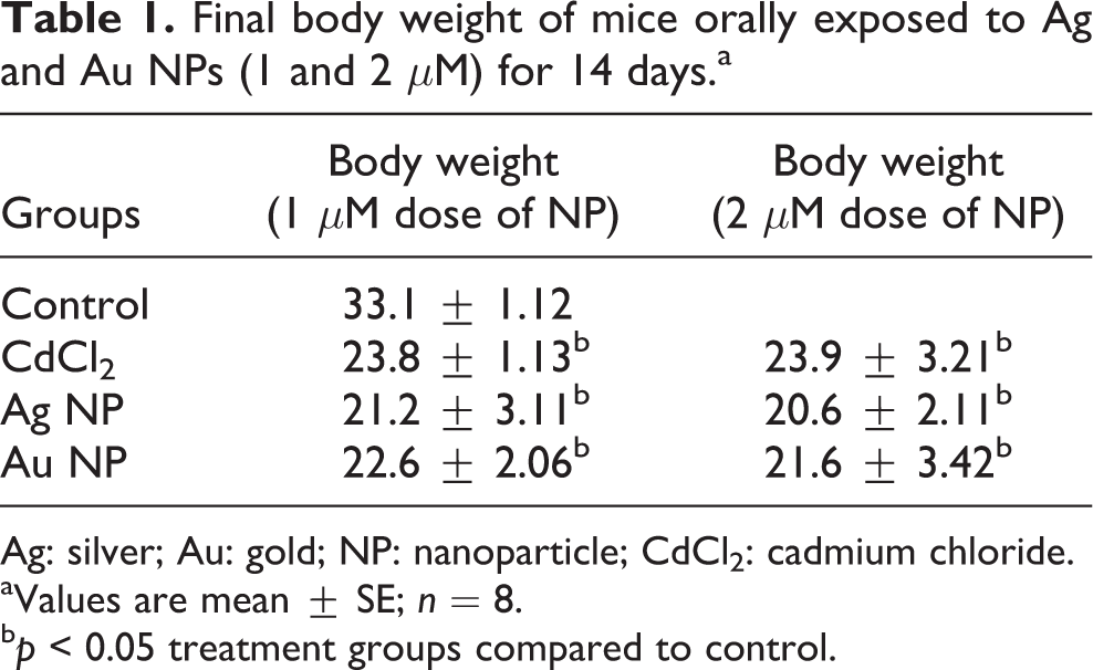
Ag: silver; Au: gold; NP: nanoparticle; CdCl2: cadmium chloride.
aValues are mean ± SE; n = 8.
b p < 0.05 treatment groups compared to control.
Effects of NPs on blood oxidative stress, status of antioxidant enzymes and urinary 8-OHdG level in mouse
Significant elevation of blood ROS level was observed on exposure to both 1 μM and 2 μM dose of NP compared to normal group (Table 2). GSH, GPx, and GST activities showed depletion in both Ag and Au NPs exposed groups (low and high concentrations) compared to the normal group. However, the alterations were more pronounced in the group exposed to 2 μM Ag NP compared to all other groups. Among the two NPs, Ag NP exhibited more pronounced toxicity in blood than Au, particularly at the higher concentration.
Effects of 14 days’ oral exposure to Ag and Ag NPs (1 μM and 2 μM) on various blood and urinary biochemical variables indicative of oxidative stress, and status of antioxidant enzymes in mouse.a
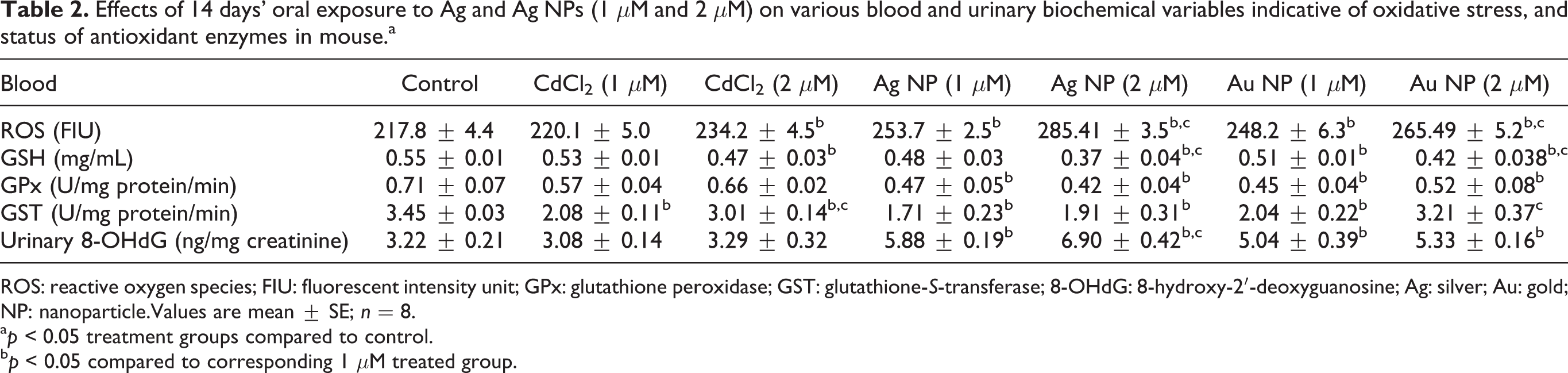
ROS: reactive oxygen species; FIU: fluorescent intensity unit; GPx: glutathione peroxidase; GST: glutathione-S-transferase; 8-OHdG: 8-hydroxy-2′-deoxyguanosine; Ag: silver; Au: gold; NP: nanoparticle.Values are mean ± SE; n = 8.
a p < 0.05 treatment groups compared to control.
b p < 0.05 compared to corresponding 1 μM treated group.
8-OHdG is an important biomarker for assessing DNA damage. Its level significantly increased in group exposed to a higher concentration of Ag NP (2 μM), compared to normal and other exposed groups (Table 2). Exposure to Au NPs also led to a significant elevation in the levels of 8-OHdG, but the increase was less pronounced compared to the effects in Ag NPs exposed animals.
Effects of NPs on tissue oxidative stress and status of antioxidant enzymes
Effects of Ag and Au NPs on the tissue oxidative stress in mice are presented in Figures 1 to 3. The levels of ROS increased significantly in all the tissues (brain, liver, kidney, and spleen) on exposure to Ag and Au NPs compared to normal animals (Figure 1). GSH:GSSG ratio also decreased significantly in all the tissues (data not shown). Altered activity of antioxidant enzymes (GPx and GST) was observed in the exposed groups, signifying toxicity produced by both the NPs. GPx activity decreased in brain and liver following exposure to Ag and Au NP exposure, while it increased in kidney and spleen (Figure 2). On the other hand, GST activity increased in brain, while showed depletion in other soft tissue. The changes were more pronounced in the group exposed to 2 μM Ag NP. In the case of spleen, Au NPs were more toxic (Figure 3).
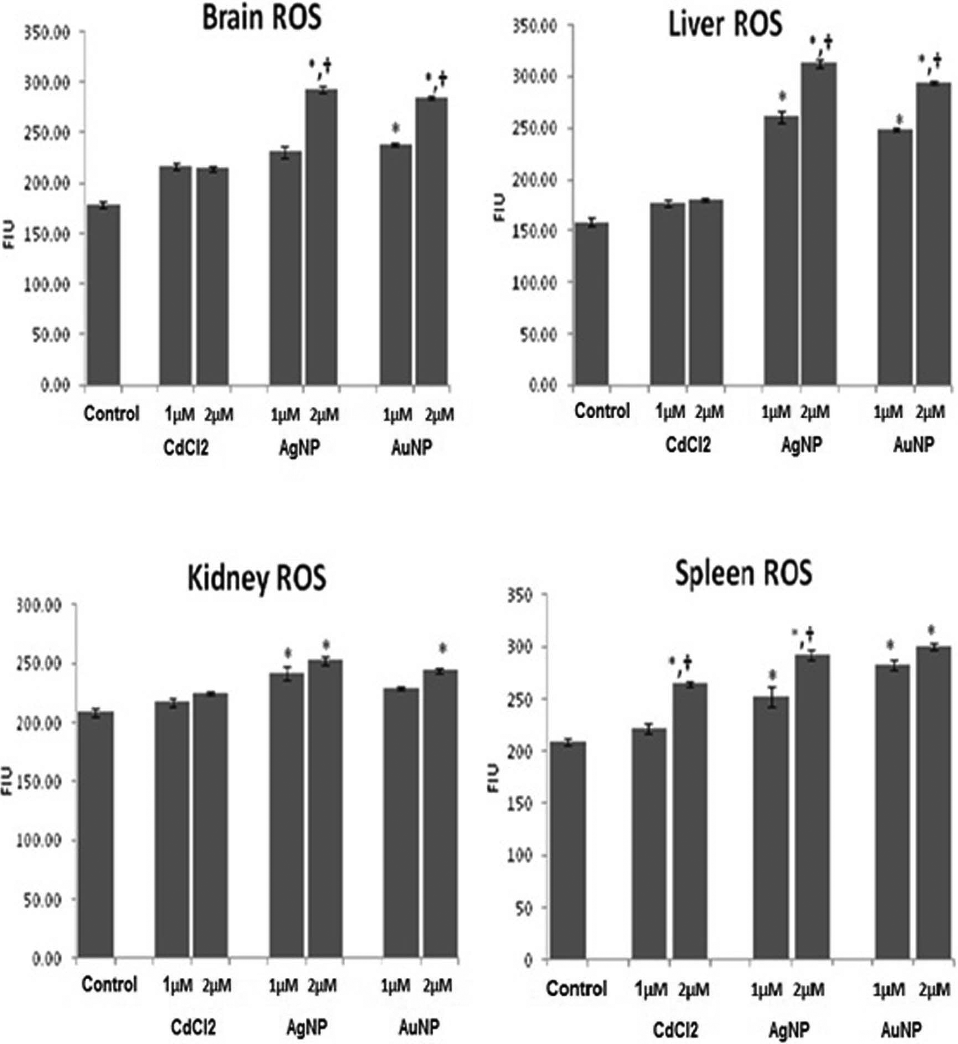
Effects of 14 days’ oral exposure to Ag and Au NPs (1 and 2 μM on the generation of ROS in mouse soft tissues. ROS is given in units of FIU. Values are mean ± SE; n = 8. *p < 0.05 treatment groups compared to control, † p < 0.05 compared to corresponding 1 μM treated group. ROS: reactive oxygen species; FIU: fluorescent intensity unit; Ag: silver; Au: gold; NP: nanoparticle.
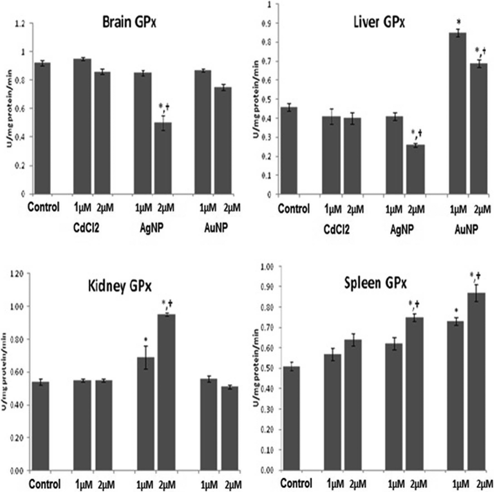
Effects of 14 days’ oral exposure to Ag and Au NPs (1 μM and 2 μM) on the GPx activity in mouse soft tissues. GPx is given in units of units per milligram protein per minute. Values are mean ± SE; n = 8. *p < 0.05 treatment groups compared to control, † p < 0.05 compared to corresponding 1 μM treated group. GPx: glutathione peroxidase; Ag: silver; Au: gold; NP: nanoparticle.
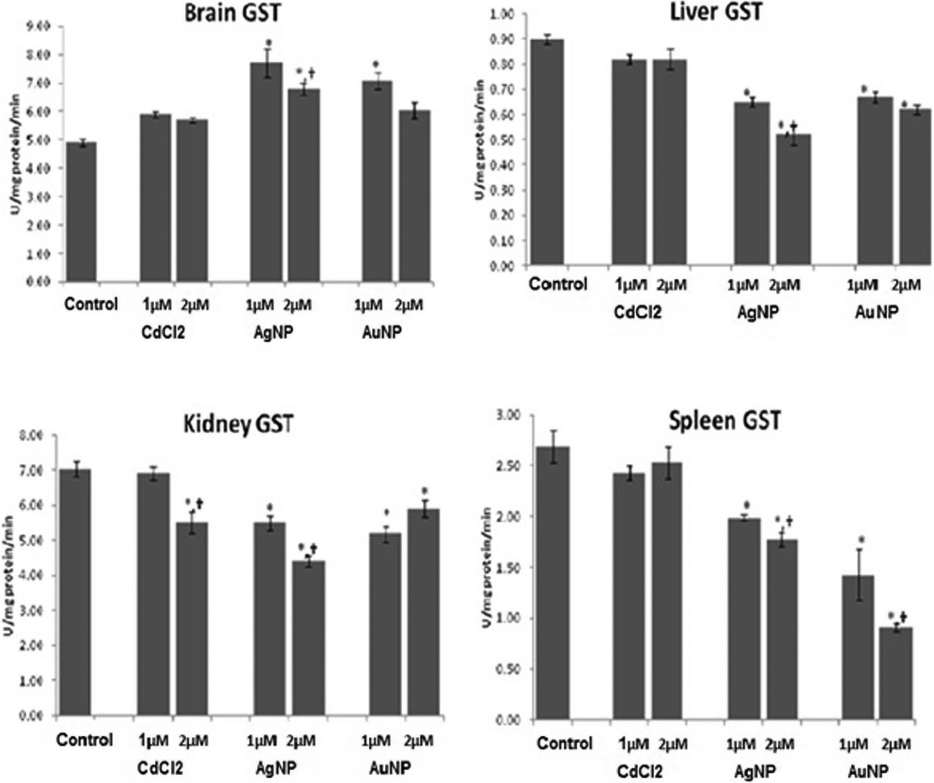
Effects of 14 days’ oral exposure to Ag and Au NPs (1 μM and 2 μM) on the GST activity in mouse soft tissues. GST: is given in units of units per milligram protein. Values are mean ± SE; n = 8. *p < 0.05 treatment groups compared to control, † p < 0.05 compared to corresponding 1 μM treated group. GST: glutathione-S-transferase; Ag: silver; Au: gold; NP: nanoparticle.
Effects of NPs on AST and ALT activities
Significantly increased AST activity was noted on exposure to both the NPs (1 μM and 2 μM), suggesting their hepatotoxic potential, whereas no change in ALT activity was noted (Table 3). Changes were noted in urea and creatinine levels. Ag NPs at the higher concentration were found to be more toxic than Au NPs.
Effects of 14 days oral exposure to Ag and Ag NPs (1 and 2 μM) on various biochemical variables suggestive of liver and kidney damage in mouse.
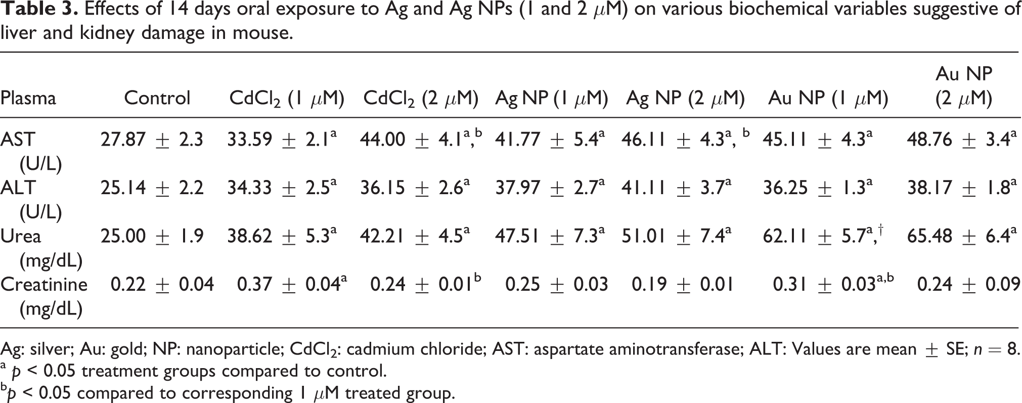
Ag: silver; Au: gold; NP: nanoparticle; CdCl2: cadmium chloride; AST: aspartate aminotransferase; ALT: Values are mean ± SE; n = 8.
a p < 0.05 treatment groups compared to control.
b p < 0.05 compared to corresponding 1 μM treated group.
Effects of NPs on liver and kidney MT induction, inflammatory markers (IL-6), and generation of NO synthetase (NOS) in plasma
Figure 4 depicts the effect of Ag and Au NPs on the hepatic and renal MT induction, inflammatory markers and NOS activity in mouse plasma. For MT induction, a separate group was challenged with cadmium as a positive control. Both the NPs induced MT. The levels, however, were more significant in kidney than liver. Among the two NPs, Ag NP at the higher concentration (2 μM) was able to induce more pronounced MT in kidneys than Au NP.
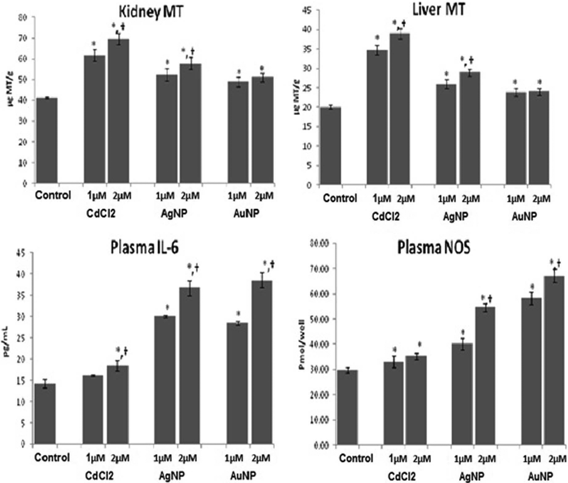
Effects of 14 days’ oral exposure to Ag and Au NPs (1 μM and 2 μM) on the induction of renal and hepatic MT (μg MT/g), plasma IL-6 (pg/mL), and plasma NOS (picomole per well) in mouse. Values are mean ± SE; n = 8. *p < 0.05 treatment groups compared to control, † p < 0.05 compared to corresponding 1 μM treated group. MT: metallothionein; Ag: silver; Au: gold; NP: nanoparticle; IL-6: interleukin-6; NOS: nitric oxide synthetase.
A significant elevation in plasma IL-6 and NOS level was observed on exposure to Ag and Au NPs at both the doses (1 μM) and higher (2 μM) (Figure 4). However comparatively higher increase in both the variables was observed in case of Ag NPs, particularly at concentration of 2 μM compared to normal and other exposed groups.
Discussion
Ag and Au NPs have recently attracted scientific and technological interest due to their various biomedical applications in chemical sensing, biological imaging, drug delivery, and cancer treatment. Despite their widespread use, not much information concerning their toxicity to humans and the underlying cellular actions is available. This toxicity information is necessary before their utilization in various applications. In the present investigation, we studied the effect of oral administration of Ag and Au NPs utilizing two doses in mice on biochemical variables indicative of oxidative damage, inflammation, and organ damage. Exposure to Ag and Au NPs at the equimolar concentrations of 1 μM and 2 μM for 14 consecutive days, significantly altered most of the biochemical variables, indicative of oxidative stress in blood and tissues. The effects were more pronounced in case of Ag NPs, at the higher dose of 2 μM.
Ag and Au NPs tend to release free Ag+ and Au+ ions and aggregate in aqueous environments (Schrand et al., 2010). The inherent instability of Ag and Au NPs might be one of the contributory factors to the toxicity and thus is of major concern (Choi et al., 2010). Ag has received much attention because of its toxicity at the high ionic concentrations. Ag NPs have also been used on a number of occasions to evaluate the cellular uptake and tissue distribution of particles, due to their ease of detection (Johnston et al., 2010). The increased blood ROS level suggests free radical generations by Ag and Au NPs. Within blood, Ag binds to albumin, enabling its transportation throughout the body leading to its toxic manifestation (Wadhera and Fung, 2005). In addition, depletion of endogenous antioxidant GSH, following exposure to Ag and Au NPs, suggests their binding to GSH, or related enzymes involved in its synthesis or its depletion due to enhanced free radical generation (Hussain et al., 2005; Schrand et al., 2010).
Increased ROS level was also noted in liver following Ag and Au NPs exposure, with the effects of Ag being more pronounced at the higher concentration. Significant depletion in GSH:GSSG ratio supports the generation of oxidative stress. Following oral exposure, Ag and Au NPs translocate from the gut, enter the bloodstream, and get distributed to various organs (Johnston et al., 2010). The primary site of Ag and Au particulate accumulation has consistently been demonstrated to be the liver (Kim et al, 2009). This hypothesis is further supported by another study by Piao et al., (2011) which suggests that Ag NPs induce oxidative cell damage in human liver cells through inhibition of reduced glutathione and induction of mitochondria-involved apoptosis. Despite these findings and the current observations, there is still a lack of information regarding the in vivo effects of NPs on this potential target organ.
Only few studies are available which report the ability of Ag and Au NPs to induce MT (Choi et al., 2010; Renault et al., 2005). In this study, we observed significant induction of MT following exposure to these NPs. The induction was highest in case of Ag NPs, particularly at the concentration of 2 μM. Cadmium, which is a known inducer of MT, produced significant hepatic and renal MT expression. MTs are the low molecular weight proteins, which are capable of sequestering metallic ions due to their cysteine residue. This may decrease metal’s ability to exert their toxic effects on other cellular ligands. MTs are also known to be inducible by oxidative stress (Mehta and Flora, 2001), one of the principal mechanisms involved in NP induced toxicity. However, it is well known that the toxicity of Ag and Au NPs is concentration dependent, thus the particular concentration of Ag NPs (2 μM) is more effective in inducing MT, in comparison with other dose. It, however, may be concluded that MT induction is not directly related to NP toxicity but might be correlated to tissue oxidative stress. This is an interesting observation which requires further investigations.
The available reports indicate that NPs can cross blood–brain barrier and enter the central nervous system (Lockman et al., 2004; Sharma and Sharma, 2007 ; Wang et al., 2007). The rapid utilization of NPs and their potential risk to central nervous system (CNS) has elicited much concern recently. Exposure to both NPs led to an elevated level of ROS and also altered GSH:GSSG ratio in brain. It is thus important to suggest that toxicity of NP in infants or fetus by their CNS sensibility or induction of chronic oxidative stress could derive in CNS degenerative diseases. An increase in the activities of antioxidant enzymes, GPx in liver, kidney and spleen, and GST in all the four organs, is one of the interesting observations of this study. It is recognized that the increase in the antioxidant enzyme activities due to the exposure to toxicants is indicative of a defensive response to protect the body against the toxic effects of these toxicants. It has been considered that the low levels of oxidative stress induce the expression of protective mechanisms, whereas larger doses result in the activation of pro-inflammatory mechanisms, and cell death at the extreme levels (Nel et al., 2001). To determine if NPs produce hepatic and renal toxicity, we determined the activities of AST, ALT, urea, and creatinine in plasma, which are metabolites associated with the functionality of liver and kidney (Reeves et al., 2010). Among all these metabolites in plasma of animals treated with both the NPs, only an increase in the activity of AST was noted following exposure to Ag NP (at the higher dose of 2 μM).
It is also recognized that Ag NPs have high affinity towards thiol (–SH) groups within proteins, and thus promote their denaturation (Chen et al., 2008; Wadhera and Fung, 2005). This is of particular concern, due to the high availability of such functional groups within antioxidants, which plays a key role in cellular antioxidant defense system. Ag NPs may thus alter cellular antioxidants and promote the development of an oxidant-mediated response due to the accumulation of ROS within cells (Chen et al., 2008). Au NPs have also been demonstrated to promote ROS production within cells (Hussain et al., 2005), which would further promote oxidative stress. Ag NPs are well documented in their ability to bind DNA, which may not only be exploited during the treatment of a disease (including anticancer agents or within gene therapy), but may also contribute to genotoxicity, or block transcription (Pan et al., 2007).
Genotoxicity was observed in response to exposure to both Ag and Au NPs. Enhanced formation of 8-OHdG adduct, suggesting the formation of DNA lesions, was noted in case of exposure to a higher concentration of Ag NP (2 μM). Oxidative stress leading to the generation of ROS in metabolizing cells could attack DNA base guanine forming 8-OHdG lesions, which is known to have mutagenic potential and hence used routinely as a biomarker for carcinogenesis (Ng et al., 2010). Studies reported in the past also indicate the genotoxic potential of both Ag and Au NPs; however, a more pronounced effect was seen in case of Ag NPs (Choi et al., 2010; Girgis et al., 2012; Siddiqi et al., 2012; Tiwari et al., 2011). Ag and Au NPs have the abilities to interact with the cellular membrane and produce free radicals, leading to mitochondrial permeability (Panyala et al., 2008). Song et al., (2012) also recently reported that 8-OHdG levels increased significantly in the liver DNA of the copper oxide NP treated group. Authors further concluded that these effects were dose-dependent and that metal NPs are capable of causing genotoxicity, and oxidative stress might be the causative factor for the toxicity elicited by these metal NPs. Ag and Au NPs induced generation of oxidative damage is in agreement with our findings, with the toxicity of Ag NPs being more prominent than Au NPs. These free radicals, particularly superoxide anions, react with NO, generated in response to oxidative stress, leading to the formation of harmful peroxynitrite species, which in turn oxidizes lipids, DNA, and proteins (Jia et al., 2009). Our results demonstrate an increased generation of NOS on NPs exposure; however, a marked elevation in case of Au NPs at the higher concentration (2 μM) was observed. These results are in agreement with previous reports signifying radical mediated damages ranging from subtle modulations of cell signaling to overwhelming oxidative injury leading to enhanced toxicity (Stamler, 2004; Yang et al., 2006). Toxicity of NPs is manifested by inflammation resulting from oxidative stress. In our study, increased expression of inflammatory marker IL-6 observed in case of high concentrations (2 μM) of both Ag and Au NPs is in agreement with previous findings (Folkmann et al., 2009; Park et al., 2010). The increase in the concentration of IL-6 demonstrates the inflammatory potential of Ag and Au NPs at the higher concentration and can be an early sign of NP toxicity. Xenobiotic, metals or accumulated nanomaterial-induced stress may lead to the production of many chemokines, cytokines, and antibodies. The size of these NPs allows them to pass directly through cell membranes, penetrate through and between cells, aggregate proteins on their surfaces, and even translocate to cells within the blood–brain barrier (Oberdorster et al., 2005). In these cases, the NPs accumulate within cells and must be expelled before an immune response, inflammation, is activated as phagocytes and leukocytes (as well as other immune cells) encounter the expelled NPs (Park et al., 2010). An interesting recent study also supports our observations that Ag NPs demonstrate higher propensity in inducing inflammation, mediated by ROS and nuclear factor signaling pathways and leading to the induction of cytochrome c oxidase-2, tumor necrosis factor-α, and IL-6. No such effects were noted with Au NP (Nishanth et al., 2011).
Though toxicity of both Ag and Au NPs has been established, toxic effects of Ag NPs are found to be more pronounced, particularly at the higher dose of 2 μM. Bar-Ilan et al., (2009) reported the toxicity of Ag and Au NPs and demonstrated that both these NPs exhibited significantly different toxic profiles, with Ag being toxic and Au inert in all the exposed group. They also concluded that toxicity and biocompatibility of NPs and the types of abnormalities observed in vivo and in vitro system are highly dependent on the concentration and dose of NPs.
Conclusion
Our study, thus, demonstrates that Ag and Au NPs elicited a dose-dependent and oxidant-mediated response. The results also suggest a more pronounced toxicity of Ag NPs over Au NPs. It is, however, important that the toxicity of NPs must be related to the dose and time response, as at the comparatively lower dose levels, they may stimulate a protective antioxidant driven response. Thus, the dose dependency of particle toxicity is an important issue. More extensive investigations in the future are therefore required to fully understand the tissue distribution and fate of Ag and Au particles inside the body, and in addition, consequences of particle accumulation both in vivo and in vitro.
Footnotes
Acknowledgment
Authors thank the Director of the Establishment, for his support and encouragement. Rupal Shrivastava thanks MP Council of Science and Technology (MPCOST) for the award of Junior Research Fellowship.
Funding
This research received no specific grant from any funding agency in the public, commercial, or not-for-profit sectors.