






Abstract
The present study was undertaken to investigate the toxic effect of ground water fluoride (F) on motility, abnormality, and antioxidant status of spermatozoa. Treatment of ground water F with epididymal sperm suspension caused complete loss of sperm motility and decrease in the activities of superoxide dismutase (SOD) and catalase (CAT) and increase in the concentration of malondialdehyde (MDA) and abnormality of spermatozoa at 15 and 30 min time intervals. Further, incubation of spermatozoa with ground water F for 5, 10, and 15 min time intervals significantly reduced the sperm motility and activities of SOD and CAT and increased the concentration of MDA and abnormality of spermatozoa. The study revealed that F-induced effect on sperm motility and antioxidant status is time dependent. Increase in oxidative stress and concomitant decrease in motility of spermatozoa in ground water F clearly indicates that F-induced oxidative stress affected the sperm motility. The present study for the first time demonstrated the toxic effect of ground water F on spermatozoa at shorter duration of exposure, which affects the capability of spermatozoa in fertilization.
Introduction
Pure drinking water is one of the major growing concerns in today’s society. Ground water constitutes 97% of global fresh water and is an important source of drinking water in many regions of the world. Sodium, calcium, magnesium, potassium, chloride, bicarbonate, sulfate, arsenic, cadmium, lead, mercury, and so on, are the major elements of ground water. Fluoride (F) is one such element present universally in all sources of water and the higher concentration of F is found in ground water. The main source of F in ground water is F bearing rocks such as fluorspar, fluorite, cryolite, fluorapatite, and hydroxyl apatite (Meenakshi et al., 2004). F is also present in most of everyday needs viz., tooth pastes, drugs, cosmetics, chewing gums, mouthwashes, and so on (Rao et al., 1997; Thatte, 1994). F is often called as two-edged sword. Though a small amount of F is beneficial for human health for preventing dental caries, it is harmful when present in excess. World Health Organization recommends that F content in drinking water should be in the range of 1.0 to1.5 ppm. In India, Andhra Pradesh, Tamilnadu, Karnataka, Gujarat, Rajasthan, Punjab, Haryana, Bihar, and Kerala are the F endemic regions where sever fluorosis is reported (Susheela, 1999).
Research carried out in different parts of world shows a positive correlation between F levels and dental (Aoba and Fejerskov, 2002; Shivashankara et al., 2000) and skeletal fluorosis (Hileman, 1988; Susheela, 2003). Numerous studies in rat and animal models have shown the adverse effect of F on physiological systems (Gadallah, 2016; Gessner et al., 1994; Gupta et al., 1993; Shashi and Thapar, 2000; Singh et al., 2001; Zhao et al., 2014). Studies have reported the incidences of kidney stones (Doull et al., 2006; Juuti and Heinonen, 1980; Whitford, 1996), gastrointestinal effects, nausea, vomiting, diarrhea, abdominal pain (Gessner et al., 1994; Penman et al., 1997; Sidhu and Kimmer, 2002), myocardial infarction, impaired signaling, bone disorders (Pratheebaa et al., 2013), and diminished cognitive abilities (Lu et al., 2000; Wang et al., 2007; Xiang et al., 2003; Zhao et al., 1996) after the chronic ingestion of ground water F.
Studies on effects of F on reproduction began in the 1970s. Oral administration of different doses of sodium fluoride (NaF) dissolved in drinking water caused significant decrease in weight of testis, epididymis, and ventral prostate; sperm motility and its density in cauda epididymis; number of primary spermatocyte, secondary spermatocyte, and spermatids; sertoli cell and activities of testicular Delta(5), 3β-hydroxysteroid dehydrogenase (HSD), and 17β-HSD; serum concentration of testosterone and number of mature Leydig cells in rats and mice (Ghosh et al., 2002; Giri et al., 2013; Gupta et al., 2007; Kumar et al., 2012; Wan et al., 2006). In addition, F is known to cause histological alterations in reproductive organs (Kumar and Susheela, 1995; Wan et al., 2006). Disorganization and denudation of germinal epithelial cells of seminiferous tubules with the absence of spermatozoa in the lumen (Chinoy and Sequeira, 1989) are the severe effect of F on testis. Further, F increases the abnormalities of spermatozoa and decreases the ability of epididymal spermatozoa to capacitate by decreasing the sperm head tyrosine phosphorylation and actin polymerization (Dvorakova-Hortova et al., 2008). These studies clearly indicate the adverse effect of F on male reproduction. Further, studies have reported the effect of ground water F in rodent models and human beings (Ozsvath, 2009; Reddy et al., 2007; Vega et al., 1988). Studies have shown a decrease in weight of body and testis, sperm motility, and sperm count in rats after the ingestion of ground water F (Singh et al., 2012). A single study reported the effect of ground water F on hormonal profile of human beings residing in F endemic areas. Exposure of ground water F at 3–27 mg/day caused significant increase in serum level of FSH and decrease in inhibin-B, testosterone, and prolactin in male individuals (Ortiz-Perez et al., 2003). These in vivo studies indicate that ground water F do have adverse effect on male reproduction. However, it is not known whether ground water F has any direct effect on sperm motility in in vitro. In addition, whether the effect of ground water F on spermatozoa is dependent on duration of exposure is also not known.
The antioxidant system plays a vital role in metabolism, and its imbalance leads to oxidative stress. Oxidative stress is one of the major causes of male infertility due to the elevated levels of reactive oxygen species (ROS) (Lanzafame et al., 2009). Oxidative stress affects metabolites viz., DNA, lipids, proteins, and enzymatic systems, which lead to decline in the semen parameters (Agarwal et al., 2014). Spermatozoa are unable to restore the damage induced by oxidative stress because they lack the necessary cytoplasmic enzyme repair systems (Saleh and Agarwal, 2002). Earlier studies have shown an association of oxidative stress with reproductive impairment (Aitken et al., 1993, 1998). High levels of ROS cause adenosine triphosphate (ATP) depletion, loss of motility, increase in abnormal spermatozoa and lipid peroxidation, and decrease in fertilizing ability of spermatozoa (Aitken et al., 1992; de-Lamirande and Gagnon, 1995; Kobayashi and Suda, 2012). F exposure increases oxidative stress by increasing the level of ROS (Bisht et al., 2017; Hassan and Yousef, 2009; Izquierdo-vega et al., 2008; Kumari and Rao, 1991; Rzeuski et al., 1998; Shanthakumar et al., 2004; Shivarajashankara et al., 2001; Zhang et al., 2007). Studies have reported decrease in activities of antioxidant enzymes and increase in lipid peroxidation in testis and spermatozoa due to NaF exposure (Chlubek, 2003; Ghosh et al., 2002; Izquierdo-vega et al., 2008). However, there are no reports on ground water F-induced oxidative stress in male reproductive system. In the view of these lacunae, the present study was conducted to find out whether ground water F affect motility of spermatozoa in in vitro at different time intervals and if so whether depletion of sperm motility is due to oxidative stress induced by ground water F.
Materials and methods
Animals
Adult male Wistar rats weighing 180–200 g were obtained from the Central Animal Facility, University of Mysore, Mysore, Karnataka, India. The rats were provided standard rat chow and water ad libitum and were kept in 27 ± 2°C under 12 h light/dark cycles in polypropylene cages. Approval for the proposed animal experiments was obtained from Institutional Animal Ethics Committee of University of Mysore, India (Reference number-UOM/IAEC/04/2017), and all procedures were performed in accordance with the ethical standards of the Committee for the Purpose of Control and Supervision of Experiments on Animals, India.
Collection of water sample
The F water was collected from Mandya district of Karnataka, India as usual water sources like hand pumps in polypropylene bottles. The concentration of F in the water was estimated by ion selective method and it was 5 ppm/ml.
In this experiment, F sensitive electrode and a saturated calomel electrode (external reference electrode) were used to measure the F ion content in the water. The NaF solid was dried for 1 h at 100°C, and standard solutions of NaF were prepared by dissolving 0.42 g of NaF in deionized water and diluting it to 100 ml in a volumetric flask. The solution was further diluted to 10−2 F. The instrument setup consists of F ion-selective electrode, saturated calomel electrode (reference electrode), and multimeter/pH meter. The electrode was suspended off the bottom of the beaker that holds the sample. The unknown sample (1 ml) and KCL (10 ml) was mixed in 100 ml of volumetric flask and diluted to 100 ml with deionized water. The potential in millivolts of the F ion-selective electrode versus the reference electrode for standards and known water sample were measured.
Experimental design
The study was undertaken in two parts referred to as part A and part B. Part A was undertaken to analyze the effect of ground water F on different parameter of spermatozoa at intervals of 0, 15, and 30 min of exposure. Based on results from part A, part B was undertaken to analyze the effects of ground water F at shorter time intervals of 0, 5, 10, and 15 min.
i) Part A:
Epididymal sperm suspension (60 million/ml) was incubated in phosphate buffered saline (PBS; pH 7.5) along with ground water F at 0, 15, and 30 min time intervals in room temperature. After the completion of each incubation period, sperm motility, abnormality, and biochemical estimations were analyzed and compared with control (without ground water F).
ii) Part B:
Epididymal sperm suspension (60 million/ml) was incubated in PBS (pH 7.5) along with ground water F at 0, 5, 10, and 15 min time intervals in room temperature. Different sperm parameters were analyzed at the end of each treatment period and compared with control (without ground water F).
Isolation of spermatozoa
Each part of cauda epididymis of adult male rats was minced in 1 ml of PBS and a suspension was obtained. The suspension was filtered through muslin cloth, and the resultant filtered epididymal sperm suspension was subsequently treated with ground water F and different parameters of spermatozoa were assessed at regular time intervals.
Estimation of sperm motility
An aliquot of sperm suspension was taken after the completion of each incubation period in ground water F and without ground water F. The number of motile and non-motile spermatozoa was counted from different fields until a total 200 spermatozoa were counted and the mean of counts were calculated at each time point. The percentage of motility was calculated at different time intervals (Saalu et al., 2010).
Estimation of sperm abnormality
A uniform smear of spermatozoa was made on a glass slide after staining the spermatozoa with eosin. One thousand spermatozoa per sample were observed using a light microscope under higher magnification (400×) from randomly selected areas of smear. The number of spermatozoa showing head and tail abnormalities were counted. The sum of counts of different abnormalities was expressed as total abnormal sperm count/1000 spermatozoa (Narayana et al., 2002; Vega et al., 1988).
Biochemical estimations
i) Activity of superoxide dismutase (SOD)
The activity of SOD was assayed following the method of Marklund and Marklund (1974) (sensitivity 16 unit/mg protein) with few modifications. The spermatozoa sample was homogenized in 3 ml of phosphate buffer (pH 7.0), and it was centrifuged at 7500 × g for 10 min. The pellet was discarded and the resulting supernatant was used for the assay. The assay medium in a total volume of 1 ml contained 100 µl of sample, 0.9 ml of 0.1 M tris HCL buffer (pH 8.2), and 100 µl of 0.2 mM pyrogallol. Autoxidation of pyrogallol was monitored at 420 nm for 2 min in the absence or presence of added enzymes. One unit of the enzyme activity is defined as the amount that produced 50% inhibition of pyrogallol auto-oxidation. The activity was expressed as unit/mg protein.
ii) Activity of catalase (CAT)
The CAT activity was assayed following the method of Aebi (1984) (sensitivity 0.25–4 nmol/min/ml). The sample was prepared by homogenizing the spermatozoa in 3 ml of phosphate buffer (pH 7.0) and centrifuged at 10 min. The pellet was discarded and the resulting clean supernatant was used for the enzyme assay. Briefly, 1 ml of the reaction mixture contained 100 µl sample, 915 µl of 0.1 M phosphate buffer (pH 7.0) and 25 µl of 8.8 mM H2O2. The absorbance was read at 240 nm, and the decrease in absorbance was followed for 3 min at room temperature using a UV-Visible spectrophotometer. The enzyme activity was expressed as µmol H2O2 consumed/min/mg protein.
iii) Malondialdehyde (MDA) concentration
The concentration of MDA was assayed according to the method of Ohkawa et al. (1979) (sensitivity 4 nmol/mg protein). The MDA is the end product of lipid peroxidation of poly unsaturated fatty acids, and it is estimated utilizing its reaction with thio barbituric acid (TBA). The spermatozoa was homogenized with 3 ml of phosphate buffer (pH 7.0) and centrifuged at 7500 × g for 10 min. The pellet was discarded, and the resulting clean supernatant was used for the enzyme assay. The sample (0.1 ml) was added to a reaction mixture of 0.2 ml of 8.1% sodium dodecyl sulphate, 1.5 ml of 20% acetic acid, and 1.5 ml of 0.8% of TBA. The reaction mixture was made up to 4 ml with distilled water and then heated in a boiling water bath at 95°C for 60 min. After incubation, it was cooled to room temperature, shaken vigorously, and optical density was measured at 533 nm. The concentration of MDA was expressed as nmol/mg protein.
Statistical analysis
The mean ± SE of each parameter was computed considering the data, and mean values of each parameter of different treatment groups were compared using independent samples t test and judged significant if p < 0.05.
Results
Effect of ground water F on sperm parameters and antioxidant status at 0, 15, and 30 min of incubation
Motility
Incubation of ground water F for 15 and 30 min time intervals caused complete loss of sperm motility compared to their respective controls. No significant differences were observed in sperm motility of control and ground water F at 0 min of incubation (Figure 1).

Mean percentage of sperm motility in control and ground water F-treated spermatozoa at 0, 15, and 30 min time intervals. Note the decreased sperm motility in ground water F-treated groups compared with controls. *Significantly (p < 0.05) different compared with controls as judged by independent sample t test (*p < 0.05, **p < 0.01, and ***p < 0.001). F: fluoride.
Abnormality
A significant increase in abnormality was observed in spermatozoa treated with ground water F at 15 and 30 min compared to their respective controls. However, percentage of abnormal spermatozoa of control and ground water F-treated groups was not different from each other at 0 min of incubation (Table 1).
Effect of ground water F on abnormality and activities of antioxidant enzymes of spermatozoa at 0, 15, and 30 min of incubation.

SOD: superoxide dismutase; CAT: catalase; F: fluoride.
ap < 0.05.
bp < 0.01.
cp < 0.001.
Concentration of MDA
There was a significant increase in concentration of MDA in spermatozoa treated with ground water F for 15 and 30 min compared to their respective controls. No significant differences were observed in concentration of MDA in spermatozoa treated with control and ground water F at 0 min of incubation (Figure 2).

Mean MDA concentration of spermatozoa in control and ground water F-treated spermatozoa at 0, 15, and 30 min time intervals. Note the increased level of MDA in ground water F-treated groups compared with controls. *Significantly (p < 0.05) different compared with controls as judged by independent sample t test (*p < 0.05, **p < 0.01, and ***p < 0.001). MDA: malondialdehyde; F: fluoride.
Activities of SOD and CAT
The activities of SOD and CAT were significantly decreased in spermatozoa treated with ground water F for 15 and 30 min compared to their respective control. However, there were no significant differences in the activities of SOD and CAT of control and ground water F-treated groups at 0 min of incubation (Table 1).
Effect of ground water F on sperm parameters and antioxidant status at 0, 5, 10, and 15 min of incubation
Motility
A significant decrease in sperm motility was observed in ground water F at 5 and 10 min of incubation compared to their respective controls, whereas that of 15 min caused complete loss of sperm motility. No significant differences were observed in sperm motility of control and ground water F at 0 min of incubation (Figure 3).
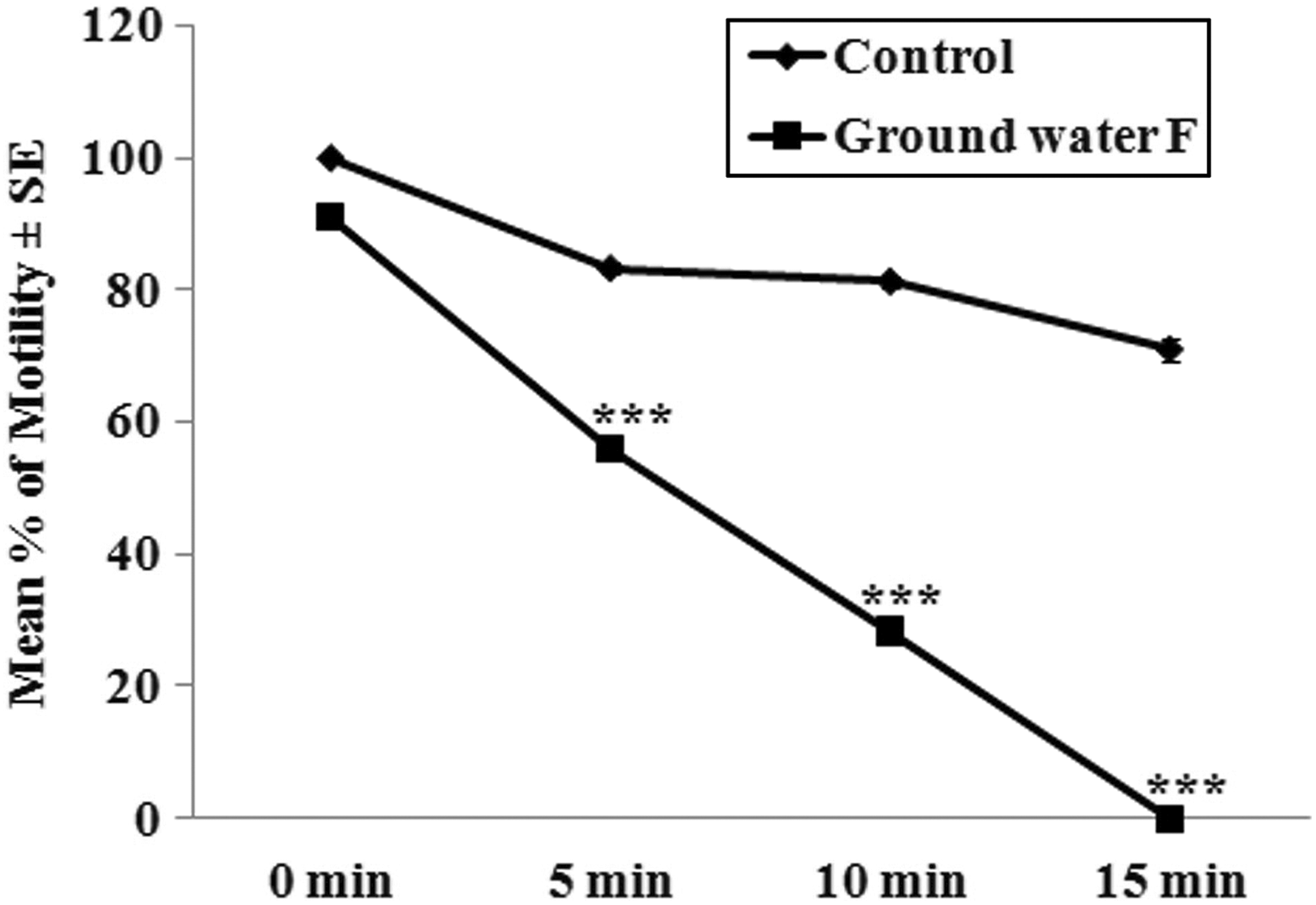
Mean percentage of sperm motility in control and ground water F-treated spermatozoa at 0, 5, 10, and 15 min time intervals. Note the decreased sperm motility in ground water F-treated groups compared with controls. *Significantly (p < 0.05) different compared with controls as judged by independent sample t test (*p < 0.05, **p < 0.01, and ***p < 0.001). F: fluoride.
Abnormality
A significant increase in abnormality was observed in spermatozoa treated with ground water F at 5, 10, and 15 min compared to their respective controls. However, percentage of abnormal spermatozoa of control and ground water F-treated groups did not differ from each other at 0 min of incubation (Table 2).
Effect of ground water F on abnormality and activities of antioxidant enzymes of spermatozoa at 0, 5, 10, and 15 min of incubation.

SOD: superoxide dismutase; CAT: catalase; F: fluoride.
ap < 0.001.
bp < 0.01.
cp < 0.05.
Concentration of MDA
There was a significant increase in concentration of MDA in spermatozoa treated with ground water F at 5, 10, and 15 min compared to their respective controls. No significant differences were observed in concentration of MDA in spermatozoa treated with control and ground water F at 0 min of incubation (Figure 4).

Mean MDA concentration of spermatozoa in control and ground water F-treated spermatozoa at 0, 5, 10, and 15 min time intervals. Note the increased level of MDA in ground water F treated groups compared with controls. *Significantly (p < 0.05) different compared with controls as judged by independent sample t test (*p < 0.05, **p < 0.01, and ***p < 0.001). MDA: malondialdehyde; F: fluoride.
Activities of SOD and CAT
The activities of SOD and CAT were significantly decreased in spermatozoa treated with ground water F at 5, 10, and 15 min compared to their respective controls. However, there were no significant differences in the activities of SOD and CAT of control and ground water F-treated groups at 0 min of incubation (Table 2).
Discussion
Sperm motility is one of the major factors needed for successful fertilization. Any factor that affects sperm motility could result in failure of fertilization. The present study revealed the toxic effect of ground water F on motility, abnormality, and antioxidant status of spermatozoa as treatment of ground water F caused significant decreases in motility and activities of SOD and CAT and increase in abnormality and lipid peroxidation of spermatozoa. The present study for the first time revealed that the adverse effect of ground water F on spermatozoa is dependent on duration of exposure. There was a time-dependent decrease in motility and activities of antioxidant enzymes and increase in abnormality and lipid peroxidation of spermatozoa due to treatment of ground water F. These results were similar to our previous study where treatment of NaF caused time-dependent depletion of motility of spermatozoa in in vitro (Chaithra et al., 2018). The present investigation involved two different in vitro experiments where spermatozoa were treated with ground water F at 0, 15, and 30 min time intervals. In the first in vitro experiment, ground water F caused complete loss of sperm motility at the 15-min time interval. To further investigate this, a second in vitro experiment was conducted where a spermatozoa suspension was treated with ground water F for 0, 5, 10, and 15 min time intervals. Interestingly, at the -15min time interval, ground water F caused complete loss of sperm motility. These results clearly indicate that F exposure caused adverse effects even at short durations of exposure.
F is known to cause oxidative stress. Earlier studies have reported increased oxidative stress in testis and spermatozoa on exposure to F (Ghosh et al., 2002; Izquierdo-vega et al., 2008). Spermatozoa are particularly susceptible to oxidative stress because of the presence of large quantities of polyunsaturated fatty acids (Alverz and Storey, 1995) as well as limited stores of antioxidant enzymes (Aitken and Fisher, 1994; Baker and Aitken, 2005; de- Lamirande and Gagnon, 1995; Sanocka and Kurpisz, 2004; Sharma and Agarwal, 1996). In the present study, significant increases in lipid peroxidation and decreases in the activities of SOD and CAT of spermatozoa due to ground water F treatment clearly indicate F-induced oxidative stress in spermatozoa. Ground water F-induced oxidative stress severely affects the functional ability of spermatozoa, which is evident by the drastic reduction in motility of spermatozoa. Further, oxidative damage can seriously compromise the ability of spermatozoa to engage in sperm–oocyte fusion (Aitken et al., 1993). F-induced reproductive defects have been reported previously in animal models and human beings (Elbetieha et al., 2000; Ortiz-Perez et al., 2003; Verma and Guna Sherlin, 2002). The effects were observed in heavily F-exposed experimental models with concentration of more than 10 mg/ml (Giri et al., 2013; Tiwari and Pande, 2011). However, in the present study, a 5 mg/ml concentration of F in ground water caused severe damage in sperm motility and induced oxidative stress indicating that lower concentrations of F has a toxic effect.
The mechanism of F-induced depletion of sperm motility has not been clearly elucidated. There are different possible mechanisms through which F affects sperm motility. F-induced reduction in sperm motility is either due to the depletion of intracellular ATP (Kim et al., 2015), alterations in the intracellular concentration of ions viz., Mg, Ca, Zn, and Se (Darszon et al., 1999), reduced level of fructose (Chinoy et al., 1995) or decreased level of androgen carrier proteins involved in sperm motility (Bataineh and Nusier, 2006; Chinoy et al., 1997). In addition, F-induced oxidative stress is one of the factors that affects sperm motility. Studies have reported F-induced oxidative stress in spermatozoa, which is accompanied by decreases in sperm motility. For instance, treatment of F in rats caused a significant decrease in the activities of peroxidase and CAT in spermatozoa, with decreases in sperm motility (Ghosh et al., 2002). Similarly, Izquierdo-vega et al. (2008) reported increased oxidative stress in spermatozoa of rat exposed to F.
Cellular oxidative stress may occur as a result of overproduction of ROS or deficiency in the antioxidant system. F affects the redox system either by increasing ROS production or by decreasing the activities of antioxidant enzymes (Izquierdo-vega et al., 2008). F decreases the activities of antioxidant enzymes by binding to their divalent cofactors at the active site of the enzyme (Lawson and Yu, 2003; Wilde and Yu, 1998) as well as by decreasing the production of mRNA (Zhan et al., 2006). In addition, F causes mitochondrial injury by decreasing the mitochondrial membrane potential (MMP). Decline in MMP promotes increased production of ROS (Wang et al., 2003). Spermatozoa fertility mainly depends on maintenance of MMP by the electron transport chain. Thus, F induced either an increase in ROS production or decrease in activities of antioxidant enzyme resulting in increased abnormal spermatozoa and decreased sperm motility.
The present study for the first time demonstrated that exposure to ground water F decreased the motility of spermatozoa, because of F induced increased oxidative stress and the effect is time dependent. The study gains importance in the human society where males are exposed to high level of F through drinking water from ground water sources. The investigation appears to be direct evidence for F induced toxicity on male reproduction, which reflects on reproductive complication in male individuals in long-term exposure to ground water F.
Conclusion
The present study for the first time demonstrated the toxic effect of ground water F on spermatozoa at a shorter duration of exposure and that F-induced decreased sperm motility is associated with increased oxidative stress. The present study is direct evidence of F-induced toxicity on male reproduction in human beings residing in areas where there is high level of F in drinking water.
Footnotes
Declaration of Conflicting Interests
The author(s) declared no potential conflicts of interest with respect to the research, authorship, and/or publication of this article.
Funding
The author(s) disclosed receipt of following financial support for the research, authorship, and/or publication of this article: The first author is thankful to University Grant Commission, India for financial assistance under National fellowship for scheduled caste scheme.
