
Abstract
Cerium (IV) oxide (CeO2), which is used as a biomaterial, has wide application in areas such as the biomedical, glass polishing, electronic, automotive, and pharmacology industries. Comparing with the literature, in this study, the genotoxic effects of cerium (IV) oxide microparticles (COMPs) and cerium (IV) oxide nanoparticles (CONPs) were investigated for the first time in human peripheral blood cultures at concentrations of 0.78, 1.56, 3.125, 6.25, 12.5, 25, and 50 ppm for 72 h under in vitro conditions. Particle sizes of COMPs and CONPs were determined using scanning electron microscopic analysis. Micronucleus and chromosome aberration tests were used to determine the genotoxicity of COMPs and CONPs. The average particle sizes of COMPs and CONPs were approximately 148.25 and 25.30 nm, respectively. It was determined that CeO2 particles in both micro and nano sizes were toxic at all concentrations compared to the negative control group (distilled water). Importantly, COMPs and CONPs were genotoxic even at the lowest concentration (0.78 ppm). Comparing particle sizes, the data indicated that COMPs were more toxic than CONPs. The results suggest that genotoxicity of COMPs and CONPs may be a function of applied concentrations and particle sizes.
Introduction
With the recent expansion of nanotechnological products, people, other living things, and receiving environments have become more exposed to these nanomaterials. The quantity, transport, decay, transformation, and final fate of nanomaterials released into environments are not fully known. Nanoparticles that can enter the body through dermal, oral, and respiratory means can impair the integrity of genetic material by direct or indirect mechanisms (Li et al., 2005; Oberdörster et al., 2005). Although organic compounds can be converted into nontoxic compounds in nature, this is less likely for inorganic compounds. Therefore, it is more important to know the toxic effects of micro- and nanostructured inorganic compounds (Dadook et al., 2014).
Nanostructured cerium (IV) oxide (CeO2), also known as nanoceria, is the most abundant metal oxide in the rare earth family. It is used for different applications including as a catalyst, conductor, electrode, ultraviolet absorbent, high-performance luminescence devices, and glass polisher (He et al., 2015). It also has free radical scavenging capability because of the oxygen vacancies following reduction of Ce4+ to Ce3+. This featurehas been reported to reduce reactive oxygen species (ROS) and oxidative stress in biological systems (Niu et al., 2007). Cerium oxide nanoparticles have toxic effects on numerous bacteria. Furthermore, when it is used with biodegradable materials such as chitosan, it contributes to increased biocompatibility and accelerates wound healing treatment (Huang et al., 2018; Zhang et al., 2019). Among its numerous biological applications is antibacterial, antioxidant, and anti-inflammatory activities (Heckert et al., 2008; Pirmohamed et al., 2010; Varini et al., 2019; Walkey et al., 2015; Zhang et al., 2019). Due to the structural characteristics of the nanoceria, it combines with soil particles and remains in the soil, so it is also likely to participate in physical and biological cycles (Kos et al., 2017).
In addition to its widespread use, cerium (IV) oxide nanoparticles (CONPs) continue to find application in developing nanotechnology with its versatile properties. Undoubtedly, the use of nanoceria as a biomaterial will continue to be one of the highlights of biology, biomedical, and materials science in the coming years. However, this nanoparticle has been considered in the category of hazardous compounds by Organization for Economic Co-operation and Development (OECD), and OECD recommends that the toxicity profile should be investigated with further experimental studies (Naz et al., 2019). In particular, the methods of studies for determining toxicity may be varied, as well as the parameters related to the particle (size, shape, surface load, crystal structure, concentration, and application time, etc.) are suggested to be studied separately. The aim of this study was to determine and compare the genotoxicity profiles of cerium (IV) oxide microparticles (COMPs) and nanoparticles CONPs used in biomedical applications using chromosome aberration (CA) and micronucleus (MN) test methods.
Materials and methods
Chemicals
Cytochalasin B, glacial acetic acid, methanol, mitomycin-C, potassium chloride (KCl), Giemsa stain, disodium hydrogen phosphate, potassium dihydrogen phosphate, CONPs, COMPs (Sigma-Aldrich), colcemid (Roche), chromosome medium B (Gibco), and Heparin (drug name is Nevparin) (Mustafa Nevzat Medicine Company) were purchased commercially.
Determination of particle sizes
The particle sizes of CONPs and COMPs were measured by scanning electron microscopy (SEM). SEM analyses were performed using a Zeiss Sigma 300 (Carl Zeiss Microscopy GmbH, Jena, Germany) instrument at an acceleration voltage of 2.00 kV.
Cell cultures
In this study, heparinized human blood samples were provided by five male volunteers who did not use any addictive substances such as alcohol, cigarettes, and drugs, did not have any genetic or systemic disease and were not exposed to any known physical and chemical mutagens based on their professions. Informed consent forms were signed by each volunteer. The cell cultures were prepared according to our previous study (Akbaba and Türkez, 2018). The blood samples (0.5 mL) were cultured in 6 mL of culture medium (Chromosome Medium B, Biochrom, United Kingdom). Then, 0.5 mL aliquots from the solutions of test compounds were added to the tubes to obtain final concentrations of 0.78, 1.56, 3.125, 6.25, 12.5, 25, and 50 ppm. The culture tubes were incubated for 72 h at 37°C. Distilled water and mitomycin-C (10−7 M) were used as negative control group and positive control group, respectively. The blood samples remaining after the analysis of the people who applied to the hospital for routine tests were used. Blood samples of the volunteers were taken by the specialist staff of the hospital. The blood samples were not taken directly from volunteers.
CA assay
The CA tests were performed with slight modifications using the method of Evans (Evans, 1984). Chromosome medium B (6 mL) was added to labeled sterile cell culture tubes. Then, 0.5 mL of whole blood was added onto the medium. CeO2 solutions were prepared at the concentrations described above, and 0.5 mL was added to the tubes. After incubation at 37°C for 70 h and 15 min, 0.1 mL of colchemide solution was added. Later, the tubes were incubated for 1 h and 45 min at 37°C and centrifuged at 78 x g for 10 min. After removing the supernatant, 8 mL of hypotonic solution (0.075 M KCl) was added dropwise onto the remaining cell suspensions. Cells were incubated for 15–20 min at 37°C and centrifuged at 78 x g for 10 min, and the resulting supernatant was removed. Cold fixation solution (7 mL, methanol:glacial acetic acid, 3:1 v/v) was added to the remaining cell suspension and centrifuged again. The resulting supernatant was removed. This procedure was repeated twice. The remaining cell suspensions were dropped onto cold slides and allowed to dry for 3 days without exposure to daylight. The slides were stained with Giemsa stain for 15 min and allowed to dry.
MN assay
MN assays were applied according to Fenech’s method (Fenech, 2000). This method is similar to the CA method. Unlike the CA method, 0.1 mL of cytochalasin B was added to the tubes after 44 h of incubation. At the end of the incubation period of 72 h, the slides obtained by applying the procedures in the CA test were made ready for microscopic investigation.
Slide analysis
Slides were observed using a BestScope BLM-280 LCD Digital Biological Microscope (Beijing, China) light microscope at 100× magnification with immersion oil. The slides were investigated by two observers who were blinded to the obtained results. Gaps and breaks of chromatid/chromosome were classified in accordance with Environmental Health Criteria 46 (IPCS, 1985). To calculate the CA frequencies, 30 well-spread metaphases were analyzed. Taking into consideration the MN evaluation criteria (Fenech et al., 2003; Titenko-Holland et al., 1997), 1500 binucleus cells were counted at each concentration for each individual, and the MN ratios were calculated.
Statistical analysis
IBM SPSS statistics for Windows (version 18.0, IBM Corp., Armonk, NY, USA) package program was used for the statistical evaluation of the data obtained from the experimental study. To evaluate statistical differences between the groups, analysis of variance (ANOVA) was used considering criteria of ANOVA. The results were given as mean ± standard deviation (X ± SD), and p values less than 0.05 were considered statistically significant.
Results
Characterization of particles
The average particle sizes for COMPs and CONPs were 148.25 and 25.30 nm, respectively. The results of SEM analysis are shown in Figures 1 and 2.
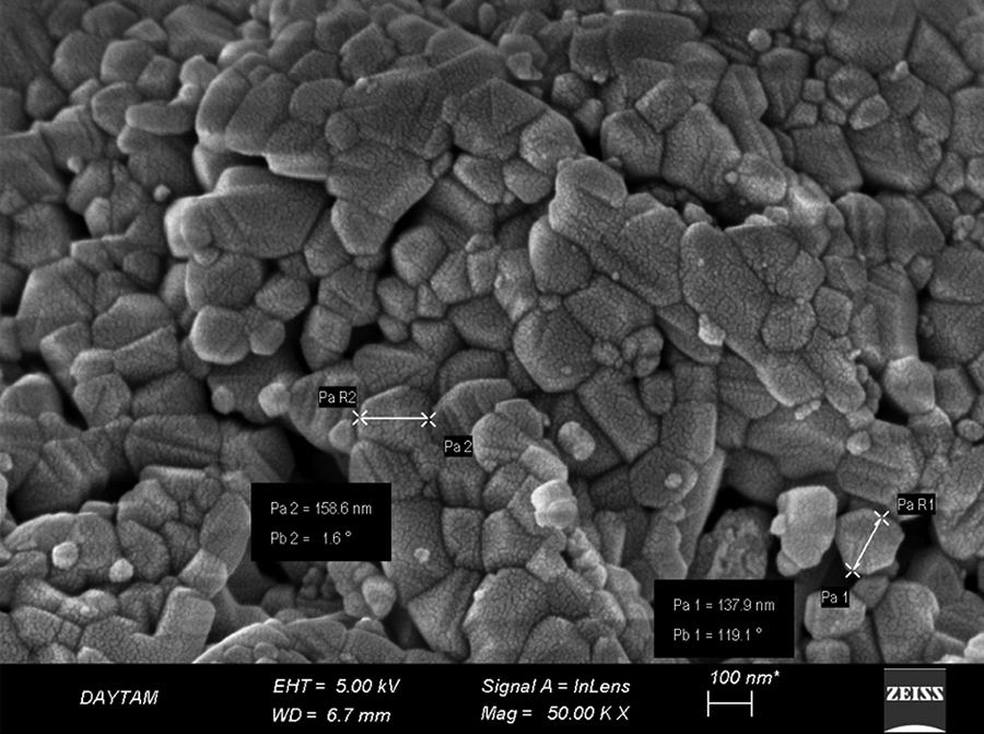
SEM image of COMPs. SEM: scanning electron microscope; COMPs: cerium (IV) oxide microparticles.
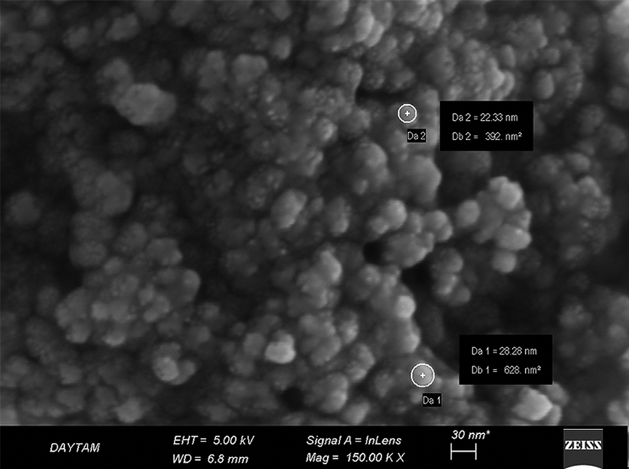
SEM image of CONPs. SEM: scanning electron microscope; CONPs: cerium (IV) oxide nanoparticles.
CA data
When CA frequencies were evaluated, it was found that cerium oxide particles of both micro and nano dimensions were genotoxic in all concentrations compared to the negative control group. The results are given in Table 1 and Figure 3.
Statistical distribution of CA frequencies of COMPs and CONPs between groups.a
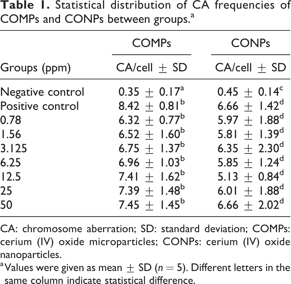
CA: chromosome aberration; SD: standard deviation; COMPs: cerium (IV) oxide microparticles; CONPs: cerium (IV) oxide nanoparticles.
a Values were given as mean ± SD (n = 5). Different letters in the same column indicate statistical difference.
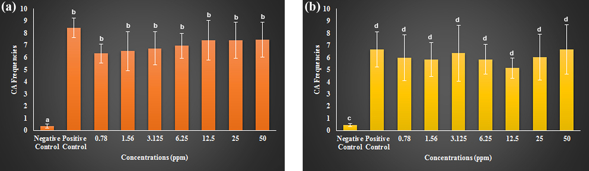
CA frequencies caused by COMPs (a) and CONPs (b) at different concentrations. COMPs: cerium (IV) oxide microparticles; CONPs: cerium (IV) oxide nanoparticles; CA: chromosome aberration. Different letters on the columns were significantly different at p < 0.05.
Furthermore, the genotoxicity of COMPs and CONPs at the same concentrations were compared, and no statistically significant difference was observed between them. As shown in Table 2 and Figure 4, although there is no difference between their toxicity, the toxicity of COMPs is higher than CONPs.
Statistical distribution of CA frequencies generated by cerium (IV) oxide compound according to particle size.a
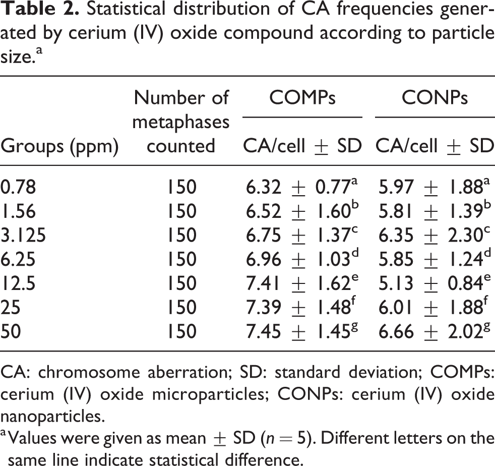
CA: chromosome aberration; SD: standard deviation; COMPs: cerium (IV) oxide microparticles; CONPs: cerium (IV) oxide nanoparticles.
a Values were given as mean ± SD (n = 5). Different letters on the same line indicate statistical difference.
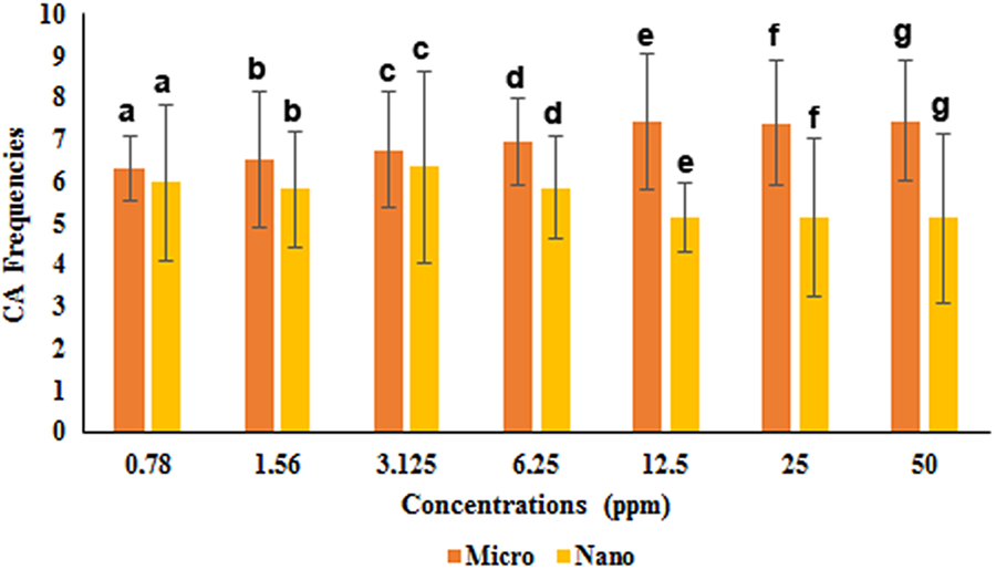
Comparison of CA frequencies caused by COMPs and CONPs at the same concentrations. COMPs: cerium (IV) oxide microparticles; CONPs: cerium (IV) oxide nanoparticles; CA: chromosome aberration. Different letters on the columns were significantly different at p < 0.05.
MN test results
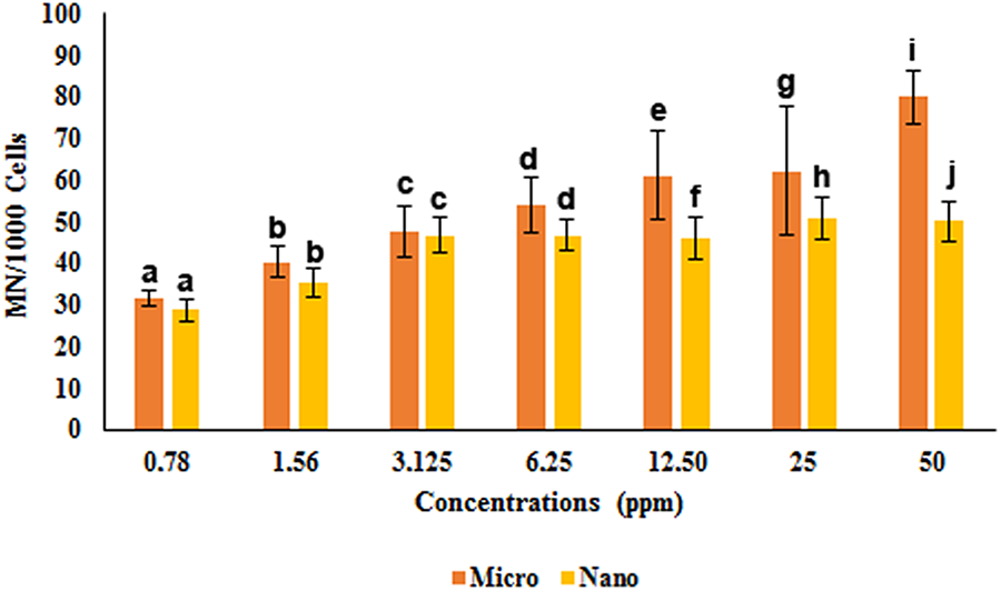
Bases on the results of MN tests, all studied concentrations of CeO2 in nano and micro dimensions were found to be genotoxic compared to the negative control group, similar to CA test results. MN ratios caused by COMPs did not show any statistically significant alteration at concentrations ranges of 3.125–25 ppm. Similarly, no statistically significant difference was found between the toxicities at 0.78 and 1.56 ppm. Remarkably, COMPs at 50 ppm, which is the highest concentration, had the same toxic effect as the positive control group. The toxicity of CONPs at concentrations of 0.78 and 1.56 ppm was determined to be statistically similar to each other but different from other concentrations. The toxicities at 3.125, 6.25, 12.5, 25, and 50 ppm also were statistically similar to each other, at these concentrations, CONPs exhibited the toxicity close to the toxicity of the positive control group. When the MN results were compared, it was determined that toxicity increased with increasing concentration. All results are presented in Table 3 and Figure 5.
Statistical evaluation of the micronuclei ratios of caused by COMPs and CONPs between the groups.a
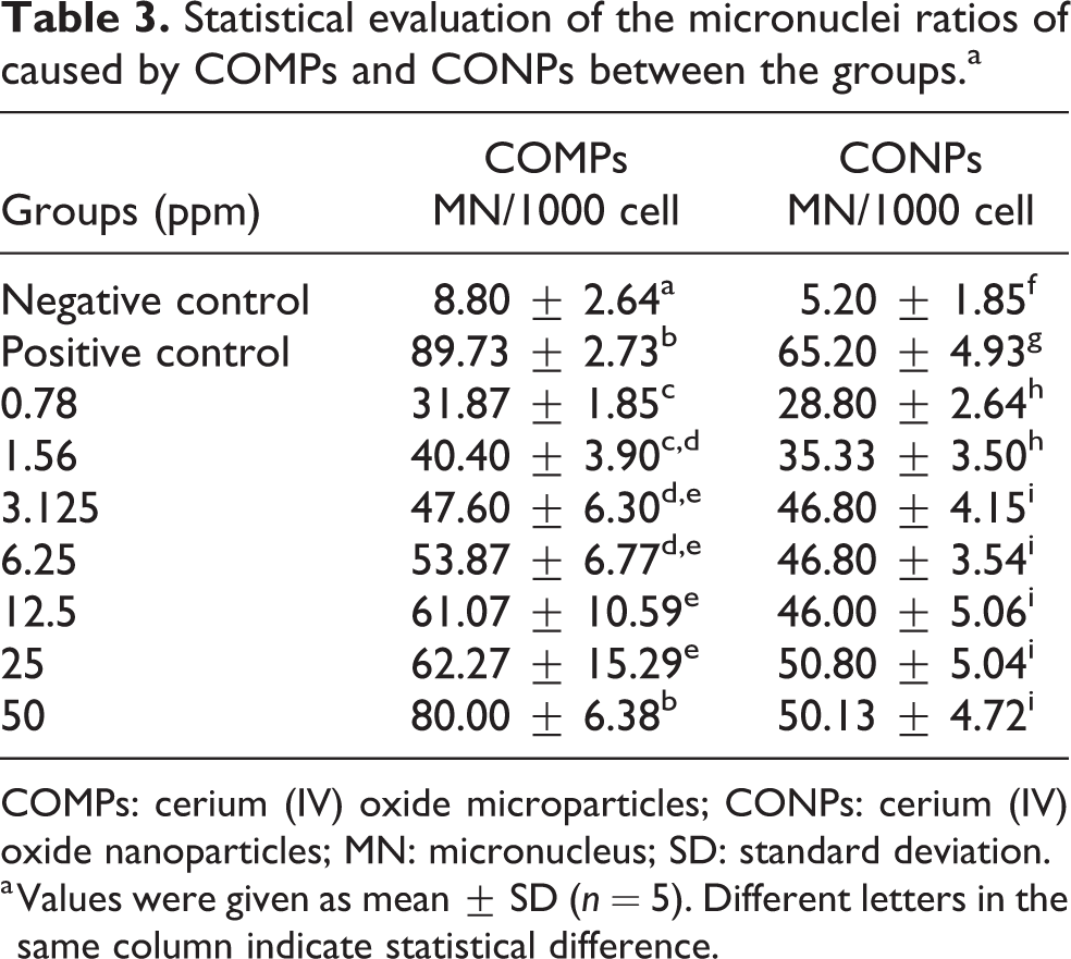
COMPs: cerium (IV) oxide microparticles; CONPs: cerium (IV) oxide nanoparticles; MN: micronucleus; SD: standard deviation.
a Values were given as mean ± SD (n = 5). Different letters in the same column indicate statistical difference.
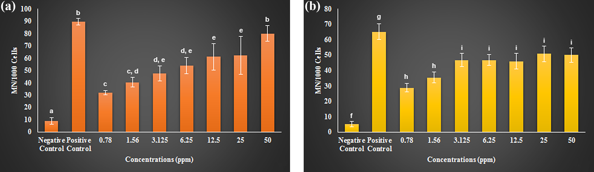
MN test results of COMPs (a) and CONPs (b) at different concentrations. COMPs: cerium (IV) oxide microparticles; CONPs: cerium (IV) oxide nanoparticles; MN: micronucleus. Different letters on the columns were significantly different at p < 0.05.
The toxicities of both COMPs and CONPs at 0.78, 1.56, 3.125, and 6.25 ppm were found to be similar. However, the toxic effect of concentrations of 12.5, 25, and 50 ppm was found to be different from each other. In general, when the toxicity of CeO2 was evaluated, it was observed that COMPs were more toxic than CONPs (Table 4 and Figure 6).
Statistical distribution of MN ratios formed by cerium (IV) oxide compound according to particle size.a
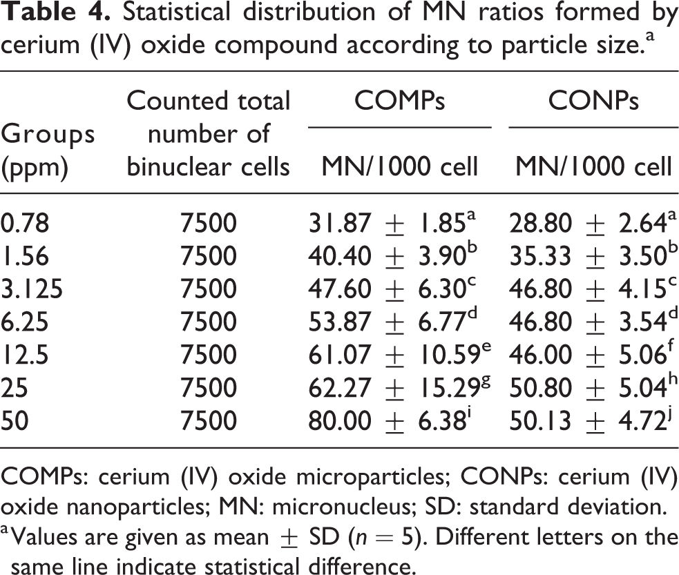
COMPs: cerium (IV) oxide microparticles; CONPs: cerium (IV) oxide nanoparticles; MN: micronucleus; SD: standard deviation.
a Values are given as mean ± SD (n = 5). Different letters on the same line indicate statistical difference.
Comparison of MN ratios caused by COMPs and CONPs at the same concentrations. COMPs: cerium (IV) oxide microparticles; CONPs: cerium (IV) oxide nanoparticles; MN: micronucleus. Different letters on the columns were significantly different at p < 0.05.
Discussion
The genotoxic effects of COMPs and CONPs, which have a wide range of use, especially in biomedical applications, were investigated using CA and MN methods. In the in vitro experimental studies, human peripheral blood lymphocyte cultures were used. The statistical results showed that CeO2 was genotoxic even at the lowest concentration (0.78 ppm) in both micro and nano sizes.
Although CONPs were reported to be toxic in previous studies, it has also been reported in several studies that it is not toxic and may even exhibit antioxidant and antigenotoxic properties at low concentrations. For example, CONPs (<25 nm) were reported to have antigenotoxic effects on the BEAS-2B lung cell line at the concentration range of 2.5–7.5 µg/mL in vitro conditions and have the antioxidant effect by eliminating oxidative stress. The antigenotoxic and antioxidant properties of CONPs had been reported at a concentration range of 2.5, 5, and 7.5 µg/mL (Rubio et al., 2016). But it should be taken into account that the toxicity of the nanoparticle may change related to the exposed dose. In another study, the cytotoxicity of CONPs was investigated on L929 cells by 3-(4,5-dimethylthiazol-2-yl)-2,5-diphenyltetrazolium bromide (MTT) test and no toxicity was found at a concentration of 800 µg/mL (Kargar et al., 2015).
Unlike these data, there were many studies reporting that CONPs were cyto-genotoxic depending on concentrations, cell lines, toxicity tests used, experimental conditions (in vitro–in vivo), and varying exposure times. De Marzi et al. (2013) investigated the cytotoxic and genotoxic properties of CeO2 at a size of 40 nm at the concentration range of 0.5–5000 µg/mL. The cytotoxicity and genotoxicity of the nanoparticle were evaluated on A549, CaCo2, and HepG2 cell lines by the MTT and Comet methods, respectively. While no toxicity was observed in the cell lines exposed to the nanoparticle for a period of 24 h, it was determined that the nanoparticle was both cytotoxic and genotoxic as a result of the 10-day exposure (De Marzi et al., 2013). Similarly, the genotoxic effect of CONPs (<25 nm) was investigated on human lymphocyte cultures at concentrations range of 6, 12, and 18 µg/mL. It was reported that DNA damage occurred in the H2AX and Comet tests at 3 h and in the MN method at 24 h. There was no significant difference between concentrations when dose-related genotoxicity was evaluated, and the genotoxicity increased significantly at 72 h of exposure in all used tests (Könen-Adıgüzel and Ergene, 2018).
In another study, the toxic effects of long-term and short-term exposure to CONPs used as additives in diesel fuels were assessed. After combustion in the diesel engine, the size of CeO2 nanoparticles increases to over 20 nm. For this reason, CeO2 nanoparticles with a particle size smaller (7 nm) and larger (25 nm) were used to compare. As a result of biochemical and histopathological evaluations, when CONPs were transnasally instilled into mice, it was found to cause pulmonary toxicity and oxidative stress in both dimensions. It has also been stated that these nanoparticles can cross the blood-brain barrier and reach the organs and central nervous system. It was reported that CONPs caused lipid peroxidation in the liver, spleen, kidney, and brain as a result of systemic accumulation. In addition, while CONPs at a size of 7 nm also caused more severe pulmonary damage, its effects on systemic toxicity were similar to those of CONPs at a size of 25 nm (Wu et al., 2019). Even the use of CeO2 nanoparticles as a diesel fuel additive is not recommended, and its use as biomedical material should be discussed. When the oxidation state of cerium changes in biological systems, cerium causes an increase in the release of ROS. In human skin fibroblast cells, DNA lesions and chromosomal damage take place due to oxidative stress (Auffan et al., 2009). The toxicity of CONPs at 5–70 nm sizes was evaluated using the RAW264.7 cell line. Effects of particle size, morphological structure, and concentration (15, 30, 60, and 120 mg/mL) of CeO2 on the production of lactate dehydrogenase (LDH), ROS, and tumor necrosis factor alpha (TNF-α) were investigated. Forest et al reported that the crystal structure of the nanoparticle was an important parameter for toxicity. While CeO2 with cubic and octahedral crystal structures had been reported to cause no toxicity, nanoparticles with rod-shaped crystal structures increased LDH release and TNF-α production depending on dose (Forest et al., 2017). In another study, it was determined that CONPs were genotoxic, whereas COMPs did not cause toxic effects on Wistar rats by using the comet, MN, and CA assays. In this study, Kumari et al. (2014) reported that CONPs showed genotoxic and histopathological effects by lowering glutathione levels in different organs (liver, kidney, and brain). In another study where cyto-genotoxicity of COMPs and CONPs was evaluated also found similar results to our results. According to the results of this research, CeO2 was a cyto-genotoxic compound by using Comet and Allium tests at a concentration range of 12.5–100 ppm (Liman et al., 2019). According to our study results, COMPs were found to be toxic even at the lowest concentration (0.78 ppm) on human lymphocyte cells. In contrast, it has been reported that COMPs have no toxic effects on primary human skin fibroblasts in the concentration range of approximately 10–1000 ppm (Benameur et al., 2015). We think that this difference may be due to the cell lines and particle size used. The fact that CONPs have a clastogenic effect on primary human skin fibroblasts cells supports our findings. In addition, CONPs caused increased lipid peroxidation and decreased glutathione levels, thereby inducing the genetic damage (Benameur et al., 2015).
In recent years, the need for material science has increased due to technological developments. In this context, it is important to investigate the properties of both synthesized new materials and existing materials. Nanoparticles are widely used in biotechnology and biomedical applications. While the release of CeO2 from automobile exhausts creates a risk, there is an uncertainty about its use as a biomaterial. Studies investigating the genotoxic effect of this compound have proposed that new researchers need to study more parameters using different methods, different cell lines, and a broad range of particle sizes. To contribute to the literature, the genotoxic effects of COMPs and CONPs were determined on human blood lymphocytes at concentrations of 0.78, 1.56, 3.125, 6.25, 12.5, 25, and 50 ppm using the CA method for the first time. When the obtained results were assessed both COMPs and CONPs were found to be genotoxic. Considering the genotoxic effects of COMPs and CONPs on human lymphocyte cells, it is recommended that their use in biomedical and medical applications as well as cosmetic, glass, and automotive industries should be limited.
Footnotes
Acknowledgements
This study was produced from the master thesis of the first author. The authors thank Dr Füreya Elif Öztürkkan for her support.
Declaration of conflicting interests
The author(s) declared no potential conflicts of interest with respect to the research, authorship, and/or publication of this article.
Funding
The author(s) received no financial support for the research, authorship, and/or publication of this article.