
Abstract
This study explored biochemical parameters of blood in workers operating in the main workshops at the Ust-Kamenogorsk Titanium and Magnesium Plant: magnesium (Mg) workshop (furnace operator, chlorinator operator, and electrolyzer operator), titanium tetrachloride workshop (mill operator and titanium (Ti) production operator), and Ti sponge workshop (reduction furnace operator, knockout operator, and crushing machine operator). The control group consisted of 112 male workers, whose duties were not related to similar occupational hazards (plumbers, electricians, janitors cleaning the administrative building, security guards, and carpenters). The activity of gamma-glutamyl transferase, aspartate aminotransferase, alanine aminotransferase, creatine phosphokinase, lactate dehydrogenase, cholinesterase, alkaline phosphate, and α-amylase and the concentration of serum calcium, Mg, phosphorus, and chloride ion were measured. To clarify the nature of pathological changes that occur in the body of mammals under the influence of toxic gases and dust, this study also included 130 sexually mature, white female rats. Animals and workers were exposed to examination of the same indicators. Changes in test results of enzymes and minerals indicated a negative impact that harmful production factors may have had on the bodies of workers. Findings showed significant fluctuations in enzyme and mineral blood profiles of workers as compared to controls. In the test animals, changes in the enzyme activity and mineral blood composition were as diverse as in the workers. These findings will be useful when identifying markers of a negative impact of harmful substances in an industrial workplace and when developing measures to prevent employees from developing an occupational disease.
Keywords
Introduction
The Ust-Kamenogorsk Titanium and Magnesium Plant (Republic of Kazakhstan) is one of the largest enterprises in the metallurgy industry and a world leader in titanium (Ti) sponge production. Ti and Mg facilities expose workers to a variety of health hazards.
The common complex of adverse production factors influencing workers engaged in Ti and Mg production is conditionally broken into three groups: dust, chemicals, and physical influences such as temperature, electromagnetic fields, noise, and vibration. These factors produce a cumulative negative effect on the workers’ health that may be considered extreme (Sultanbekov and Karabalii, 2006).
The majority of operational areas have, to different extents, chemicals and aerosols in the workplace air, and these substances are complex dust and gas mixtures, which have fibrogenic, irritating, general toxic, carcinogenic, allergenic, and other adverse effects on the human body (Vlasova et al., 2015). Dust is emitted during all, except for hydro-chemical, metallurgical processes, especially in rooms where the finely dispersed source material is produced, poured, loaded, transported, and unloaded. Dust settles on the floor, walls, and equipment (Roslaya et al., 2001; Zaytseva et al., 2013).
At Ti and Mg plants, workers operating at different process stages are exposed to dust from processing Ti, titanium dioxide (TiО2), Mg, as well as highly toxic chemical compounds. Among the latter, gaseous titanium tetrachloride (TiC14), products of its hydrolysis, chlorine vapor (Cl2), hydrogen chloride (HCl), phosgene (COCl2), and sulfur dioxide (SO2).
The effect of dust from melting Ti on the human body is aggravated by the presence of manganese, chromium, vanadium, and chlorine in the Ti (Ivanov, 2002; Sanyer et al., 2006). Studies confirm a greater toxicity from complex dusts compared to pure Ti dust or TiО2 dust, which is related to chlorine ions adsorbed on the particles of dust. Because of this, highly soluble HCl can penetrate into lungs deeper than it usually does (Chebotarev and Prokhorov, 2012; Ivanov, 2002; Prokopenko et al., 2012; Sanyer et al., 2006).
The workers may also be exposed to electromagnetic fields, noise, and vibration, which can negatively affect their health causing both occupational and nonspecific diseases to develop (Kurilov et al., 2003).
The adverse effect of noise and vibration combined is aggravated by the thermal influence (Afanasyev, 2005; Vasilyev, 2012). High air temperature and intense thermal radiation cause significant stress to the thermal control system, the central nervous system, and the neuromuscular tissues. A hot microclimate exacerbates the negative effects of not only industrial noise but also gases and toxic substances in the air, especially when performing intense muscular exertion (Afanasyev, 2005; Vasilyev, 2012).
Despite significant improvements in technology, mechanization, and automation that have been introduced in recent years, completely preventing direct contact between workers and toxic substances in vapor, gas, and dust, as well as other negative work-related hazards, is not achievable (Erubaev et al., 2004; Paltsev et al., 2003; Pavlov et al., 2002; Sidorov and Vishnevskaya, 1996).
Thus, the existing working conditions in Ti–Mg production have a negative impact on the health of workers. The literature provides information about the increased frequency of injury to the respiratory system of workers in Ti–Mg production (Alekseev et al., 2016; Chuchalin and Bobkov, 2008). Exposed workers commonly experience sub-atrophy and atrophy of the nasal mucosa, pharynx and larynx, as well as catarrhal and hypertrophic abnormalities. Hypertrophic changes and atrophy are accompanied by impaired functions of the nose, olfactory sensitivity, and by the impaired motor function of the ciliated columnar epithelium of the upper airway (Beloskurskaya, 2001; Budkar et al., 2010).
Some studies report on immune system abnormalities in furnace operators, such as changes in the immunoglobulin (A, M, and G) levels and with IgA overproduction against IgM and IgG deficiency. Inflammatory responses are a typical findings in workers exposed to chemicals (Vlasova et al., 2015). Workers in Ti production are more likely to have nonspecific diseases, such as influenza, acute upper respiratory tract catarrh, tonsillitis, stomach and intestinal diseases, neuralgia, radiculitis, arthritis, furuncles, eczema, dermatitis, and myositis (Beloskurskaya, 2001).
Workers, who come into contact with toxic gases and dust during Mg production, often report having pain in the chest, sleep disorders, and dyspeptic disorders. Apparently, this indicates a general resorptive effect of Mg, which is also evidenced by an increase in the serum level of Mg (Beloskurskaya, 2001).
There are a number of studies describing the sanitary and hygienic working conditions of and providing data on the health status of workers operating in the main workshops of the Pipe Metallurgical Company (TMK) (Amanbekov et al., 2000; Atchabarov, 2008; Tarasova et al., 2001), but these studies are limited in scope.
The issues surrounding the influence of harmful production factors on the internal environment of workers are relevant and not yet studied. Studying them is necessary to develop measures that would enable the improvement of working conditions and the health status of workers exposed to occupational hazards. The findings will also allow the early diagnosis of biochemical changes to prevent nosological diseases from developing. The literature does not describe the correlation between the main metabolic processes in the body of workers placed in a poor working environment and the disruption of homeostasis (i.e. the potential cause of occupational somatic diseases). This necessitates the most promising and objective methods of forecasting and early diagnosis, and the blood enzyme markers seem to meet these objectives, as elevated specific enzymes in blood tests indicate changes in the functional state and damage to different organs. The mineral metabolism is no less important because specific mineral substances are essential for many cells and tissues, are integral in metabolic processes and enzyme activity, and affect the regulatory systems of the body.
Materials and methods
Location of study
The study was conducted in three main workshops at the Ust-Kamenogorsk Titanium and Magnesium Plant (hereinafter referred to as the Ust-Kamenogorsk Plant): (1) Mg workshop, (2) TiC14 workshop, and (3) Ti sponge workshop.
Brief description of sanitary and hygienic working conditions across workshops
Findings of the air quality assessment that was conducted by an in-house laboratory showed the presence of toxic chemicals in the workplace air, such as SO2, HCl, chlorine, magnesium chloride (MgCl2), TiC14, as well as Mg aerosols, Ti slag dust, TiО2 dust, and the Ti sponge particles (Table 1). The concentration of these substances often significantly exceeded the permissible level.
The main workplace air pollutants in the Ust-Kamenogorsk Plant.
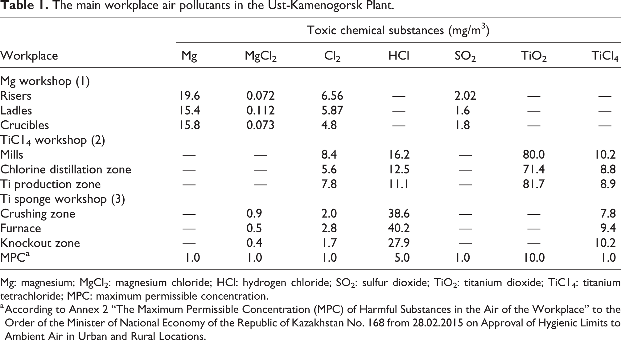
Mg: magnesium; MgCl2: magnesium chloride; HCl: hydrogen chloride; SO2: sulfur dioxide; TiO2: titanium dioxide; TiC14: titanium tetrachloride; MPC: maximum permissible concentration.
a According to Annex 2 “The Maximum Permissible Concentration (MPC) of Harmful Substances in the Air of the Workplace” to the Order of the Minister of National Economy of the Republic of Kazakhstan No. 168 from 28.02.2015 on Approval of Hygienic Limits to Ambient Air in Urban and Rural Locations.
Workshop no. 1(Mg workshop) exposures
A significant concentration of Mg particles in the air of the work place was found. Mg particles in the workshop were highly dispersed (2 µm and smaller) and accounted for 27–60% of dust in the workplace, with the highest degree of dispersion in areas where carnallite was unloaded. During carnallite dehydration, workers were exposed to heat, low humidity, and chlorine gas (Cl2). In addition, the level of carbon dioxide (CO2) in the workplace was increased.
Workers standing in close proximity to the electrolyzers were exposed to extreme heat and the flowing particles of Mg, magnesium oxide (MgO), and MgCl2. Filling electrolyzers with MgCl2 and melted carnallite was physically demanding. In addition, manual sampling of the slime was a time-consuming operation, not only associated with significant physical stress but also performed under exposure to thermal radiation, sometimes to elevated chlorine, and high air temperature during the summer period. Not to mention low humidity and low oxygen level in the workplace.
In the refining facility, the metal surface was dusted with sulfur powder. Metal casting was fraught with infrared radiation, with a risk of burns, and with a release of Mg and MgO particles, sulfuric acid vapor, and SO2 into the workplace air.
Workshop no. 2 (TiC14workshop) exposures
During the extraction of Ti slag by reduction of ore-thermal smelting of ilmenite concentrates (FeTiO3), the electric furnace of ilmenite was linked to the generation of extreme heat and low humidity. Mill operators were exposed to a high level of noise and vibration as well as to high concentrations of TiO2 in the workplace.
Industrial TiC14 was produced by chlorinating Ti-containing a charge in molten salt using a salt chlorinator and by further condensation of the vapor–gas mixture that escapes from the condensation system. TiC14 was further purified from vanadium as well as from low- and high-boiling admixtures. Vanadium was removed with lower titanium chlorides in a first distillation column cube. Workers in these production zones were exposed to high concentrations of HCl.
Ti slag dust in the workshop was highly dispersed and its particles had a polygonal irregular shape. According to the in-house laboratory, the content of Ti slag dust was as follows: 84.7% TiO2, 6.38% iron (Fe), 5.5% silicon dioxide (SiO2), 2.8% aluminum oxide, 0.99% chromium oxide, under 0.2% calcium oxide, 0.54% MgO, 1.0% manganese oxide, 0.20% vanadium pentoxide, and 0.20% zirconium dioxide. The remaining compounds were present in micro concentrations. In addition to TiC14, chlorination units produced aluminum, vanadium, silicon, Fe, zirconium, and carbon chlorides.
Workshop no. 3 (Ti sponge workshop) exposures
Mg reduction of TiC14 was carried out in the reduction furnace. This zone was characterized by extremely high temperatures. Ti sponge aerosol contained Ti metal and the same admixtures as the Ti slag dust but in lower quantities. The degree of dust dispersion was very high—85% of particles were 2 µm in size or smaller and were shaped as irregular polygons.
In Ti sponge production, the most significant workplace hazards occurred in the: store room (e.g. TiО2 dust containing 95.7% of TiO2, traces of manganese, chromium, vanadium, etc. and under 3.7% of SiO2); chlorination zone (e.g. max concentrations of chlorine and HCl, which are produced through TiCl4 hydrolysis); refining zone (e.g. HCl, TiCl4, Cl2, and COCl2, which is the most specific product); rectification zone (e.g. TiC14, HCl, and chlorine, which are the most frequent hazards, can be found in significant quantities); Ti recovery zone. The average levels of chlorine, HCl, TiC14, and MgCl2 in the air usually fell within the permissible range but during different seasons of the year, they could significantly exceed the maximum permissible concentration (MPC) (Minister of National Economy of the Republic of Kazakhstan, 2015); Ti sponge knockout and processing zone, where the amount of Ti dust significantly exceeded the MPC (Minister of National Economy of the Republic of Kazakhstan, 2015). Workers, who knocked Ti sponge out, have been spending more than half of their time performing hard physical labor while being exposed to intense noise and vibration.
Study design and sample size
The field study involved people working in the main workshops of the Ust-Kamenogorsk Plant, those listed earlier. These jobs had a high and a moderate risk of occupational diseases according to Sultanbekov and Karabalii (2006): Workshop no. 1—furnace operator, chlorinator operator, and electrolyzer operator; workshop no. 2—mill operator and Ti production operator; workshop no. 3—reduction furnace operator, knockout operator, and crushing machine operator. The sample included 196 male workers (females did not work in these workshops) aged 25–45 years (mean age, 36.25 ± 7.37), with work experience not less than 4 years (mean work experience, 7.42 ± 3.19 years), recruited at the time of annual health screening. Demographic data (living conditions), medical history data (past illnesses, previous surgery/operations, injuries, and allergies), habits and addictions (smoking and alcohol consumption), and information about working conditions (the use of personal protective equipment (PPE) at work) were collected for each worker. Health screening involved general examination by health-care specialists and common laboratory tests (complete blood count and urine analysis). Smokers were considered individuals who smoked at least one cigarette per day.
The control group consisted of 112 male workers, whose duties were not related to similar occupational hazards (plumbers, electricians, janitors cleaning the administrative building, security guards, and carpenters). The mean age and work experience in the control group were comparable with those in the study group (37.45 ± 6.37 and 7.12 ± 5.56, respectively).
Exclusion criteria: chronic diseases of the respiratory system, cardiovascular system, liver, kidneys, and neurological diseases.
Laboratory analysis: fasting samples of blood were taken from the vein in the forearm in the morning. The common tests described below measured the activity of gamma-glutamyl transferase (GGT), aspartate aminotransferase (AST), alanine aminotransferase (ALT), creatine phosphokinase (CPK), lactate dehydrogenase (LDH), cholinesterase (CHE), alkaline phosphate (ALP), α-amylase, as well as the concentration of serum calcium (Ca), Mg, phosphorus (P), and chloride ion (Cl).
Methods applied to laboratory animals
To clarify the nature of pathological changes that occur in the body of animals under the influence of toxic gases and dust (TiО2 aerosol, Ti metal dust, TiC14 and related products of hydrolysis, as well as chlorine and COCl2), this study also included 130 sexually mature, 10- to 12-month-old, white female Wistar rats, obtained from the vivarium of the Almazov National Medical Research Centre. This approach enabled the creation of the most efficient experimental model to draw parallels between clinical and experimental findings and thus obtain complete information. Therefore, animals were placed within the territory of workshops no. 1 (electrolysis zone), no. 2 (milling zone), and no. 3 (chlorination zone) in special cages (25–26 rats), at the breathing level. The animals were not removed from these zones during the study period and were exposed to harmful factors constantly considering the continuity of production. The control group of animals included 28 units kept at a considerable distance from the main workshops, in a separate, clean, well-ventilated room. The experimental and control animals were killed at the same time (2, 4, and 12 weeks). The animals within groups weighted 200–220 g before the experiment. The temperature and humidity levels were monitored at all locations where cages have been placed. The controls were kept in a room with 50–60% relative humidity at the temperature 20–22°C. Animals in the workshops were exposed to elevated temperatures, 25–30°C, and to low humidity levels, 35–45%. In the workshop 1, the temperature could reach the short-term point of 32°C and the overall exposure time at 30°C was higher as compared to workshops 2 and 3. All animals were provided with fresh water daily. Among controls, water consumption was 20–25 mg per day per animal, and in the experimental group, it was 35–40 mg per day. Rats received 30 g of pellet food per day per animal alongside fresh fruits (apples) and vegetables (carrots). The total food consumed cannot be estimated since animals were hiding certain portions in the litter. At the time of the experiment, negative weight changes were not detected in animals from workshops 2 and 3. In workshop 1, animal weight dropped 5% after 12 weeks by contrast, most likely due to a greater loss of water caused by rougher exposure conditions. Animals were monitored for clinical signs at 3-day intervals; no abnormalities were detected. Changes in the enzyme profile and mineral blood levels were examined upon the same variables as humans after 2, 4, and 12 weeks of the experiment. The study was conducted in accordance with the ethical principles approved by the Human Experiments Ethics Committee of Sarsen amanzholov east Kazakhstan university (Protocol No 12/07/2018). The patient signed a voluntary informed agreement to participate in the study. The study was conducted in accordance with the ethical principles approved by the Animal Experiments Ethics Committee of Sarsen amanzholov east Kazakhstan university (Protocol No 5 of 14th November 2018).
Methods for profiling enzyme activities and measuring mineral blood levels
The plasma serum levels of enzymes and minerals were measured by the International Federation of Clinical Chemistry and Laboratory Medicine methods.
The GGT activity was determined from the rate of 3-carboxy-4-nitroaniline formation (Schumann et al., 2002, 2010). AST level was determined from the rate of decrease of NADH measured at 340 nm (in the reaction involving malate dehydrogenase). ALT levels were determined from the NADH reduction rate measured at 340 nm (in the reaction involving LDH) (Beleta and Gella, 1990; Bergmeyer et al., 1985). The CPK activity was determined from the rate of NADPH production, measured at 340 nm, in a series of reactions involving hexokinase and glucose-6-phosphate dehydrogenase (Schumann et al., 2002, 2010). The LDH activity was determined from the NADH reduction rate measured at 340 nm (Beleta and Gella, 1990; Bergmeyer et al., 1985; Commitee, 1982). The CHE activity was determined from the reduction rate of hexacyanoferrate(III), measured at 405 nm (DGKC, 1992). The ALP activity was determined from the rate of 4-nitrophenol formation, measured at 405 nm (Schumann et al., 2011).
The activity of α-amylase was determined from the rate of 4-nitrophenol formation, measured at 405 nm (Schumann et al., 2006, 2010).
Ca was determined using the Arsenazo III reagent. Ca reacts with Arsenazo III to form a colored complex, which can be measured spectrophotometrically (Burtis and Bruns, 2015; Michaylova and Illkova, 1971). Mg was determined by the xylidine blue method. The Mg reacts with xylidine blue in an alkaline medium, liberating a colored complex, which can be determined by the spectrophotometric analysis (Barbour and Davisdon, 1988). Fe was determined using Chromazurol. Fe ions react with chromazurol B and cetyltrimethylammonium bromide to form a colored complex that can be measured spectrophotometrically (Paris et al., 1986). P was determined using the molybdate method. Inorganic P reacts with ammonium molybdate in an acidic medium to form a phosphomolybdate complex, which can be measured spectrophotometrically (Muñoz et al., 1983). Cl activity was measured with a potentiometric cell comprising a reference electrode and an ion-selective chloride electrode, which changes its electrical potential difference in response to a change in the chemical potential of Cl in solution (Burtis and Bruns, 2015).
The testing was conducted using a COBAS-Integra 400 plus analyzer (Switzerland) and an SF-46 spectrophotometer (Russia) with the corresponding reagent kits (Erba Lachema, Czech Republic).
Data processing
The findings were processed by methods of computing means, standard and mean deviations, linear regression and correlation, and by a delta method, using the STATISTICA 6.0 software.
Results and discussion
The study involved workers with diverse alcohol consumption habits. All workers wore the required-use PPE (special garments and masks) at work. Table 2 lists more details on the sample.
Occupation, risk category, and demographic characteristics of study participants.
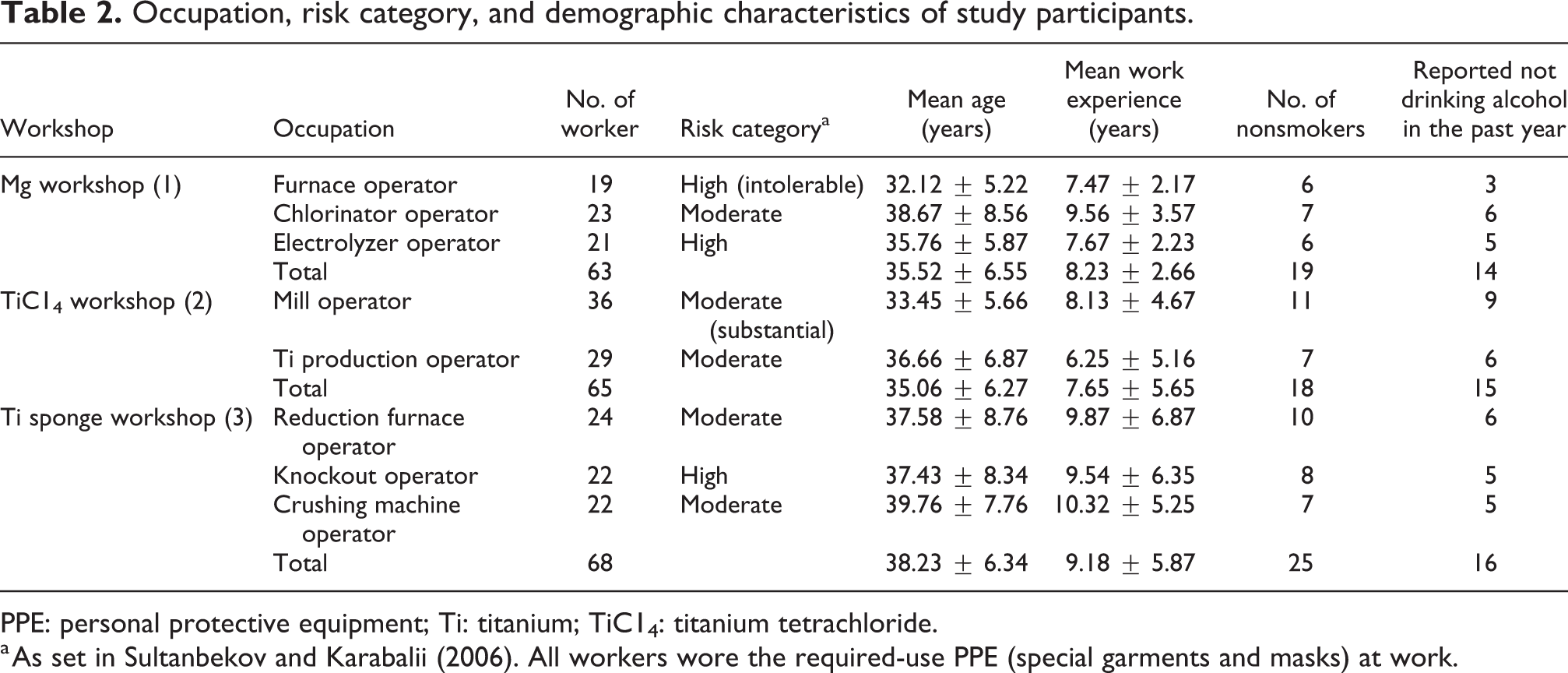
PPE: personal protective equipment; Ti: titanium; TiC14: titanium tetrachloride.
a As set in Sultanbekov and Karabalii (2006). All workers wore the required-use PPE (special garments and masks) at work.
The blood enzymes profiles of workers across workshops
According to the blood enzymes profiles of workers operating in different workshops, the activity of different enzymes tended to change and these changes varied depending on the workshop (Table 3).
The blood enzymes profiles of workers of the Ust-Kamenogorsk Plant by workshops.a
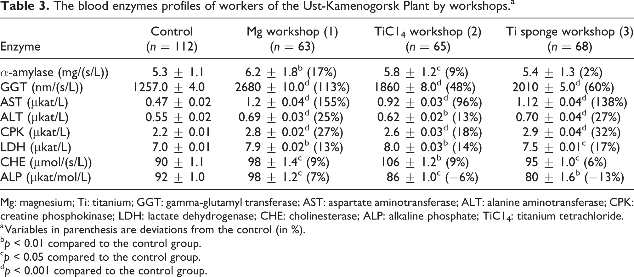
Mg: magnesium; Ti: titanium; GGT: gamma-glutamyl transferase; AST: aspartate aminotransferase; ALT: alanine aminotransferase; CPK: creatine phosphokinase; LDH: lactate dehydrogenase; CHE: cholinesterase; ALP: alkaline phosphate; TiC14: titanium tetrachloride.
a Variables in parenthesis are deviations from the control (in %).
b p < 0.01 compared to the control group.
c p < 0.05 compared to the control group.
d p < 0.001 compared to the control group.
The findings revealed significant fluctuations in enzyme blood profiles of workers as compared to controls. The activity of most enzymes, barring ALP, increased to varying degrees. The ALP activity in workshops 2 and 3 was slightly decreased. In workshop 1, it was slightly increased. The significant increase of AST and GGT activities was detected in all workshops. Elevation of ALT and CPK was found in all workshops. Changes in the activity of the remaining enzymes were less prominent. The α-amylase activity was elevated in workshop 1, slightly increased in workshop 2, and normal in workshop 3. The LDH activity was most elevated in workshop 3. The CHE activity was slightly increased in all workshops. Thus, the degree of change in enzyme activity varied across workshops, exhibiting the greatest changes in workshop 1, probably because of a greater negative effect of hazards.
Normally, aminotransferase can be found in various tissues of the body. The maximum concentration of AST is found in the liver and heart muscle, while the maximum ALT concentration is found in the liver and kidneys. Determining the activity of AST and ALT is of particular importance in the diagnosis of liver diseases, as serum concentrations tend to rise in hepatitis and other liver diseases associated with necrosis. Serum AST is also elevated after myocardial infarction, in skeletal muscle diseases, and in acute pancreatitis or hemolytic disease, while the ALT levels increase in diseases of the skeletal or cardiac muscles (Burtis and Bruns, 2015; Young, 2000; Young and Friedman, 2001).
Increased GGT activity is seen in any and all forms of liver disease, with the greatest icnreases seens in intrahepatic or posthepatic biliary obstruction. Significant higher values are also detected in metastatic neoplasm of the liver. In pancreatitis, as well as when the specific pancreatic neoplasms occur, enzymatic activity may be moderately increased.
Serum CPK concentration increases in patients with some diseases of the skeletal muscles, central nervous system, and thyroid gland (Burtis and Bruns, 2015; Young and Friedman, 2001).
The plasma LDH concentration is increased in patients with liver or kidney diseases, myocardial infarction, malignant neoplasms, progressive muscular dystrophy, and almost any case of hemolysis (Burtis and Bruns, 2015; Young and Friedman, 2001). Serum and urine α-amylase activity assays are widely used in the diagnosis of pancreatic diseases, such as acute and chronic pancreatitis (Young, 2000; Young and Friedman, 2001). CHE is synthesized in the liver and can serve as an indicator of its function. CHE activity decreases due to reduced synthesis (Burtis and Bruns, 2015; Young and Friedman, 2001). In adults, ALP present in normal serum originates primarily from liver and the bone tissue. High values are found in patients with bone diseases that are associated with increased osteoblast activity. In patients with hepatobiliary diseases, a drop in the level of ALP activity may indicate a lack of Mg and zinc in the body (Burtis and Bruns, 2015; Young and Friedman, 2001).
Mineral blood levels in workers by workshops
The analysis of mineral substances in the blood of workers in the main occupations at the Ust-Kamenogorsk Titanium–Magnesium Combine JSC, broken down by workshops, shows an uneven dynamic of changes when compared to the control group (Table 4).
Mineral blood levels in workers of the Ust-Kamenogorsk Plant by workshops.a
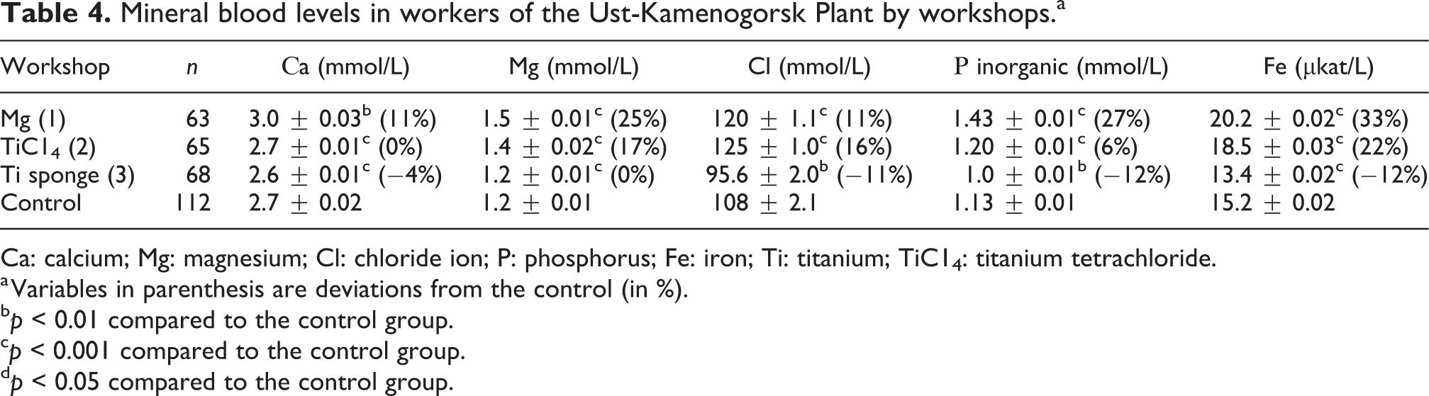
Ca: calcium; Mg: magnesium; Cl: chloride ion; P: phosphorus; Fe: iron; Ti: titanium; TiC14: titanium tetrachloride.
a Variables in parenthesis are deviations from the control (in %).
b p < 0.01 compared to the control group.
c p < 0.001 compared to the control group.
d p < 0.05 compared to the control group.
The most significant changes were discovered in the Mg production workshop in which the level of Fe, inorganic P, and Mg was substantially increased compared to the control group. In the blood of the workshop 2 workers there were increased levels of Fe, Mg, Cl, and inorganic P compared to the control group. Moderate changes in mineral substances were found in the blood of workers operating in workshop 3: the concentrations of Fe, inorganic P, and Cl decreased, compared to the control group. The blood level of Ca tended to decrease, while the level of Mg remained within the reference range.
Thus, there were diverse changes in the blood concentrations of mineral substances. While workers stayed at their workplaces within the workshops, they were exposed to diverse negative factors. Blood concentrations of minerals were influenced by factors such as temperature, humidity, concentrations of CO2 and oxygen, dust level, and the presence of toxic gases. These negative factors varied depending on the workshop, so did the intensity of exposure to them. For this reason, it was challenging to identify the causes of abnormal concentrations of minerals in the blood. Note that findings of the present study apply only to those workplaces (Table 2) that are associated with the greatest risks according to Sultanbekov and Karabalii (2006). Other workshops were not considered. One of the present findings is that the blood concentrations of Fe were higher in workers in workshops 1 and 2. As it is known, Fe is a part of hemoglobin. Thereby, the blood concentration of Fe directly depends on the hemoglobin level. A significant increase in hemoglobin causes blood viscosity to increase and thus can result in strokes, myocardial infarction, and thrombosis. High hemoglobin is detected in people exposed to low oxygen levels alongside high levels of CO2. The body will try to compensate for the shortage of oxygen by increasing hemoglobin and thus improve the transport of oxygen into the cell. This is the case in workshops addressed in this study.
Thickening of the blood occurs with a significant loss of water, and decreased plasma volume leads to the increased concentration of red blood cells. In the workshop conditions, workers are exposed to thermal radiation, which can result in dehydration, but the natural feeling of thirst is perceived differently, that is, one can feel slaked and therefore drink much less water than necessary. For this reason, science-based guidelines for protection from heat exposure were developed. In particular, it was recommended to add salt to water and to monitor the urine color of workers in order to detect dehydration (Jacklitsch et al., 2016).
The situation is aggravated for smokers, since carbon monoxide that originates from smoking strongly binds to hemoglobin. Smokers thus receive less oxygen.
In workshop 3, a moderate decrease in the blood concentration of Fe was detected but to determine the causes of such a change, a separate study is needed.
Workers who are in contact with toxic gases and dust during Mg production often have abnormally high levels of serum Mg (Beloskurskaya, 2001; Chernitskiy et al., 2019), as confirmed by the present study on workers from workshop 1, where large amounts of Mg dust particles were found floating in the air. The workshop 2 area also contained a small amount of MgO, which was found in the Ti slag dust.
The high concentrations of chlorine ion and inorganic P in the blood of workers from workshops 1 and 2 as well as an increased level of Ca in the workshop 1 workers can be linked to dehydration. It is not clear what caused these elements to decrease in workshop 3 workers and for this reason, a separate study is required.
The blood enzymes profiles of experimental animals
Table 5 provides data on the levels of enzyme activity in animals placed in different workshops (at three different intervals: 2, 4, and 12 weeks, respectively).
The blood enzymes profiles of experimental animals.a
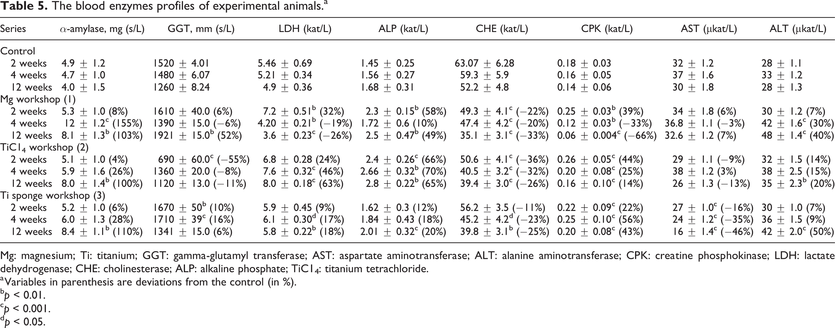
Mg: magnesium; Ti: titanium; GGT: gamma-glutamyl transferase; AST: aspartate aminotransferase; ALT: alanine aminotransferase; CPK: creatine phosphokinase; LDH: lactate dehydrogenase; CHE: cholinesterase; ALP: alkaline phosphate; TiC14: titanium tetrachloride.
a Variables in parenthesis are deviations from the control (in %).
b p < 0.01.
c p < 0.001.
d p < 0.05.
A significant increase in α-amylase activity was detected in all workshops. In workshop 1, the α-amylase activity was found to sharply increase at week 4 of the experiment and slightly fall by the end of the experiment. In workshops 2 and 3, a strong increase in α-amylase activity was detected at week 12 of the experiment. The activity of this enzyme characterizes the function of the pancreas.
The GGT activity varied between workshops. While in workshop 1, it showed a significant increase at week 12 of the experiment, in workshop 2, an upward trend followed a significant reduction at week 2. The GGT activity, however, remained slightly reduced until the end of the experiment. In workshop 3, animals had their GGT activities slightly increased throughout the experiment.
The activity of LDH also varied between workshops, moderately increasing in workshop 1 at week 2 and subsequently decreasing after 4 and 12 weeks. The activity of ALP in the blood of animals was increased in all workshops throughout the experiment. In workshop 1, it was slightly increased at week 4 of the experiment, but the smallest increase of ALP activity was found in the workshop 3.
CHE activity in the blood of animals was decreased in all workshops throughout the experiment. The activity of CPK in animals varied between workshops. In workshop 1, it increased after 2 weeks of the experiment then significantly decreased afterward, at weeks 4 and 12. In workshop 2, CPK activity showed a significant increase at week 2. Later measurements revealed a reduction of CPK activity but it remained slightly increased by the end of the experiment. In workshop 3, it was increased throughout the entire observation period, reaching the lowest point at week 2 and the highest point at week 4.
The most noticeable changes in AST activity were detected in workshop 3, where it constantly decreased during the 12-week time frame. The ALT activity showed an upward trend in all workshops during the observation period, more prominently in workshops 1 and 3.
Changes that were discovered in the enzyme spectrum were apparently a result of the workplace factors that affected enzyme activity and biosynthesis in the liver tissue.
Blood concentrations of minerals in experimental animals
The analysis of mineral substances in the blood of animals revealed the elevation of the following blood concentrations in different workshops: Ca, Cl, and inorganic P. In addition to these changes, there was a decrease in the levels of Mg and Fe in the workshop 1 (Table 6).
Blood concentrations of minerals in experimental animals.a
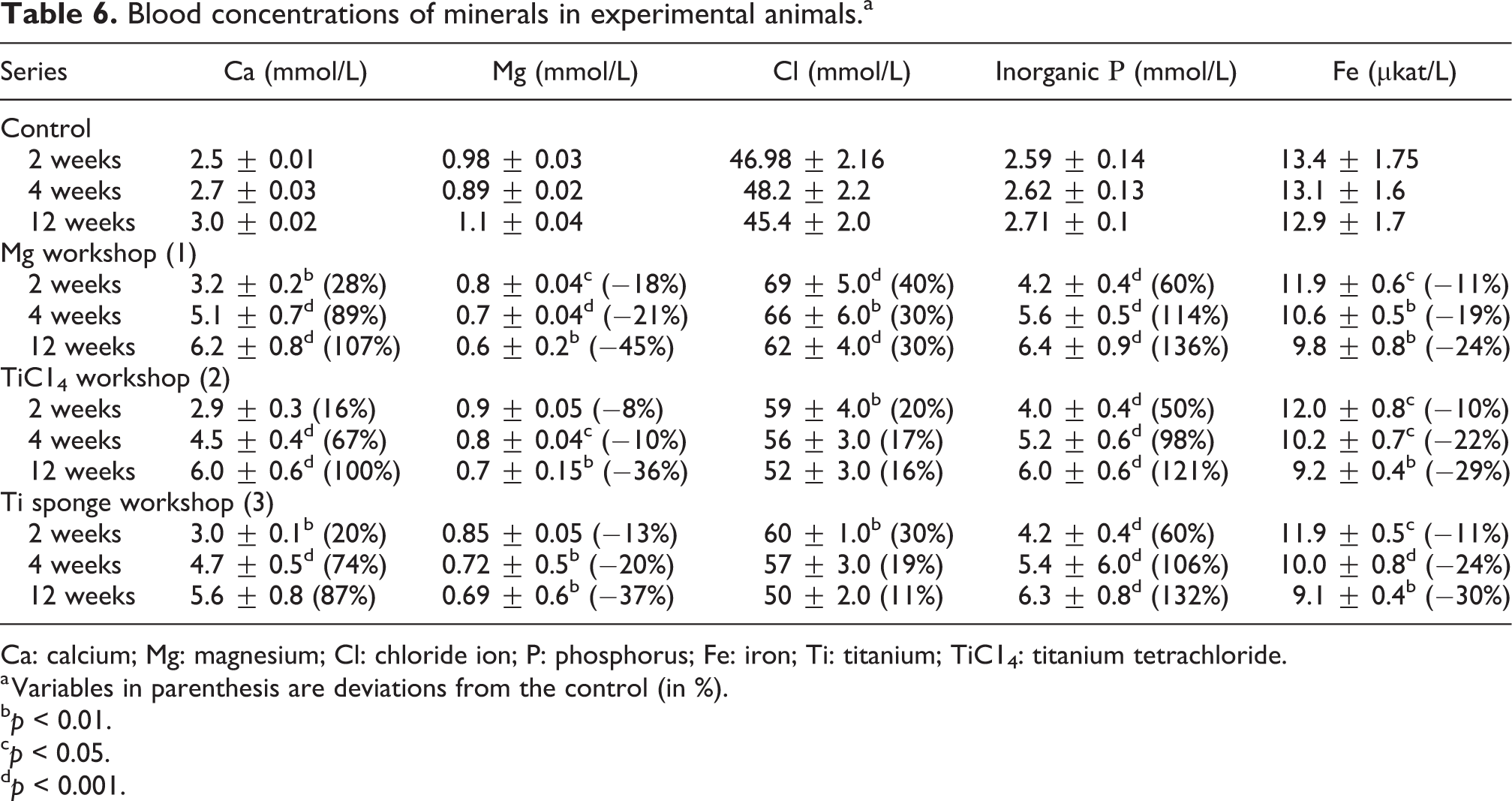
Ca: calcium; Mg: magnesium; Cl: chloride ion; P: phosphorus; Fe: iron; Ti: titanium; TiC14: titanium tetrachloride.
a Variables in parenthesis are deviations from the control (in %).
b p < 0.01.
c p < 0.05.
d p < 0.001.
The Ca concentration was increased in all workshops compared to controls, constantly rising during the observation period of 12 weeks. The serum Mg concentration of experimental animals was reduced in all workshops as compared to controls, steadily declining during the observation period. The serum level of Cl in all animals remained elevated throughout the experiment compared to controls. The serum level of inorganic P was increased in all workshops and continued to increase throughout the observation period. The blood level of Fe in experimental animals was reduced in all workshops compared with controls, constantly decreasing from measurement to measurement. Thus, a downward trend for blood concentrations of Mg and Fe was observed in all animals despite their locations. Similarly, an upward trend was equally observed for other trace elements. Throughout the experiment (after 2, 4, and 12 weeks), the changes in the mineral spectrum were most evident in workshop 1, probably due to a greater intensity of exposure to negative factors, mainly temperature and humidity. Given the peculiarities of production, slightly higher temperature and lower humidity levels were observed in workshop 1 where animal cages were placed. As it is known, the increases in Ca, Cl, and inorganic P in the blood as well as decreases in Mg are often results of dehydration.
In contrast to workers, animals demonstrated a steady reduction of Mg and Fe concentration in the blood, while workers in workshops 1 and 2 experienced an increase in the level of Mg. However, the comparative analysis of blood content in workers and animals can not be completely compared because of the different duration of exposure to harmful factors and less extreme temperature and humidity conditions to which animals were exposed. Cages were placed at a considerable distance from the workshops because higher temperatures could cause heat stroke and death of the animals.
Conclusions
The adverse work factors in Ti–Mg production (gas release, high-temperature differences, heat, electromagnetic fields, dust, etc.) negatively affect all body systems and thereby the health and performance of workers. These factors have a varying degree of impact, which depend on the job that a worker has. Recently, when assessing health risks, hygienists address the quantitative assessment methods, which enable the selection of the most effective preventive measures. The present study reported on changes in the enzymes profiles and mineral blood levels of high- and moderate-risk category workers and laboratory animals. Among workers, the most indicative changes were increases in the activity of ALT, GGT, and ALT, which may indirectly indicate an increased levels of liver stress as well as CPK activity (increased levels of stress on the muscles and nervous system). Among the laboratory animals, changes in α-amylase activity (associated with the pancreas) were the most profound. Workers in workshops 1 and 2 had increased mineral blood concentrations, while in workshop 3, these variables decreased by contrast. In laboratory animals, the concentration of Ca, Cl, and inorganic Р increased. Meanwhile, the Mg concentrations decreased due to water loss, as did the blood concentrations of Fe.
Thus, enzyme and mineral blood tests are promising tools for the early diagnosis of pathological abnormalities in the body that will lead to the development of occupational diseases. They can signal the need for a thorough medical examination. However, this approach needs additional refinement.
Footnotes
Declaration of conflicting interests
The author(s) declared no potential conflicts of interest with respect to the research, authorship, and/or publication of this article.
Funding
The author(s) received no financial support for the research, authorship, and/or publication of this article.